This website uses cookies to ensure you get the best experience on our website.


- Table of Contents
Plan paraffin-section ITPKC staining around cytoplasmic and nuclear signal (HPA tissue IHC). Use medium-staining adrenal glandular cells as a reference (HPA tissue IHC), and start the catalog antibody at 1:100–1:300 (datasheet A30549).

Expected localisation, validated protocols, controls and antibodies — the at-a-glance facts below, then the full design guide.
| Expected localisation | Cytoplasmic and nuclear tissue staining (HPA tissue IHC) | |
| Staining pattern | General cytoplasmic and nuclear staining across tissues (HPA tissue IHC) | |
| Antigen retrieval | Tris-EDTA pH 8.0 HIER, heat-mediated (datasheet A30549) | |
| Positive control | Adrenal gland+4 more · see all | |
| Negative control | None in HPA (detected in all 44 tissues); use no-primary + isotype controls |
| Fixation | Keep fixation conditions consistent across sections. (standard IHC practice; not target-specific) | |
| Caveat | High pancreatic expression, but low exocrine-cell staining (UniProt; HPA tissue IHC) | |
| Regulation | No expression regulator annotated (UniProt) | |
| Isoform / epitope | No annotated isoforms; one chain with no transmembrane segment (UniProt) |
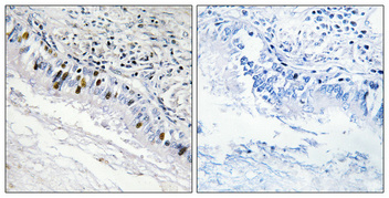
The catalog antibody IHC-P protocol (datasheet: A30549) is paired with a published cerebral cortex IHC protocol (PMC13343476).
| Sample | Paraffin-embedded Human lung cancer tissue; fixative not specified (datasheet A30549) |
| Fixation | Image fixative and duration unreported (datasheet A30549); verify before use. |
| Sectioning | 4–5 µm sections on charged slides (standard) |
| Deparaffinisation | Xylene, graded ethanol series to water (standard) |
| Antigen retrieval | Heat retrieval: Tris-EDTA pH 8.0 (datasheet A30549); 20 min, 95–100 °C (standard) |
| Peroxidase block | 3% H2O2, 10 min, room temperature (standard) |
| Blocking | 10% normal serum of the secondary host, 30 min, room temperature (standard) |
| Primary antibody | Rabbit anti-ITPKC, 1:100 - 1:300 (datasheet A30549) |
| Primary incubation | Overnight at 4 °C (standard) |
| Detection | HRP-polymer secondary, DAB chromogen 5–10 min (standard) |
| Counterstain | Hematoxylin, blue, dehydrate and mount (standard) |
| Expected result | ITPKC-positive staining in glandular cells of adrenal gland (HPA tissue IHC: Medium). HPA tissue profile: General cytoplasmic and nuclear expression. No signal in the no-primary control. |
ITPKC can localise to both nucleus and cytoplasm and actively shuttles between them; it has no transmembrane segment (UniProt Q96DU7). In paraffin-section IHC, expect cell-associated nuclear and cytoplasmic staining, including medium staining in appendix glandular cells, bone marrow hematopoietic cells, and cerebellar Purkinje cells (HPA tissue IHC). HPA calls the tissue IHC profile Approved, with medium consistency between antibody staining and RNA expression (HPA tissue IHC).
| Distinct nuclear and cytoplasmic chromogen in appendix glandular cells, with medium intensity (HPA tissue IHC). | This fits the reported cell type and intensity. Both compartments are plausible because ITPKC shuttles between nucleus and cytoplasm (UniProt Q96DU7). Judge the pattern against neighboring tissue and the negative control; brown color alone does not identify its source (general IHC practice). |
| Staining is restricted to membranes or extracellular material, with no convincing cellular nuclear or cytoplasmic signal. | This does not fit the reported general nuclear and cytoplasmic profile (HPA tissue IHC). ITPKC has no transmembrane segment or annotated signal peptide (UniProt Q96DU7). Check morphology, the negative control, and whether chromogen has deposited outside cells before assigning a compartment (general IHC practice). |
| The strongest staining is in a different cell population from the reported positive cells in the same tissue. | For example, HPA reports medium staining in bronchial respiratory epithelial cells (HPA tissue IHC). An unexpected dominant pattern warrants checking cell identification, nonspecific antibody binding, and endogenous detection activity (general IHC practice). HPA's broad tissue profile does not establish that every other cell must be negative (HPA tissue IHC). |
| Brown haze covers stroma and cells without discernible cellular boundaries or nuclear detail. | Diffuse deposition cannot establish ITPKC localisation; the reference profile is cellular, with general nuclear and cytoplasmic expression (HPA tissue IHC). Review the negative control and detection background, then assess whether blocking, washing, or chromogen development needs adjustment (general IHC practice). |
| No signal appears in an appendix section expected to contain glandular cells with medium staining (HPA tissue IHC). | First confirm that the relevant cells are present and that the positive-control run developed (general IHC practice). A blank slide may reflect an assay failure or an unsuitable section; it does not, by itself, overturn the reported HPA observation (HPA tissue IHC). |
| Compartment readout | HPA tissue IHC describes general cytoplasmic and nuclear expression, consistent with active shuttling in the UniProt record (HPA tissue IHC; UniProt Q96DU7). HPA's supported nuclear-speckle localisation comes from ICC-IF; routine chromogenic IHC need not resolve speckles (HPA subcellular ICC-IF; general IHC practice). |
| Choice of reference cells | HPA reports medium staining in appendix glandular cells, bone marrow hematopoietic cells, and cerebellar Purkinje cells, but low staining in pancreatic exocrine glandular cells and liver cholangiocytes (HPA tissue IHC). Select the reference by its annotated cell population, not solely by the organ name (HPA tissue IHC). |
| Tissue-level expression versus cellular staining | UniProt describes high expression in pancreas and liver, while HPA records low IHC staining in the specified pancreatic exocrine cells and liver cholangiocytes (UniProt Q96DU7; HPA tissue IHC). These statements address different measurements or cell populations; neither predicts strong staining in those named cells (UniProt Q96DU7; HPA tissue IHC). |
| Strength of the IHC evidence | The HPA tissue profile is Approved with medium staining–RNA consistency; both listed rabbit polyclonal antibodies have Approved IHC status (HPA tissue IHC; HPA antibodies). This supports a reference pattern but leaves an unexpected specimen-specific result to be evaluated with controls and morphology (general IHC practice). |
| Situation | Likely cause | Next action |
|---|---|---|
| Known-positive appendix glandular cells remain blank (HPA tissue IHC). | The expected cells may be absent from the section, or the IHC run may have failed (general IHC practice). | Confirm cell identity and section quality; check the concurrent positive control, then review retrieval and detection steps used for the validated IHC assay (general IHC practice). No ITPKC-specific retrieval condition is supplied. |
| Signal appears only at membranes or outside cells. | That location conflicts with the reported nuclear and cytoplasmic pattern and the absence of a transmembrane segment (HPA tissue IHC; UniProt Q96DU7). | Inspect the negative control and tissue morphology, and repeat scoring using identifiable cells and compartments (general IHC practice). Do not count isolated extracellular chromogen as a positive cell. |
| A broad brown haze obscures the counterstain. | Nonspecific binding, endogenous detection activity, or excessive chromogen development can produce background in chromogenic IHC (general IHC practice). | Compare with the negative control; review blocking, washes, detection reagents, and development time under the laboratory's IHC workflow (general IHC practice). |
| Unexpected cells stain more strongly than the HPA-reported population. | Cell misidentification, cross-reactivity, or endogenous detection activity are possible explanations (general IHC practice). | Recheck morphology and controls, then compare the exact tissue and cell population with the HPA profile. Treat an unlisted cell as unresolved rather than automatically negative (HPA tissue IHC; general IHC practice). |
| Pancreatic exocrine cells or liver cholangiocytes give only weak signal. | HPA records low IHC staining in those cells, although UniProt describes high expression in pancreas and liver (HPA tissue IHC; UniProt Q96DU7). | Score the specified cells against their HPA IHC levels; use a reported medium-staining cell population when assessing assay performance (HPA tissue IHC; general IHC practice). |
| IF/ICC: should a positive result show nuclear speckles? | HPA supports nuclear-speckle localisation in ICC-IF images, whereas tissue IHC has a broader nuclear and cytoplasmic profile (HPA subcellular ICC-IF; HPA tissue IHC). | For IF/ICC interpretation, compare nuclear signal with the supported speckle pattern (HPA subcellular ICC-IF). Assess chromogenic tissue IHC by its reported cellular pattern without requiring visible speckles (HPA tissue IHC; general IHC practice). |


Comprehensive Human Protein Atlas IHC scoring per tissue (reliability: Approved — Medium consistency between antibody staining and RNA expression data.). Rows are taken directly from the HPA tissue chart — click any row's HPA link to view the source.
| Tissue | Cell type | Level | Evidence | Source |
|---|---|---|---|---|
| Adrenal gland | Glandular cells | Medium | Protein (IHC) | HPA → |
| Appendix | Glandular cells | Medium | Protein (IHC) | HPA → |
| Bone marrow | Hematopoietic cells | Medium | Protein (IHC) | HPA → |
| Breast | Glandular cells | Medium | Protein (IHC) | HPA → |
| Bronchus | Respiratory epithelial cells | Medium | Protein (IHC) | HPA → |
| Tissue | Cell type | Level | Evidence | Source |
|---|---|---|---|---|
| None in HPA: ITPKC is detected in all 44 scored tissues. Use a no-primary (secondary-only) and an isotype control instead. | ||||
Troubleshoot ITPKC staining in paraffin sections by checking retrieval, controls, cell identity and compartment before interpreting chromogenic signal.
Two anti-ITPKC antibodies list human, mouse and rat reactivity (catalog: A07845-1, A30549). A30549 includes a human paraffin-section IHC image; IF/ICC is listed without an image (A30549 catalog and image caption).
A07845-1 lists IHC and human, mouse and rat reactivity; no IHC image is supplied (A07845-1 catalog). A30549 lists IHC, IF and ICC with human, mouse and rat reactivity, and its IHC image shows human lung cancer paraffin sections (A30549 catalog and image caption).
Which to pick: For tissue IHC, choose A30549 for its documented human paraffin-section image with Tris-EDTA, pH 8.0 retrieval; the fixative is unreported (A30549 image caption). For IF/ICC, choose A30549 because both applications are listed, although no IF image is supplied (A30549 catalog). Both SKUs list mouse and rat reactivity, but the supplied IHC image documents human tissue only; validate staining in mouse or rat sections for the intended experiment (catalog reactivity; A30549 image caption).