This website uses cookies to ensure you get the best experience on our website.


- Table of Contents
Plan JAK2 paraffin IHC around cytoplasmic staining, with strong signal in lung macrophages and gallbladder glandular cells (HPA tissue IHC). This guide covers the catalog antibody’s 1:50 dilution, consistent fixation, chromogenic detection, and scoring by cell type (datasheet: M00027; standard IHC practice).

Expected localisation, validated protocols, controls and antibodies — the at-a-glance facts below, then the full design guide.
| Expected localisation | Cytoplasmic staining in several tissues (HPA tissue IHC) | |
| Staining pattern | Cytoplasmic staining, strongest in subsets of immune cells (HPA tissue IHC) | |
| Antigen retrieval | EDTA pH 8.0 HIER, heat-mediated (datasheet M00027) | |
| Positive control | Gallbladder+4 more · see all | |
| Negative control | Caudate+4 more · see all |
| Fixation | Keep fixation consistent across sections (standard IHC practice; not target-specific) | |
| Caveat | Immune-cell-rich regions may dominate the signal (HPA tissue IHC) | |
| Regulation | Expression regulation not specified (UniProt) | |
| Isoform / epitope | 0 annotated isoforms; no processing or membrane span (UniProt) |
Compare the catalog antibody’s IHC-P protocol (datasheet M00027) with four published JAK2 IHC workflows; one measures phospho-JAK2 (PMC4084704).
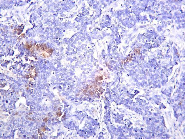
| Sample | Paraffin-embedded human prostate cancer tissue; fixative not specified (datasheet M00027) |
| Fixation | Image fixative and duration unreported (datasheet M00027); verify before use. |
| Sectioning | 4–5 µm sections on charged slides (standard) |
| Deparaffinisation | Xylene, graded ethanol series to water (standard) |
| Antigen retrieval | Heat retrieval: EDTA pH 8.0 (datasheet M00027); 20 min, 95–100 °C (standard) |
| Peroxidase block | 3% H2O2, 10 min, room temperature (standard) |
| Blocking | 10% goat serum (datasheet M00027) |
| Primary antibody | Rabbit monoclonal (clone COG-10) anti-JAK2, 1:50 (datasheet M00027) |
| Primary incubation | Overnight at 4 °C (datasheet M00027) |
| Detection | HRP-conjugated secondary, DAB chromogen (datasheet M00027) |
| Counterstain | Hematoxylin, blue, dehydrate and mount (standard) |
| Expected result | JAK2-positive staining in glandular cells of gallbladder (HPA tissue IHC: High). HPA tissue profile: Cytoplasmic expression in several tissues, most abundant in subsets of immune cells. No signal in the no-primary control. |
In paraffin-section IHC, expect predominantly cytoplasmic JAK2 staining, especially in subsets of immune cells (HPA: tissue IHC). Lung macrophages and lymph-node and tonsil non-germinal center cells show high staining (HPA: High). JAK2 has no transmembrane segment, although it participates in receptor signaling and is also annotated in the nucleus (UniProt O60674: topology, function, subcellular location). HPA rates the tissue IHC pattern Approved, with medium consistency against RNA data (HPA: reliability).
| Cytoplasmic staining is strong in lung macrophages or non-germinal center cells of lymph node or tonsil. | This matches the reported high-staining cell populations and provides a practical positive-pattern check; it does not imply that every cell in the section should stain (HPA: tissue IHC). |
| An IHC section shows predominantly nuclear staining with little cytoplasmic staining in expected positive cells. | Recheck specificity and detection before calling it the expected tissue pattern: HPA describes tissue IHC as cytoplasmic, while UniProt also lists nuclear localization (HPA: tissue IHC; UniProt O60674: subcellular location). |
| Strong staining appears mainly in cell types listed as not detected, such as skeletal-muscle myocytes. | Treat this as a specificity or detection concern, not proof that JAK2 is absent there: HPA reports a staining observation, and its tissue IHC reliability has medium RNA consistency (HPA: Not detected in myocytes; reliability). |
| Brown deposit spreads across tissue and obscures cell boundaries. | The pattern cannot be scored confidently by cell type or compartment. Check a no-primary control for detection background, then compare staining with the cytoplasmic, cell-restricted HPA pattern (general IHC practice; HPA: tissue IHC). |
| No stain is visible in an included lung macrophage-rich area. | A technical failure is possible because HPA reports high staining in lung macrophages. First establish that the expected cells are present and the detection run worked; an individual negative section is not decisive (HPA: High in lung macrophages; general IHC practice). |
| Tissue and cell selection | JAK2 is broadly expressed, but IHC intensity varies by cell population; use the named high-staining cells as pattern references and interpret listed not-detected cells at the cell level (UniProt O60674: tissue specificity; HPA: tissue IHC). |
| Membrane-associated signal | JAK2 lacks a transmembrane segment and acts with receptors through their intracellular signaling machinery. A crisp membrane-only IHC pattern should therefore be checked against the reported cytoplasmic tissue pattern (UniProt O60674: topology, function; HPA: tissue IHC). |
| Antibody and epitope scope | HPA lists two IHC antibodies as Approved, but the supplied record gives no epitope or phospho-specificity for the antibody used here. JAK2 has annotated phosphorylated residues, so staining alone cannot establish activation state (HPA: antibody validation; UniProt O60674: modified residues). |
| IF/ICC Q&A: why might nuclear staining appear? | HPA reports supported nucleoplasmic localization in ICC-IF, whereas its tissue IHC profile is cytoplasmic. Interpret each assay against its own evidence; nuclear IF signal does not make nuclear-only chromogenic IHC an established tissue pattern (HPA: ICC-IF subcellular; tissue IHC). |
| Situation | Likely cause | Next action |
|---|---|---|
| Expected high-staining cells are present, but IHC signal is absent. | The run may lack sensitivity or have a detection problem; the supplied sources give no JAK2-specific retrieval condition (HPA: High in lung macrophages and lymphoid non-germinal center cells). | Check the run controls and detection steps; optimize retrieval and antibody concentration within the validated IHC workflow, then reassess the expected cells (general IHC practice). |
| Staining is widespread and poorly confined to cells. | Detection background may obscure the reported cytoplasmic pattern; HPA tissue observations alone cannot identify the chemical source of that background (HPA: tissue IHC). | Review the no-primary control, blocking and washes, and compare the signal with the expected cellular distribution (general IHC practice; HPA: tissue IHC). |
| Macrophage-rich areas show granular brown signal even without primary antibody. | Endogenous detection activity is a possible source of chromogenic background in such areas (general IHC practice). | Use the no-primary control to distinguish detection activity from antibody-dependent stain; review the detection-system blocking step before scoring macrophages (general IHC practice). |
| Nuclear staining dominates a paraffin section. | This differs from HPA's cytoplasmic tissue IHC profile, although nuclear JAK2 is annotated and nucleoplasmic ICC-IF is supported (HPA: tissue IHC, ICC-IF; UniProt O60674: subcellular location). | Compare cytoplasmic signal in expected positive cells and review controls before accepting a nuclear IHC interpretation; keep the IF observation assay-specific (HPA: tissue IHC, ICC-IF; general IHC practice). |
| Cells listed as not detected stain strongly. | Cross-reactivity or detection background is possible; a single discordant section cannot resolve specificity (HPA: Not detected in listed cell types; reliability). | Check the no-primary control and repeat the comparison alongside an HPA high-staining cell population using the same IHC conditions (HPA: tissue IHC; general IHC practice). |
| Staining intensity varies among cells within one tissue. | This can reflect the cell-specific distribution: HPA reports high, medium, low and not-detected staining across named populations (HPA: tissue IHC). | Score the identified cell type and its cytoplasmic pattern separately from nearby cells; avoid treating whole-section intensity as a single JAK2 result (HPA: tissue IHC; general IHC practice). |




Comprehensive Human Protein Atlas IHC scoring per tissue (reliability: Approved — Medium consistency between antibody staining and RNA expression data.). Rows are taken directly from the HPA tissue chart — click any row's HPA link to view the source.
| Tissue | Cell type | Level | Evidence | Source |
|---|---|---|---|---|
| Gallbladder | Glandular cells | High | Protein (IHC) | HPA → |
| Lung | Macrophages | High | Protein (IHC) | HPA → |
| Lymph node | Non-germinal center cells | High | Protein (IHC) | HPA → |
| Tonsil | Non-germinal center cells | High | Protein (IHC) | HPA → |
| Adrenal gland | Glandular cells | Medium | Protein (IHC) | HPA → |
| Tissue | Cell type | Level | Evidence | Source |
|---|---|---|---|---|
| Caudate | Glial cells | Not detected | Protein (IHC) | HPA → |
| Cervix | Glandular cells | Not detected | Protein (IHC) | HPA → |
| Esophagus | Squamous epithelial cells | Not detected | Protein (IHC) | HPA → |
| Prostate | Glandular cells | Not detected | Protein (IHC) | HPA → |
| Skeletal muscle | Myocytes | Not detected | Protein (IHC) | HPA → |
Troubleshoot JAK2 staining in paraffin sections by checking retrieval, compartment, cell type and detection controls against the available tissue evidence.
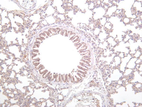
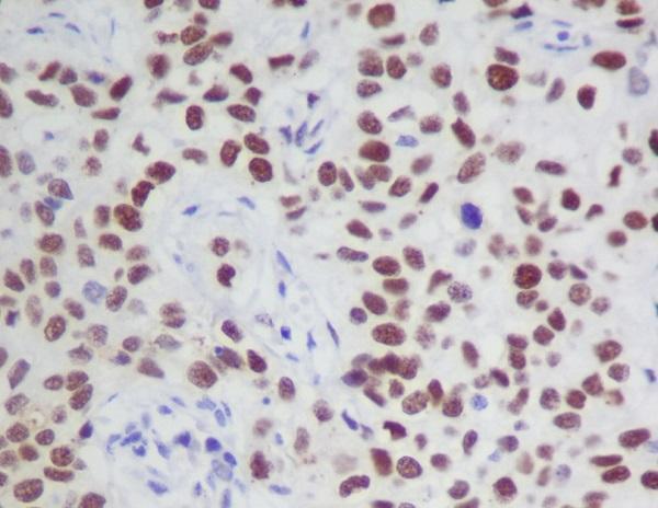
The catalog includes JAK2 antibodies with IHC images from human, mouse, and rat tissues (catalog IHC captions) and IF images from human cells (catalog IF captions).
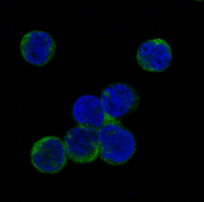
M00027 has IHC data from a paraffin-embedded human prostate cancer section and IF data from A549 cells (M00027 image captions). M00027-3 has IHC images from human lung carcinoma, mouse lung, and rat lung; MP00027 has phospho-JAK2 IHC data from paraffin-embedded human cervix cancer and IF data from treated Jurkat cells (respective image captions).
Which to pick: For tissue IHC, choose M00027 when its documented paraffin-section procedure is useful: EDTA retrieval at pH 8.0 and a 1:50 primary dilution (M00027 IHC caption); the fixative is unreported (M00027 IHC caption). For IF/ICC, M00027 has an A549 IF example and lists both IF and ICC, while MP00027 targets phospho-JAK2 Y1007/Y1008 and has a Jurkat IF example (catalog applications and IF captions). For cross-species tissue IHC, M00027-3 has human, mouse, and rat lung images, although their captions do not specify tissue processing or fixative (M00027-3 IHC captions).