This website uses cookies to ensure you get the best experience on our website.


- Table of Contents
Plan chromogenic JCHAIN IHC in paraffin sections using the catalog antibody at 2–5 μg/ml (datasheet A02644-2). Use the documented cytoplasmic and plasma staining pattern to guide control selection and scoring (HPA tissue IHC).

Expected localisation, validated protocols, controls and antibodies — the at-a-glance facts below, then the full design guide.
| Expected localisation | Cytoplasmic staining; additional plasma positivity (HPA tissue IHC) | |
| Staining pattern | Enterocyte cytoplasm; additional plasma positivity (HPA tissue IHC) | |
| Antigen retrieval | EDTA pH 8.0 HIER, heat-mediated (datasheet A02644-2) | |
| Positive control | Appendix+4 more · see all | |
| Negative control | Adipose tissue+4 more · see all |
| Fixation | Keep paraffin-section fixation consistent (standard IHC practice; not target-specific) | |
| Caveat | As a secreted protein, RNA and protein locations may differ (HPA tissue IHC) | |
| Regulation | Expression regulation not annotated (UniProt) | |
| Isoform / epitope | 0 annotated isoforms; signal peptide is cleaved (UniProt) |
The catalog antibody protocol uses EDTA retrieval (datasheet A02644-2). The published IHC options below report JCHAIN staining in tissue sections (PMC12561060; PMC11904056; PMC10552693).
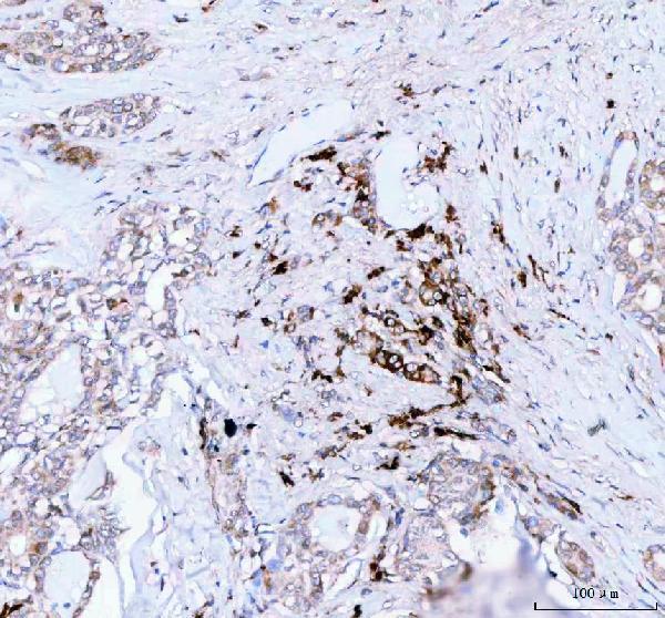
| Sample | Paraffin-embedded human thyroid cancer tissue; fixative not specified (datasheet A02644-2) |
| Fixation | Image fixative and duration unreported (datasheet A02644-2); verify before use. |
| Sectioning | 4–5 µm sections on charged slides (standard) |
| Deparaffinisation | Xylene, graded ethanol series to water (standard) |
| Antigen retrieval | Heat retrieval: EDTA pH 8.0 (datasheet A02644-2); 20 min, 95–100 °C (standard) |
| Peroxidase block | 3% H2O2, 10 min, room temperature (standard) |
| Blocking | 10% goat serum (datasheet A02644-2) |
| Primary antibody | Rabbit anti-JCHAIN, 2-5 μg/ml (datasheet A02644-2) |
| Primary incubation | Overnight at 4 °C (datasheet A02644-2) |
| Detection | HRP-conjugated secondary, DAB chromogen (datasheet A02644-2) |
| Counterstain | Hematoxylin, blue, dehydrate and mount (standard) |
| Expected result | JCHAIN-positive staining in endocrine cells of appendix (HPA tissue IHC: High). HPA tissue profile: Cytoplasmic expression in several tissues, most abundant in gastrointestinal tract and lymphoid tissues. Additional plasma positivity. No signal in the no-primary control. |
JCHAIN is secreted and has no transmembrane segment (UniProt P01591: subcellular location and topology). On paraffin sections, expect predominantly cytoplasmic staining, with additional plasma positivity (HPA: tissue IHC profile). Staining is most abundant in gastrointestinal and lymphoid tissues; reported High cell populations include intestinal enterocytes, appendix and rectal endocrine cells, breast glandular cells, and lung macrophages (HPA: tissue IHC, Enhanced reliability).
| Distinct cytoplasmic chromogen in colon or duodenal enterocytes, with a readable counterstain. | This matches an HPA High cell population and the reported cytoplasmic profile (HPA: colon and duodenum tissue IHC). Compare cells within the section: a positive call should rest on identifiable stained cells, not overall brown color. Additional plasma positivity can occur (HPA: tissue IHC profile). |
| A sharply nuclear or continuous cell-border pattern dominates the positive cells. | Treat this as a localization mismatch: HPA reports cytoplasmic expression, while UniProt describes a secreted protein without a transmembrane segment (HPA: tissue IHC profile; UniProt P01591: topology). Review morphology, counterstain, and detection controls before attributing that compartmental pattern to JCHAIN (general IHC practice). |
| Strong chromogen appears in adipocytes or adrenal glandular cells. | HPA lists both cell populations as Not detected (HPA: adipose tissue and adrenal gland tissue IHC). A discrepant result warrants a check for cross-reactivity or endogenous detection activity; it alone cannot identify the cause (general IHC practice). Assess whether staining follows cell boundaries or appears as nonspecific deposits. |
| A diffuse brown haze covers stroma, empty spaces, and many unrelated cell types. | This is difficult to reconcile with the reported cell-associated cytoplasmic pattern, although HPA also notes plasma positivity for this secreted protein (HPA: tissue IHC profile; UniProt P01591: subcellular location). Compare a no-primary control and inspect washes, blocking, and chromogen development as general IHC checks; do not score haze as positive cells. |
| No convincing signal appears in an adequately sampled colon section. | Colon enterocytes are reported High, so absent staining makes this run uninformative until technical failure and tissue identification are checked (HPA: colon tissue IHC). Check the antibody's IHC-P instructions and the run controls; HPA tissue data do not establish a JCHAIN-specific retrieval, fixation, or dilution requirement (general IHC practice). |
| Cell population and tissue selection | HPA reports High staining in colon, duodenal, and small-intestinal enterocytes, but Not detected staining in adipocytes; choose controls by the named cell population, not tissue label alone (HPA: tissue IHC). |
| Secretion and compartment | JCHAIN lacks a transmembrane segment and participates in secretory IgA and IgM complexes (UniProt P01591: topology and subunit). Cytoplasmic staining plus additional plasma positivity is reported; extracellular color needs morphological context (HPA: tissue IHC profile). |
| Protein processing | UniProt records a signal peptide at residues 1–22 and a mature chain at 23–159 (UniProt P01591: processing). The payload gives no antibody epitope, so processing cannot predict this antibody's staining intensity or a retrieval condition. |
| Evidence and validation scope | HPA rates tissue IHC reliability Enhanced and lists Enhanced IHC status for HPA044132 and CAB034436 (HPA: tissue IHC and antibodies). Its note says secreted proteins may have different RNA and protein locations; RNA enrichment is not a cell-level staining map (HPA: reliability note and RNA specificity). |
| IF/ICC evidence | HPA summarizes JCHAIN as secreted but provides no main ICC-IF location or cell-line images (HPA: subcellular record). That evidence cannot establish a specific IF compartment or serve as an IF protocol; evaluate IF/ICC on its separate guide page. |
| Situation | Likely cause | Next action |
|---|---|---|
| Positive-control colon enterocytes are unstained. | A run or specimen problem is plausible because HPA reports High staining in these cells (HPA: colon tissue IHC). The supplied sources do not assign a JCHAIN-specific failure mechanism. | Confirm enterocytes are present, review the IHC-P antibody instructions, and check detection and run controls (general IHC practice). Record any retrieval change as empirical; no target-specific fixation or retrieval sensitivity is supplied. |
| Only weak staining appears in a candidate positive section. | Expected levels differ by cell population: bone marrow hematopoietic cells are Medium, whereas listed intestinal enterocytes are High (HPA: tissue IHC). Weak color alone cannot separate biology from a technical issue. | Compare matched cell populations and an HPA-listed High control in the same run (HPA: tissue IHC). Then review antibody dilution and detection against the catalog IHC-P instructions (general IHC practice). |
| Adipocytes or adrenal glandular cells look strongly positive. | Those populations are listed Not detected; cross-reactivity or endogenous detection activity is possible, but the image alone cannot distinguish them (HPA: tissue IHC; general IHC practice). | Run a no-primary control and inspect the pattern for deposits or broad background; review endogenous activity blocking for the detection chemistry used (general IHC practice). Keep the result provisional until controls resolve the discrepancy. |
| Staining is nuclear or outlines most cell membranes. | That dominant pattern conflicts with HPA's cytoplasmic profile and the UniProt secreted, non-transmembrane annotation (HPA: tissue IHC profile; UniProt P01591: topology). It is a warning sign rather than proof of one artefact. | Check counterstain and tissue morphology, compare the positive control, and examine a no-primary control (general IHC practice). Report the unusual compartment separately instead of scoring it as the expected JCHAIN pattern. |
| Brown haze obscures otherwise identifiable cells. | Diffuse background can result from detection or wash conditions in chromogenic IHC (general IHC practice). Some plasma positivity is reported for JCHAIN, so extracellular color by itself does not prove nonspecific staining (HPA: tissue IHC profile). | Compare no-primary and positive controls; review blocking, washing, and chromogen development (general IHC practice). Score only localized staining that can be assigned confidently to the relevant cells. |
| Can the IHC pattern be used as an IF/ICC localization prediction? | HPA has no ICC-IF cell-line images and gives no main subcellular location beyond a secreted summary (HPA: subcellular record). Its tissue IHC profile describes chromogenic tissue staining, not an IF validation. | Use the separate IF/ICC guide for that application. If interpreting an IF image, require its own controls and cell-level evidence before assigning a compartment (general IF practice); do not treat this IHC pattern as an IF protocol. |


Comprehensive Human Protein Atlas IHC scoring per tissue (reliability: Enhanced — High consistency between antibody staining and RNA expression data. Secreted protein, tissue location of RNA and protein is expected to differ.). Rows are taken directly from the HPA tissue chart — click any row's HPA link to view the source.
| Tissue | Cell type | Level | Evidence | Source |
|---|---|---|---|---|
| Appendix | Endocrine cells | High | Protein (IHC) | HPA → |
| Breast | Glandular cells | High | Protein (IHC) | HPA → |
| Colon | Enterocytes | High | Protein (IHC) | HPA → |
| Duodenum | Enterocytes | High | Protein (IHC) | HPA → |
| Lung | Macrophages | High | Protein (IHC) | HPA → |
| Tissue | Cell type | Level | Evidence | Source |
|---|---|---|---|---|
| Adipose tissue | Adipocytes | Not detected | Protein (IHC) | HPA → |
| Adrenal gland | Glandular cells | Not detected | Protein (IHC) | HPA → |
| Caudate | Glial cells | Not detected | Protein (IHC) | HPA → |
| Cerebellum | Cells in granular layer | Not detected | Protein (IHC) | HPA → |
| Cerebral cortex | Endothelial cells | Not detected | Protein (IHC) | HPA → |
Troubleshoot JCHAIN chromogenic IHC by checking retrieval, cellular and extracellular staining, background, and scoring against the supplied tissue evidence.
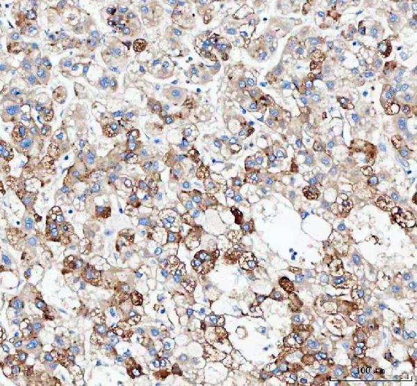
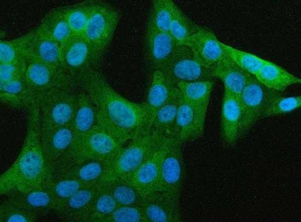
A02644-2 has IHC images from paraffin sections of human thyroid cancer, human liver cancer and rat colon, plus an IF image from U2OS cells (catalog image captions).
A02644-2 is listed for IHC and shows staining in paraffin sections of human thyroid cancer, human liver cancer and rat colon (catalog applications; IHC image captions). A02644-2 is also listed for IF/ICC and shows IF staining in U2OS cells (catalog applications; IF image caption).
Which to pick: Choose A02644-2 for paraffin-section IHC: its thyroid cancer image documents EDTA retrieval at pH 8.0 and a primary antibody concentration of 2 μg/ml; the fixative is unreported (A02644-2 IHC image caption). For IF/ICC, A02644-2 has a U2OS cell image using 5 μg/ml primary antibody (A02644-2 IF image caption). For work across species, A02644-2 lists human, mouse and rat reactivity, with IHC images for human and rat; it is rabbit hosted, and clonality is unreported (catalog reactivity, host and clone fields; IHC image captions).