This website uses cookies to ensure you get the best experience on our website.


- Table of Contents
Plan KAT5 staining in paraffin sections using the nuclear tissue pattern and high-staining cell types reported by HPA (HPA tissue IHC). This guide covers fixation consistency, antigen retrieval, the catalog antibody’s 1:100–1:300 IHC dilution range, and localisation changes reported for KAT5 (standard IHC practice; datasheet: 1:100–1:300; UniProt).

Expected localisation, validated protocols, controls and antibodies — the at-a-glance facts below, then the full design guide.
| Expected localisation | Nuclear in most tissues (HPA tissue IHC) | |
| Staining pattern | Nuclear staining in cells across most tissues (HPA tissue IHC) | |
| Antigen retrieval | Tris-EDTA pH 9.0 HIER, 95–98 °C, 20 min (rule: nuclear antigen) | |
| Positive control | Caudate+4 more · see all | |
| Negative control | None in HPA (detected in all 45 tissues); use no-primary + isotype controls |
| Fixation | Keep fixation consistent across sections (standard IHC practice; not target-specific) | |
| Caveat | EDN1 stimulation can shift KAT5 to the perinuclear region (UniProt) | |
| Regulation | PDCD5 stabilizes KAT5 (UniProt) | |
| Isoform / epitope | 4 isoforms; check antibody epitope coverage (UniProt) |
The catalog antibody’s IHC-P protocol is accompanied by published KAT5 staining procedures for heart tissue (PMC10985082) and tissue sections (PMC13079337).
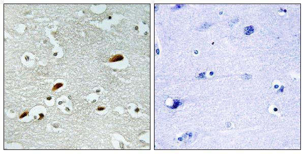
| Sample | Paraffin-embedded human brain tissue; fixative not specified (datasheet A30543) |
| Fixation | Image fixative and duration unreported (datasheet A30543); verify before use. |
| Sectioning | 4–5 µm sections on charged slides (standard) |
| Deparaffinisation | Xylene, graded ethanol series to water (standard) |
| Antigen retrieval | Heat-induced epitope retrieval in Tris-EDTA buffer, pH 9.0, 20 min at 95–98 °C (standard rule: nuclear antigen) |
| Peroxidase block | 3% H2O2, 10 min, room temperature (standard) |
| Blocking | 10% normal serum of the secondary host, 30 min, room temperature (standard) |
| Primary antibody | Rabbit anti-KAT5, 1:100 - 1:300 (datasheet A30543) |
| Primary incubation | Overnight at 4 °C (standard) |
| Detection | HRP-polymer secondary, DAB chromogen 5–10 min (standard) |
| Counterstain | Hematoxylin, blue, dehydrate and mount (standard) |
| Expected result | KAT5-positive staining in neuronal cells of caudate (HPA tissue IHC: High). HPA tissue profile: Nuclear expression in most tissues. No signal in the no-primary control. |
KAT5 should stain mainly nuclei in most tissues (HPA tissue IHC: nuclear expression in most tissues; HPA ICC-IF: supported nucleoplasm). Strong examples include neuronal cells in caudate and glandular cells in colon (HPA tissue IHC: High). KAT5 has no transmembrane segment (UniProt Q92993: topology). HPA rates its tissue IHC pattern Approved, with medium consistency between staining and RNA expression (HPA tissue IHC: reliability).
| Distinct nuclear chromogen in neuronal or glandular cells. | This fits the expected compartment and documented strong examples (HPA tissue IHC: nuclear expression; High in caudate neuronal cells and colon glandular cells). Compare the positive nuclei with neighboring tissue and the counterstain; intensity alone does not establish antibody specificity (general IHC interpretation). |
| Predominantly membranous or widespread cytoplasmic staining with little nuclear signal. | Treat this as discordant with the usual tissue pattern (HPA tissue IHC: nuclear expression in most tissues). KAT5 lacks a transmembrane segment (UniProt Q92993: topology). Some cytoplasmic localization is reported, so isolated extranuclear signal is not automatically false (UniProt Q92993: subcellular location). |
| Strong staining in a cell population expected to be weak in the chosen tissue. | Possible cross-reactivity or endogenous detection activity should be checked with appropriate controls (general IHC interpretation). HPA reports Low staining in pancreatic exocrine glandular cells and splenic red-pulp cells; Low does not mean absent (HPA tissue IHC: Low). Do not reject a slide solely because these cells stain faintly. |
| Broad, diffuse chromogen obscures nuclei and tissue boundaries. | The pattern cannot be scored confidently against the predominantly nuclear reference (HPA tissue IHC: nuclear expression in most tissues). Background can arise from detection chemistry, inadequate blocking, or excess antibody concentration (general IHC practice); a reagent control helps identify detection-derived signal (general IHC practice). |
| No nuclear signal in a documented strong-positive tissue. | First suspect an assay or specimen problem, rather than calling KAT5 absent (HPA tissue IHC: High in cerebellar Purkinje cells and cervical glandular cells). Check tissue preservation, retrieval, antibody and detection steps with a concurrent positive control (general IHC practice). HPA's Approved rating is not a guarantee for every specimen (HPA tissue IHC: reliability). |
| Tissue and cell selection | Choose a documented High population for the positive control, such as cerebellar Purkinje cells or duodenal glandular cells (HPA tissue IHC: High). Pancreatic exocrine glandular cells are Low and are a weaker sensitivity check, not an established negative control (HPA tissue IHC: Low; negative list empty). |
| Compartment and antibody evidence | The tissue reference is nuclear, and HPA016953 is IHC Approved (HPA tissue IHC: profile; HPA antibodies: HPA016953). This supports a nuclear interpretation, while the tissue reliability statement notes only medium staining–RNA consistency (HPA tissue IHC: reliability). |
| KAT5 location and processing | A soluble nuclear pattern fits the absence of a transmembrane segment, signal peptide, and propeptide; the annotated chain spans residues 1–513 (UniProt Q92993: topology and processing). These annotations give no basis for expecting a cell-surface or secreted staining pattern (UniProt Q92993: topology and processing). |
| Isoforms and epitope uncertainty | UniProt lists four KAT5 isoforms, but the supplied evidence gives no antibody epitope or isoform coverage (UniProt Q92993: isoforms; HPA antibodies: validation status only). Do not assign a tissue staining difference to a particular isoform from this IHC result alone (general IHC interpretation). |
| IF/ICC Q: where should fluorescence appear? | Mainly in the nucleoplasm (HPA ICC-IF: supported main location). Vesicle and cytosol signals are listed as uncertain, so interpret them cautiously (HPA ICC-IF: additional locations). These cell-image observations are context for localization, not an IF/ICC protocol or a substitute for the tissue IHC reference (HPA ICC-IF; HPA tissue IHC). |
| Situation | Likely cause | Next action |
|---|---|---|
| Known High tissue is completely blank. | The stain may have failed at retrieval, antibody incubation, or detection (general IHC practice); a blank slide does not overturn HPA's High observation (HPA tissue IHC: High in colon glandular cells). | Run a concurrent documented High tissue and check each assay step against the catalog antibody's IHC-P instructions (HPA tissue IHC: High; general IHC practice). |
| Signal is weak but clearly nuclear in a Low tissue. | That may match the reference rather than indicate failure (HPA tissue IHC: Low in prostate glandular cells; nuclear expression in most tissues). | Compare with a documented High tissue in the same run before adjusting conditions (HPA tissue IHC: High in cervical glandular cells; general IHC practice). |
| Chromogen covers nuclei and surrounding tissue uniformly. | Excess primary antibody, incomplete blocking, or detection background may reduce contrast (general IHC practice). | Check a reagent control and optimize blocking, primary concentration, and detection conditions according to the assay instructions (general IHC practice). |
| A strong membrane outline dominates the slide. | That location conflicts with the usual nuclear tissue pattern and KAT5 topology (HPA tissue IHC: nuclear expression; UniProt Q92993: no transmembrane segment). | Check control staining and review whether the outline follows a tissue edge or detection artefact before scoring cells (general IHC practice). |
| Cytoplasmic staining accompanies a convincing nuclear signal. | KAT5 has annotated cytoplasmic locations, although tissue IHC is mainly nuclear (UniProt Q92993: subcellular location; HPA tissue IHC: profile). | Record nuclear and cytoplasmic staining separately; compare the extranuclear component with controls rather than treating every cytoplasmic pixel as a positive cell (general IHC interpretation). |
| A control slide stains, but the study specimen does not. | Biological variation or a specimen-specific assay issue remains possible; HPA reports Low tissue specificity at the RNA level and medium staining–RNA consistency (HPA tissue IHC: RNA specificity and reliability). | Review cell identity, specimen quality, and run controls, then report the observed result without claiming KAT5 absence from one slide (general IHC practice). |
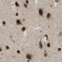
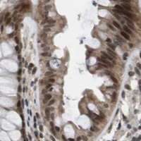


Comprehensive Human Protein Atlas IHC scoring per tissue (reliability: Approved — Medium consistency between antibody staining and RNA expression data.). Rows are taken directly from the HPA tissue chart — click any row's HPA link to view the source.
| Tissue | Cell type | Level | Evidence | Source |
|---|---|---|---|---|
| Caudate | Neuronal cells | High | Protein (IHC) | HPA → |
| Cerebellum | Purkinje cells | High | Protein (IHC) | HPA → |
| Cerebral cortex | Neuronal cells | High | Protein (IHC) | HPA → |
| Cervix | Glandular cells | High | Protein (IHC) | HPA → |
| Colon | Glandular cells | High | Protein (IHC) | HPA → |
| Tissue | Cell type | Level | Evidence | Source |
|---|---|---|---|---|
| None in HPA: KAT5 is detected in all 45 scored tissues. Use a no-primary (secondary-only) and an isotype control instead. | ||||
Use compartment-aware controls and matched processing to troubleshoot KAT5 staining in paraffin sections (UniProt Q92993; HPA tissue IHC).
A30543 has a real IHC image from paraffin-embedded human brain (IHC image caption); the catalog also lists IF/ICC applications and Human, Mouse, and Rat reactivity (catalog: applications, reactivity).
A30543 has IHC image data from paraffin-embedded human brain, including a peptide-blocked comparison (IHC image caption). A30543 lists IF/ICC applications and Human, Mouse, and Rat reactivity (catalog: applications, reactivity); no IF image is supplied (catalog: IF images).
Which to pick: Choose A30543 for tissue IHC when working with paraffin sections, as shown in human brain (IHC image caption); the fixative is unreported (IHC image caption). For IF/ICC, A30543 is the listed option (catalog: applications), though no IF image is supplied (catalog: IF images). It is also the listed cross-species option for Human, Mouse, and Rat (catalog: reactivity); its antibody is polyclonal (catalog: dilution details).