This website uses cookies to ensure you get the best experience on our website.


- Table of Contents
Plan paraffin-section KAT7 IHC around the nuclear staining seen in most tissues (HPA tissue IHC). Use testis preleptotene spermatocytes, which stain strongly, as a positive reference when assessing nuclear signal (HPA tissue IHC).

Expected localisation, validated protocols, controls and antibodies — the at-a-glance facts below, then the full design guide.
| Expected localisation | Nuclear staining in most tissues (HPA tissue IHC) | |
| Staining pattern | Nuclear staining in most tissue cell types (HPA tissue IHC) | |
| Antigen retrieval | Tris-EDTA pH 9.0 HIER, 95–98 °C, 20 min (rule: nuclear antigen) | |
| Positive control | Testis+4 more · see all | |
| Negative control | Adipose tissue+4 more · see all |
| Fixation | Keep fixation conditions consistent across sections. (standard IHC practice; not target-specific) | |
| Caveat | Staining varies by cell type within tissues (HPA tissue IHC) | |
| Regulation | Broad expression; highest in testis (UniProt) | |
| Isoform / epitope | 5 isoforms; antibody epitope coverage is unknown (UniProt) |
The catalog antibody’s IHC-P protocol is followed by two published KAT7 IHC protocols: breast tissue sections and mouse xenograft tumors (PMC13583433; PMC11844059).
| Sample | Paraffin-embedded mouse testes tissue; fixative not specified (datasheet A03896-1) |
| Fixation | Image fixative and duration unreported (datasheet A03896-1); verify before use. |
| Sectioning | 4–5 µm sections on charged slides (standard) |
| Deparaffinisation | Xylene, graded ethanol series to water (standard) |
| Antigen retrieval | Heat-induced epitope retrieval in Tris-EDTA buffer, pH 9.0, 20 min at 95–98 °C (standard rule: nuclear antigen) |
| Peroxidase block | 3% H2O2, 10 min, room temperature (standard) |
| Blocking | 10% normal serum of the secondary host, 30 min, room temperature (standard) |
| Primary antibody | Rabbit anti-KAT7, 1:50-1:100 (datasheet A03896-1) |
| Primary incubation | Overnight at 4 °C (standard) |
| Detection | HRP-polymer secondary, DAB chromogen 5–10 min (standard) |
| Counterstain | Hematoxylin, blue, dehydrate and mount (standard) |
| Expected result | KAT7-positive staining in preleptotene spermatocytes of testis (HPA tissue IHC: High). HPA tissue profile: Nuclear expression in most tissues. No signal in the no-primary control. |
KAT7 should show predominantly nuclear staining in most tissues (HPA tissue IHC: nuclear expression in most tissues; UniProt O95251: nucleus and chromosome). Testis preleptotene spermatocytes are a strong reference population (HPA: High). The HPA tissue profile is Approved, with medium consistency between staining and RNA data (HPA tissue IHC: reliability). KAT7 has no transmembrane segment (UniProt O95251: topology).
| Clear nuclear chromogen in testis preleptotene spermatocytes, with weaker staining in some other tissue cells. | This fits the reported High staining in preleptotene spermatocytes and Medium staining in several listed cell populations (HPA tissue IHC). Compare the named cell type and its nuclei; a tissue-level label does not mean every cell in that tissue should stain equally (HPA tissue IHC: cell-specific levels). |
| Predominantly membrane-like or cytoplasmic chromogen, with nuclei largely spared. | Treat this as a localisation mismatch for tissue IHC (HPA tissue IHC: nuclear expression in most tissues). KAT7 also has a reported cytosolic location (UniProt O95251; HPA subcellular: additional cytosol), so limited cytoplasmic signal alone is inconclusive. Check nuclear counterstain alignment and controls before assigning the signal to KAT7 (general IHC practice). |
| Strong colour in cells listed as Not detected, while expected nuclear staining is weak or absent. | Examples include adipocytes and prostate glandular cells (HPA tissue IHC: Not detected). Suspect off-target staining or endogenous detection activity, especially when colour does not follow nuclei (general IHC practice). 'Not detected' describes the HPA observation under its conditions; it is not proof of biological absence in every specimen (HPA tissue IHC: profile). |
| Haze covers nuclei, cytoplasm and surrounding tissue with little cell-to-cell distinction. | This is difficult to score as KAT7 because the reported tissue pattern is nuclear (HPA tissue IHC). Diffuse staining can arise from nonspecific binding or detection background (general IHC practice). Interpret it against a no-primary control and a clear positive tissue on the same run (general IHC practice). |
| No nuclear signal in testis preleptotene spermatocytes. | This conflicts with the reported High staining in that cell population (HPA tissue IHC: Testis). First confirm the cells are present and the counterstain is readable; then review the primary antibody, retrieval and detection run (general IHC practice). A blank slide by itself cannot distinguish technical failure from specimen variation. |
| Reference cell population | Testis preleptotene spermatocytes provide a High-staining reference, whereas the listed adrenal glandular and bone-marrow hematopoietic cells are Medium (HPA tissue IHC). Use cell identity as well as colour intensity when comparing fields (general IHC practice). |
| Low or undetected populations | Seminal-vesicle glandular cells, cardiomyocytes and smooth-muscle cells are listed as Low; adipocytes and prostate glandular cells are Not detected (HPA tissue IHC). These categories guide comparison under the reported conditions, but do not establish a universal negative control. |
| Antibody evidence | HPA044470 has Approved IHC status, while HPA071116 has no IHC status listed in the supplied antibody record (HPA antibodies). The tissue profile is Approved with medium staining–RNA consistency (HPA tissue IHC). Validate the actual antibody and run before treating weak or unusual staining as a biological result (general IHC practice). |
| Isoforms and epitope | Five KAT7 isoforms are listed, but this record gives no antibody epitope or isoform coverage (UniProt O95251: isoforms; HPA antibodies: supplied fields). Isoform-specific staining differences cannot be predicted from these data; avoid interpreting a negative cell population as lacking every isoform. |
| Processing and localisation | The recorded KAT7 chain spans residues 1–611, with no signal peptide, propeptide or transmembrane segment (UniProt O95251: processing and topology). This supports assessing nuclear intracellular staining; it does not predict antigen retrieval conditions or fixation sensitivity. |
| IF/ICC Q&A: may cytosolic fluorescence count? | Yes, alongside the main nucleoplasmic pattern: HPA reports supported nucleoplasm and approved additional cytosol in ICC-IF (HPA subcellular). Interpret that on the separate IF/ICC guide; the tissue IHC expectation remains predominantly nuclear (HPA tissue IHC). |
| Situation | Likely cause | Next action |
|---|---|---|
| Positive reference tissue is blank. | The expected High preleptotene-spermatocyte signal is missing (HPA tissue IHC: Testis); cell selection or an IHC run step may be at fault (general IHC practice). | Locate preleptotene spermatocytes, confirm counterstain and tissue preservation, then check the recorded primary-antibody, retrieval and detection steps against the run sheet (general IHC practice). |
| Nuclei stain, but the whole section is uniformly dark. | High background can obscure the cell-specific nuclear pattern reported for KAT7 (HPA tissue IHC: nuclear profile; general IHC practice: background assessment). | Compare with a no-primary control; review blocking, washes, primary-antibody concentration and chromogen development within the established IHC workflow (general IHC practice). |
| Colour follows cytoplasm or membranes while nuclei remain pale. | The dominant compartment conflicts with the tissue IHC profile (HPA tissue IHC: nuclear expression in most tissues). A minor cytosolic component is reported in ICC-IF (HPA subcellular). | Check alignment with the nuclear counterstain and inspect the no-primary control; score predominant nonnuclear colour cautiously (general IHC practice). |
| Strong signal appears in adipocytes or prostate glandular cells. | Those populations are listed as Not detected (HPA tissue IHC). Off-target binding or endogenous detection activity is a possible technical explanation (general IHC practice). | Compare localisation and intensity with a positive reference, then inspect no-primary and detection controls before calling the cells positive (general IHC practice). |
| The chosen antibody gives a pattern unlike the HPA tissue profile. | Validation status differs across the supplied antibodies: HPA044470 is IHC Approved; HPA071116 has no IHC status listed (HPA antibodies). | Confirm the exact antibody identity and its IHC evidence, then assess the slide against the reported nuclear profile and cell-specific reference populations (HPA antibodies; HPA tissue IHC). |
| Scattered weak nuclei are hard to classify. | The profile includes Low, Medium and Not detected cell populations, and its staining–RNA consistency is medium (HPA tissue IHC). A single weak field has limited interpretive value. | Compare the named cell type across fields with the same run's positive and no-primary controls; record ambiguous signal separately from clear nuclear positivity (general IHC practice). |
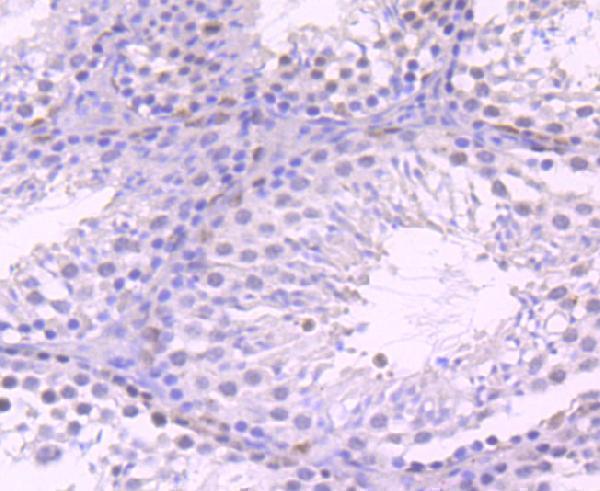
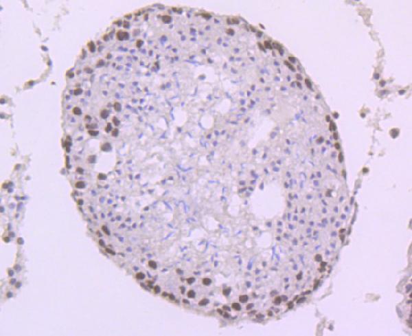
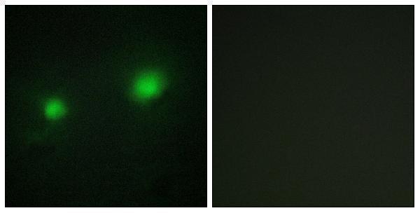


Comprehensive Human Protein Atlas IHC scoring per tissue (reliability: Approved — Medium consistency between antibody staining and RNA expression data.). Rows are taken directly from the HPA tissue chart — click any row's HPA link to view the source.
| Tissue | Cell type | Level | Evidence | Source |
|---|---|---|---|---|
| Testis | Preleptotene spermatocytes | High | Protein (IHC) | HPA → |
| Adrenal gland | Glandular cells | Medium | Protein (IHC) | HPA → |
| Bone marrow | Hematopoietic cells | Medium | Protein (IHC) | HPA → |
| Breast | Glandular cells | Medium | Protein (IHC) | HPA → |
| Bronchus | Basal cells | Medium | Protein (IHC) | HPA → |
| Tissue | Cell type | Level | Evidence | Source |
|---|---|---|---|---|
| Adipose tissue | Adipocytes | Not detected | Protein (IHC) | HPA → |
| Appendix | Glandular cells | Not detected | Protein (IHC) | HPA → |
| Cerebellum | Cells in granular layer | Not detected | Protein (IHC) | HPA → |
| Hippocampus | Glial cells | Not detected | Protein (IHC) | HPA → |
| Liver | Cholangiocytes | Not detected | Protein (IHC) | HPA → |
Troubleshoot KAT7 staining by checking nuclear localisation, processing controls and cell type before interpreting chromogenic signal.
The catalog includes anti-KAT7 antibodies for tissue IHC and cell IF/ICC (catalog applications), with images of paraffin-embedded mouse and rat testes and HUVEC cells (catalog image captions).
A03896-1 is listed for IHC in Human, Mouse, and Rat (catalog applications and reactivity); its images show paraffin-embedded mouse and rat testes (catalog IHC captions). A30463 is listed for IF/ICC in Human, Mouse, and Rat (catalog applications and reactivity); its IF image shows HUVEC cells with a peptide-blocked comparison (catalog IF caption).
Which to pick: For tissue IHC, choose A03896-1 at 1:50–1:100 (catalog dilution); its images support paraffin sections, and the fixative is unreported (catalog IHC captions). For IF/ICC, choose the polyclonal A30463 at 1:200–1:1000 (catalog dilution and clonality); its image shows HUVEC IF (catalog IF caption). For cross-species planning, both list Human, Mouse, and Rat reactivity (catalog reactivity), while the shown IHC specimens are mouse and rat testes for A03896-1 (catalog IHC captions).