KCNA1 / Potassium voltage-gated channel subfamily A member 1 · IHC design guide

Design Immunohistochemistry for KCNA1

For KCNA1 paraffin IHC, expect neuronal cytoplasmic staining in brain sections (HPA tissue IHC). Start the IHC-validated antibody at 2–5 μg/mL (datasheet A01813-1), and interpret chromogenic signal with controls because presumed off-target binding was reported (HPA tissue IHC).

Evidence assembled Oct 2026 · For research use; verify linked source records and product datasheet before use
Immunohistochemistry protocol sheet for KCNA1 (IHC for KCNA1): expected localisation Neuronal cytoplasm observed; axonal membrane annotated (HPA tissue IHC; UniProt), antibody A01813-1, validated IHC image, and IHC protocol steps
Printable KCNA1 IHC protocol sheet — expected localisation Neuronal cytoplasm observed; axonal membrane annotated (HPA tissue IHC; UniProt), antibody A01813-1, controls and protocol steps. Open the full KCNA1 IHC guide →

KCNA1 Immunohistochemistry Experimental Design Guide

Expected localisation, validated protocols, controls and antibodies — the at-a-glance facts below, then the full design guide.

Must know before staining
Expected localisation Neuronal cytoplasm observed; axonal membrane annotated (HPA tissue IHC; UniProt)
Staining pattern Neuronal cytoplasmic staining; some cerebellar membrane staining (HPA tissue IHC)
Antigen retrieval EDTA pH 8.0 HIER, heat-mediated (datasheet A01813-1)
Positive control ⓘ Cerebellum+2 more · see all
Negative control ⓘ Adipose tissue+4 more · see all
Important caveats
Reasons your staining may differ from the expected pattern.
Fixation Keep fixation conditions consistent across sections. (standard IHC practice; not target-specific)
Caveat Presumed off-target binding can confound staining (HPA tissue IHC)
Regulation KCNA2/KCNAB2 promote membrane expression (UniProt)
Isoform / epitope No annotated isoforms; epitope position matters across the membrane (UniProt)
Section 1

Recommended KCNA1 IHC & IF Protocols

Compare the catalog antibody’s IHC-P protocol with the published chromogenic IHC protocol for mouse hippocampal paraffin sections (PMC10361730).

Recommended immunohistochemistry (IHC-P) protocol parameters
SampleParaffin-embedded rat brain tissue; fixative not specified (datasheet A01813-1)
FixationImage fixative and duration unreported (datasheet A01813-1); verify before use.
Sectioning4–5 µm sections on charged slides (standard)
DeparaffinisationXylene, graded ethanol series to water (standard)
Antigen retrievalHeat retrieval: EDTA pH 8.0 (datasheet A01813-1); 20 min, 95–100 °C (standard)
Peroxidase block3% H2O2, 10 min, room temperature (standard)
Blocking10% goat serum (datasheet A01813-1)
Primary antibodyRabbit anti-KCNA1, 2-5μg/ml (datasheet A01813-1)
Primary incubationOvernight at 4 °C (datasheet A01813-1)
DetectionStreptavidin-biotin complex (SABC), DAB chromogen (datasheet A01813-1)
CounterstainHematoxylin, blue, dehydrate and mount (standard)
Expected resultKCNA1-positive staining in gLUC cells - cytoplasm/membrane of cerebellum (HPA tissue IHC: Medium). HPA tissue profile: Cytoplasmic expression in neuronal cells. No signal in the no-primary control.
💡Decision noteStart with heat-mediated EDTA retrieval at pH 8.0 (datasheet A01813-1); citrate at pH 6.0 is the published alternative (PMC10361730).
Section 2

What Is the Expected KCNA1 Staining Pattern?

KCNA1 staining should favor neuronal cells, with cytoplasmic and membrane-associated signal (HPA tissue IHC; UniProt Q09470 subcellular location). Its six transmembrane segments support membrane localization, while its annotated endoplasmic reticulum location can account for intracellular signal (UniProt Q09470 topology and subcellular location). HPA rates the tissue IHC evidence Approved but reports low agreement with RNA data and presumed off-target staining that was disregarded (HPA tissue IHC).

What am I looking at on my slide?
Neuronal cytoplasmic staining, with some membrane-associated signal, in cerebellum, cerebral cortex, or hippocampus.This fits HPA's observed Medium neuronal staining in those regions (HPA tissue IHC). Membrane and intracellular signal are compatible with KCNA1's annotated locations (UniProt Q09470 subcellular location). Judge the cell type and compartment together; HPA's Approved rating carries a low RNA-agreement caveat (HPA tissue IHC).
Predominantly nuclear staining in neurons, with little cytoplasmic or membrane-associated signal.A nuclear-dominant pattern does not match the reported neuronal cytoplasmic pattern or KCNA1's annotated locations (HPA tissue IHC; UniProt Q09470 subcellular location). Treat it as suspect and assess specificity and chromogen background before calling cells positive (general IHC practice).
Strong staining in adipocytes or glandular cells while neurons show little signal.HPA reports no detection in adipocytes of adipose tissue and glandular cells of adrenal gland (HPA tissue IHC). Such staining raises concern for cross-reactivity or endogenous detection activity; its appearance alone cannot distinguish those causes (general IHC practice). HPA also notes presumed off-target staining in its validation summary (HPA tissue IHC).
Diffuse color across cells and surrounding tissue, without a discernible neuronal pattern.This is less convincing than HPA's neuronal cytoplasmic pattern (HPA tissue IHC). Uneven reagent distribution, nonspecific binding, or detection background can produce diffuse color (general IHC practice). Compare with a no-primary control and inspect whether signal follows tissue structure (general IHC practice).
No visible staining in a well-preserved section of cerebellum, cerebral cortex, or hippocampus.Those are useful positive-reference tissues because HPA reports Medium staining there (HPA tissue IHC). An absent signal calls for checking the IHC workflow and antibody performance; it does not, by itself, establish absent KCNA1 expression (general IHC practice).
💡Expected KCNA1 appearanceCall a result positive when Medium neuronal cytoplasmic staining, potentially with membrane-associated signal, is distinguishable in an HPA-positive brain region; isolated nuclear staining or equally strong staining in HPA-negative cell types is suspect (HPA tissue IHC; UniProt Q09470 subcellular location).
How each factor affects the staining
Cellular compartmentKCNA1 has six transmembrane segments and annotated membrane, axonal, synaptic, endoplasmic reticulum, and perikaryal locations (UniProt Q09470 topology and subcellular location). HPA describes neuronal cytoplasmic staining, so cytoplasmic signal need not imply a soluble target (HPA tissue IHC).
Channel assembly and traffickingUniProt says homotetrameric KCNA1 is primarily in the endoplasmic reticulum, while interaction with specified partner subunits promotes cell-membrane expression (UniProt Q09470 subcellular location). Thus, membrane emphasis may vary; an individual section cannot establish which channel complex is present.
Tissue and antibody evidenceHPA reports Medium staining in cerebellum, cerebral cortex, and hippocampus, with no detection in several listed non-neuronal cell types (HPA tissue IHC). Its antibody is IHC Approved, with low RNA agreement and presumed off-target staining disregarded (HPA tissue IHC; HPA antibody validation).
Epitope and retrievalKCNA1 includes cytoplasmic and extracellular regions and an annotated glycosylation site at residue 207 (UniProt Q09470 topology and glycosylation). The antibody epitope is unspecified here; these annotations cannot predict a retrieval condition or a fixation effect. Optimize retrieval as a general IHC-P workflow variable (general IHC practice).
Why is my staining missing, weak or wrong?
SituationLikely causeNext action
Positive-reference brain section is blank.HPA reports Medium neuronal staining in cerebellum, cerebral cortex, and hippocampus; a blank section may reflect a workflow or antibody problem (HPA tissue IHC; general IHC practice).Check section integrity, primary-antibody application, retrieval, and detection controls; compare with a known-working run before interpreting the blank result (general IHC practice).
Only nuclei appear positive.Nuclear-dominant staining conflicts with HPA's neuronal cytoplasmic pattern and UniProt's listed locations (HPA tissue IHC; UniProt Q09470 subcellular location).Review morphology and a no-primary control; reassess antibody specificity and detection background before scoring nuclear color as KCNA1 (general IHC practice).
Color is widespread in both expected and HPA-negative cells.Nonspecific binding or endogenous detection activity can obscure cell-specific staining (general IHC practice); HPA notes presumed off-target staining (HPA tissue IHC).Compare a no-primary control and an HPA-negative cell type, then review blocking and detection controls (HPA tissue IHC; general IHC practice).
Neuronal signal is faint and hard to score.HPA's reported level is Medium, so weak color may be difficult to separate from background; the cause cannot be assigned from intensity alone (HPA tissue IHC; general IHC practice).Compare matched positive-reference and no-primary sections, and review retrieval and primary-antibody conditions within the validated IHC-P workflow (general IHC practice).
Membrane staining is less prominent than cytoplasmic staining.HPA describes cytoplasmic neuronal expression; UniProt also places KCNA1 in the endoplasmic reticulum and perikaryon (HPA tissue IHC; UniProt Q09470 subcellular location).Score neuronal cytoplasmic signal alongside membrane-associated signal. Do not reject a result solely because the membrane outline is weak; require separation from background (HPA tissue IHC; general IHC practice).
Can ICC/IF localization confirm the IHC interpretation?HPA's subcellular summary says membrane, but supplies no ICC-IF image-bearing cell lines or main-location assignment for this record (HPA subcellular ICC-IF).Use the neuronal tissue IHC pattern and UniProt locations to interpret this section; consult the separate IF/ICC guide for that application (HPA tissue IHC; UniProt Q09470 subcellular location).

Sample controls for KCNA1 IHC & IF

🧪Run cerebellum first: GLUC cells should show cytoplasmic/membrane staining at the HPA Medium level (HPA: Cerebellum, GLUC cells - cytoplasm/membrane, Medium). Use adipose tissue as the negative tissue (HPA: Adipocytes, Not detected); on the cerebellum slide, assess cells outside the identified GLUC population as internal background rather than assuming every other cell type is KCNA1-negative (HPA: GLUC cells - cytoplasm/membrane).
Positive control tissue: Cerebellum (GLUC cells - cytoplasm/membrane, HPA Medium)
Negative control tissue: Adipose tissue (HPA Not detected)
ICC-IF cell lines (HPA subcellular resource): HPA carries no ICC-IF cell line for KCNA1; derive a cell-line control from the positive tissue's cell type (GLUC cells - cytoplasm/membrane) and confirm it by RNA or western blot first.
Technical controls: Include no-primary (secondary-only) and rabbit isotype controls, matching antibody class and clonality where known (selected-SKU caption: rabbit primary); use KCNA1 knockout tissue, if available, or peptide blocking with the matching immunogen as a biological specificity control (standard IHC practice). For chromogenic brain sections, control endogenous peroxidase and, with the caption’s avidin-biotin detection system, endogenous biotin; assess autofluorescence if using IF (selected-SKU caption: SABC with DAB; standard IHC/IF practice).
⚠️Feasibility: A target-specific fixation window or fixation effect is unreported, and the selected-SKU paraffin-section caption does not state a fixative (selected-SKU caption: fixative unreported). The reported IHC procedure uses heat retrieval in EDTA at pH 8.0, but the evidence does not establish whether retrieval is required for all KCNA1 antibodies or specimens (selected-SKU caption: heat-mediated EDTA retrieval). Frozen sections and IF are not established as easier by the supplied evidence; for brain IF, check autofluorescence when interpreting signal (HPA: cerebellum, Medium; standard IF practice).

HPA tissue IHC evidence for KCNA1

Comprehensive Human Protein Atlas IHC scoring per tissue (reliability: Approved — Low consistency between antibody staining and RNA expression data. Presumed off target binding observed and disregarded.). Rows are taken directly from the HPA tissue chart — click any row's HPA link to view the source.

Positive expression · recommended positive controls

TissueCell typeLevelEvidenceSource
Cerebellum GLUC cells - cytoplasm/membrane Medium Protein (IHC) HPA →
Cerebral cortex Neuronal cells Medium Protein (IHC) HPA →
Hippocampus Neuronal cells Medium Protein (IHC) HPA →

Undetected expression · recommended negative controls

TissueCell typeLevelEvidenceSource
Adipose tissue Adipocytes Not detected Protein (IHC) HPA →
Adrenal gland Glandular cells Not detected Protein (IHC) HPA →
Appendix Glandular cells Not detected Protein (IHC) HPA →
Bone marrow Hematopoietic cells Not detected Protein (IHC) HPA →
Breast Adipocytes Not detected Protein (IHC) HPA →
Section 3

Advanced KCNA1 IHC Tips

Troubleshoot KCNA1 staining in paraffin sections by checking retrieval, compartment, controls and scoring before interpreting chromogenic signal.

How should I retrieve KCNA1 antigen if paraffin-section staining is weak?
Start with heat-mediated retrieval in EDTA at pH 8.0 for paraffin sections (datasheet A01813-1). The selected rat brain example used that retrieval, followed by 10% goat serum blocking and 2 μg/mL primary antibody overnight at 4°C (datasheet A01813-1). If staining remains weak, compare a second retrieval condition on adjacent sections while keeping antibody concentration, detection and development constant (standard IHC practice). Judge improvement by signal in neuronal structures and by background on the same slide; stronger diffuse staining alone does not establish KCNA1 specificity (UniProt Q09470 localisation; standard IHC practice).
Can fixation account for weak KCNA1 staining in paraffin sections?
The selected paraffin-section caption does not state a fixative, so KCNA1-specific fixation sensitivity is unknown for this antibody (datasheet A01813-1). Record the fixative, fixation duration, tissue thickness and processing history for each block before comparing staining across specimens (standard IHC practice). If fixation differs between specimens, stain adjacent sections in the same run and include a consistently processed positive control, such as the documented rat brain preparation (datasheet A01813-1; standard IHC practice). Treat any fixation-related difference as a processing hypothesis until a controlled comparison separates it from retrieval, detection and tissue preservation effects (standard IHC practice).
Where should convincing KCNA1 chromogenic staining appear?
Look for staining associated with neuronal processes, synaptic regions and cell membranes, while allowing perikaryal or endoplasmic-reticulum signal (UniProt Q09470 localisation). KCNA1 has been detected beside nodes of Ranvier in spinal cord fibers, including juxtaparanodal zones and some paranodal regions (UniProt Q09470, PubMed:11086297). A purely cytoplasmic neuronal pattern also appears in the tissue atlas, but that source reports low agreement with RNA and disregarded presumed off-target staining (HPA tissue IHC). Compare compartment and cell identity with matched controls before assigning diffuse staining to KCNA1; homotetrameric KCNA1 can reside primarily in the endoplasmic reticulum, and partner subunits can promote membrane expression (UniProt Q09470; standard IHC practice).
How does KCNA1 topology affect epitope access and staining interpretation?
Check the antibody's stated immunogen or epitope before choosing controls; the supplied product caption does not identify that region (datasheet A01813-1). KCNA1 has 6 transmembrane segments, cytoplasmic amino and carboxyl termini, extracellular loops, and a glycosylation site at residue 207 (UniProt Q09470 topology and processing). Those features make epitope position relevant when interpreting access after retrieval, but they do not establish which region this antibody recognizes (UniProt Q09470 topology; standard IHC practice). The supplied record annotates 0 isoforms, so do not explain divergent staining by an asserted KCNA1 isoform without independent evidence (UniProt Q09470 isoform record).
How should I adapt KCNA1 assessment to multiplex IF?
For the separate IF assay, pair KCNA1 with a marker identifying the expected neuronal population and inspect whether their signals occupy compatible cells and compartments (UniProt Q09470 localisation; HPA tissue IHC; standard IF practice). Choose fluorophores and acquisition channels after checking tissue autofluorescence, then image single-label and no-primary controls with the same settings (standard IF practice). Set permeabilisation according to the verified epitope: intracellular termini require access through the plasma membrane, whereas an extracellular-loop epitope calls for a different access check (UniProt Q09470 topology; standard IF practice). The supplied paraffin-section staining example does not validate an IF protocol or establish this antibody's epitope (datasheet A01813-1).
What should I change when KCNA1 DAB staining is widespread?
First compare the section with a no-primary control to assess detection-system and tissue background, then inspect whether the stain follows neuronal anatomy (standard IHC practice; UniProt Q09470 localisation). The selected paraffin-section example used 10% goat serum, a biotinylated secondary antibody, a streptavidin-biotin complex and DAB, so check each detection step if diffuse color appears (datasheet A01813-1). Include a peroxidase-blocking step and control DAB development time as general chromogenic IHC measures (standard IHC practice). Be cautious with broad cytoplasmic staining: the tissue atlas describes neuronal cytoplasmic signal but also notes presumed off-target binding and low RNA agreement (HPA tissue IHC).
How can I quantify KCNA1 staining across paraffin sections? ⚠ ANSWER MARKED FOR VERIFICATION
Define the neuronal region and compartment before scoring, and apply the same threshold and imaging conditions to every section (UniProt Q09470 localisation; standard IHC practice). For cellular staining, report an H-score or percentage of positive neuronal cells; for process-rich regions, report positive signal area or density per mm² within a predefined region (standard IHC practice). Normalize each measure to the relevant neuronal cell count or tissue area, and exclude folds, damaged edges and empty space by the same rules across slides (standard IHC practice). Report membrane-associated and cytoplasmic measures separately when both are present, because KCNA1 can occupy those compartments (UniProt Q09470 localisation).
When should I doubt a positive KCNA1 IHC result?
Favor signal in neuronal cells and their processes over staining confined to an unrelated cell population or an anatomically implausible compartment (UniProt Q09470 localisation; HPA tissue IHC). Inspect whether color concentrates at section edges, folds or necrotic areas, and compare those regions with a no-primary control for nonspecific deposition (standard IHC practice). Check endogenous peroxidase background with an appropriate detection control before calling DAB signal antigen-dependent (standard IHC practice). Interpret cytoplasmic staining cautiously: KCNA1 can occupy the endoplasmic reticulum, yet the tissue atlas reports presumed off-target staining and low agreement with RNA expression (UniProt Q09470 localisation; HPA tissue IHC).
Boster reagents

Best KCNA1 / Potassium voltage-gated channel subfamily A member 1 IHC Antibodies

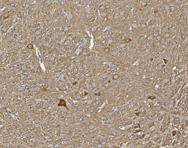
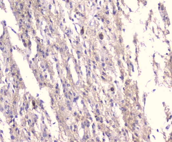
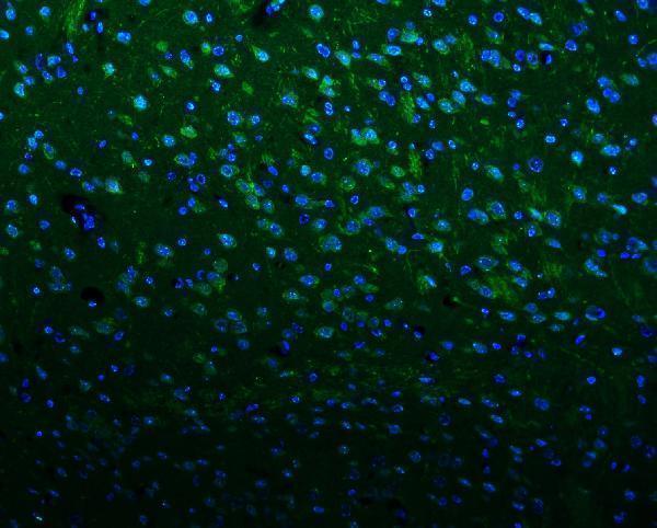
The IHC-validated antibody has images from paraffin sections of rat brain and human glioma; IF images show paraffin sections of mouse and rat brain (A01813-1 image captions).

Real IHC data IHC analysis of KCNA1 using anti-KCNA1 antibody (A01813-1). KCNA1 was detected in paraffin-embedded section of rat brain tissue. Heat mediated antigen retrieval was performed in EDTA buffer (pH8.0, epitope retrieval solution). The tissue section was blocked with 10% goat serum. The tissue section was then incubated with 2μg/ml rabbit anti-KCNA1 Antibody (A01813-1) overnight at 4°C. Biotinylated goat anti-rabbit IgG was used as secondary antibody and incubated for 30 minutes at 37°C. The tissue section was developed using Strepavidin-Biotin-Complex (SABC) (Catalog # SA1022) with DAB as the chromogen.
Anti-Kv1.1 potassium channel/KCNA1 Antibody ®
Cat # A01813-1

A01813-1 is listed for IHC and IF with human, mouse and rat reactivity (catalog: applications and reactivity). Its IHC images show rat brain and human glioma, while its IF images show mouse and rat brain (A01813-1 image captions).

Which to pick: For tissue IHC, choose A01813-1: its IHC captions document paraffin sections, and the listed IHC concentration is 2–5 μg/ml (A01813-1 IHC captions; catalog: IHC dilution). For IF, the same SKU has paraffin-section images at 5 μg/ml; ICC-specific validation and clonality are unreported (A01813-1 IF captions; catalog: clone field). For work across species, its listed reactivity covers human, mouse and rat, though the IHC images show only human and rat tissue; the IHC captions do not report the fixative (catalog: reactivity; A01813-1 IHC captions).

Each figure is that product's own IHC / IF validation image from its datasheet.

References

  1. UniProt Consortium. UniProt entry Q09470 (KCNA1_HUMAN, Potassium voltage-gated channel subfamily A member 1).
  2. Human Protein Atlas. KCNA1 tissue IHC expression (reliability: Approved).
  3. Human Protein Atlas. KCNA1 subcellular location (ICC-IF): Membrane.
  4. Human Protein Atlas. KCNA1 antibody validation summary (1 antibodies).
  5. Spontaneous seizures in Kcna1-null mice lacking voltage-gated Kv1.1 channels activate Fos expression in select limbic circuits. Journal of neurochemistry 2015 — PMC6350929.
  6. Kv1.1 subunits localize to cardiorespiratory brain networks in mice where their absence induces astrogliosis and microgliosis. Molecular and cellular neurosciences 2021 — PMC10332644.
  7. Identification of key potassium channel genes of temporal lobe epilepsy by bioinformatics analyses and experimental verification. Frontiers in neurology 2023 — PMC10361730.
  8. PubMed PMID:19912772 — UniProt-cited evidence.
  9. PubMed PMID:16541075 — UniProt-cited evidence.
  10. PubMed PMID:15489334 — UniProt-cited evidence.