This website uses cookies to ensure you get the best experience on our website.
- Table of Contents
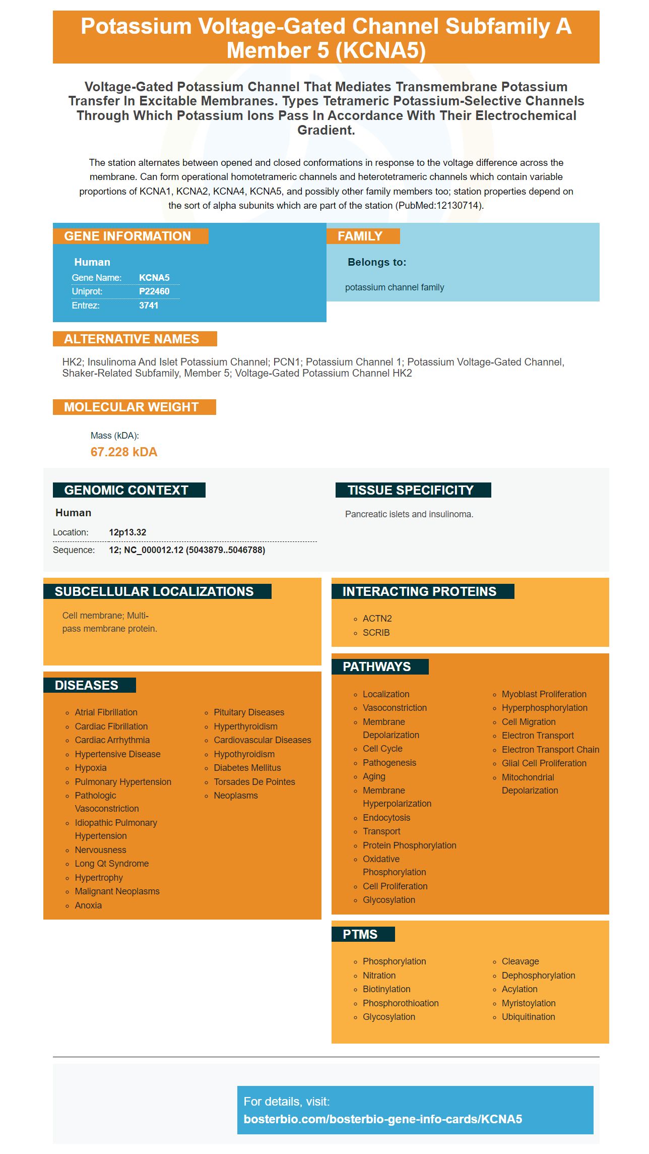
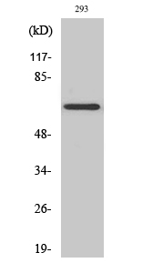
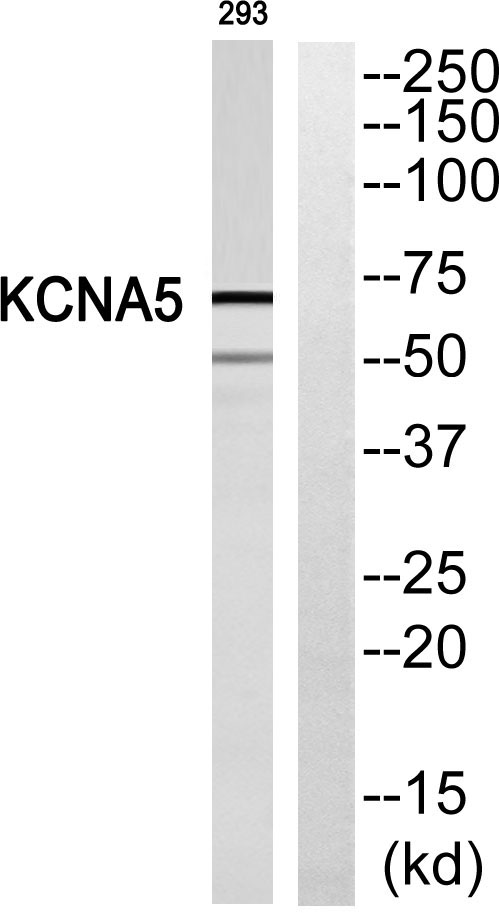
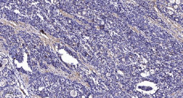
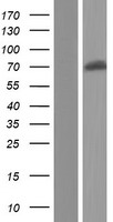
Facts about Potassium voltage-gated channel subfamily A member 5.
The station alternates between opened and closed conformations in response to the voltage difference across the membrane. Can form operational homotetrameric channels and heterotetrameric channels which contain variable proportions of KCNA1, KCNA2, KCNA4, KCNA5, and possibly other family members too; station properties depend on the sort of alpha subunits which are part of the station (PubMed:12130714).
Human | |
---|---|
Gene Name: | KCNA5 |
Uniprot: | P22460 |
Entrez: | 3741 |
Belongs to: |
---|
potassium channel family |
HK2; insulinoma and islet potassium channel; PCN1; potassium channel 1; potassium voltage-gated channel, shaker-related subfamily, member 5; voltage-gated potassium channel HK2
Mass (kDA):
67.228 kDA
Human | |
---|---|
Location: | 12p13.32 |
Sequence: | 12; NC_000012.12 (5043879..5046788) |
Pancreatic islets and insulinoma.
Cell membrane; Multi-pass membrane protein.
If you are interested in purchasing an Anti-KCNA5 Marker, you have come to the right place. Boster Bio has validated their antibodies using WB, IHC, ICC, and Immunofluorescence. Learn how to use this antibody in pulmonary vascular function. KCNA5 is a key player in pulmonary vascular function, as well as other areas of pulmonary metabolism.
When pursuing your studies on KCNA5, the Boster Bio Anti-KCNA5 Markers are an excellent choice. The primary antibodies are highly specific and have been validated using ELISA, immunohistochemistry, and Western blotting methods. Boster offers high-affinity primary antibodies for all your research needs. Read on to learn more about the Boster Anti-KCNA5 Marker.
Boster Bio Anti-PCNA Antibody is a mouse monoclonal antibody that reacts with human, mouse, and rat PCNA. This antibody is stable at -20°C for one year, and at 4°C for one month. It is manufactured using mouse ascites fluid, sodium acetate, and BSA as preservatives. It is compatible with both routine and immunohistochemistry assays, and can be stored for up to one year at a time.
Boster Bio produces high-quality ELISA kits and antibodies. Their antibody, ELISA, and ICC reagents have undergone rigorous validation and are optimized for the WB, IHC, and ICC applications. The company's research and development laboratories are equipped to manufacture the antibodies and ELISA kits in their own facilities, which is why the company stands behind their products with a Boster Quality Guarantee.
To ensure reproducibility of antibodies, the user, publisher, and vendor must collaborate. The user must validate the performance of their antibody and conduct well-designed experiments. The antibody vendor must provide high-quality antibodies and full disclosure of their methods. The publisher can enforce guidelines and enforce standards for validation data reporting. Ultimately, all researchers and scientists must work together to improve antibody reproducibility. This starts with ensuring that all antibodies used in scientific research are validated.
KO validation is considered the gold standard for Western blotting. It is used by many antibody vendors during both the development and batch testing of antibodies. KO validation is not sufficient, however, and multiple approaches are required to ensure reproducibility of results. For assay-specific validation, the antibody should be tested on a corresponding complementing assay to confirm the effect of the primary antibody.
Using this method, the antibody is compared with RNA-Seq data for the same samples. In case of a positive control, the antibody's signal must match the RNA level in the samples. This orthogonal validation method requires two tissue or cell lines with a five-fold difference in RNA expression. This confirms the antibody's specificity and provides a consistent platform for further validation.
The Boster company has made it a priority to validate all of its antibodies on Immunofluorescence, and this commitment is already paying off. Its customers validate over 650 antibodies, and the company is now aiming to validate most of them by the end of 2015. Boster validates all of its antibodies in this manner, which will help researchers avoid the need to perform multiple experiments. This approach is also cost-effective and time-efficient, and involves seeding cells of interest on 96-well plates. Images were acquired using an IN Cell microscope (GE Healthcare) and analyzed using CellProfiler 3.1.8 software.
To perform the immunofluorescence analysis, Neuro2A cells were fixed in 4% PFA at room temperature, and permeabilized in PBS with 0.1% saponin or 3% BSA. Antibodies were incubated in the cells for 16 h at 4 degC, and secondary antibodies were added. After that, cells were stained using a high-throughput microscope with five to ten fields per well.
The Boster team validates all antibodies on Immunofluorescent and is confident in its results. The Boster team tests all antibodies by the most stringent protocols and ensures that they are accurate and consistent. As a result, their products are widely used in both biomedical research. The Boster team works with researchers across the globe to provide the best quality immunofluorescence products to the scientific community.
In addition to testing and evaluating its own products, it values peer-reviewed publications by customers. The company counts over 10,000 publications with its antibodies as proof of their quality and relevance. Its goal is to provide researchers with the best antibodies to conduct research and advance knowledge. For the past decade, Boster has been a leader in the industry, and the company's mission has never wavered from this mission.
A primary motivation for the development of new antibodies is the opioid crisis. The number of people who died from overdoses last year was related to the use of opioids. A comprehensive understanding of synaptic signaling, including opioid receptor signaling, is needed to combat this crisis. By developing antibodies that target opioid receptor signaling, scientists can demonstrate dynamic modulation of ligand-directed signaling.
KCNA5 is a key component of the heart's pulmonary vascular system, and its mutations have been associated with sudden cardiac death and atrial fibrillation. Mutations in KCNA5 affect the resting membrane potential and may also cause hypoxic pulmonary vasoconstriction. However, the exact function of KCNA5 is not known. However, it is likely that it plays a critical role in pulmonary vascular function.
KCNA5 is a protein that encodes the Kv1.5 potassium channel, which controls potassium flux from the cell. Potassium balance regulates apoptosis, and KCNA5 participates in the control of apoptosis in smooth muscle cells of the pulmonary artery. KCNA5 overexpression increases K efflux and increases caspase-3 proteolytic activity.
KCNA5 is also implicated in the maintenance of membrane potential and vascular tone. It has been shown that hypoxic conditions induce specific inhibition of KCNA5 in the PASMCs, which leads to pulmonary vasoconstriction. In addition, it has been shown that KCNA5 is decreased in PASMCs in patients with primary pulmonary hypertension, which may play an important role in the pathogenesis of pulmonary hypertension.
Moreover, a decrease in cytoplasmic potassium concentration in KCNA5 may also be associated with a reduced endothelin receptor activity. Inhibitors of KCNA5 may improve the function of pulmonary artery walls and reduce pulmonary arterial pressure. Although further studies are needed to determine whether these compounds have any beneficial effects on the disease, current treatments may be a better alternative to standard pharmacological treatment.
KCNA5 is a potassium voltage-gated channel subfamily member involved in regulating vascular tone. It is involved in hypoxia-induced pulmonary vasoconstriction. The genetic association of KCNA5 with systemic sclerosis with pulmonary arterial hypertension was reported, and the current study aimed to confirm this association. A total of 2,690 matched healthy controls from five European countries were included in the study. Genotyping was performed using a TaqMan SNP genotyping assay.
Pneumonia is a multifactorial disease involving genetic predisposition and environmental factors. Mutations in KCNK3 have revived interest in potassium channels. KCNK3 is a gene that encodes two potassium (K+) channels and KCNA5 is a transcription factor that regulates these channels. In addition to enhancing pulmonary vascular function, KCNK3 also contributes to epigenetic regulation.
PMID: 2001794 by Tamkun M.M., et al. Molecular cloning and characterization of two voltage-gated K+ channel cDNAs from human ventricle.
PMID: 1986382 by Philipson L.H., et al. Sequence and functional expression in Xenopus oocytes of a human insulinoma and islet potassium channel.