This website uses cookies to ensure you get the best experience on our website.


- Table of Contents
Plan chromogenic KCNC1 IHC in paraffin sections using cerebellar synaptic glomeruli as a high-staining reference (HPA tissue IHC). Compare adipose tissue adipocytes, where staining was not detected, and optimize catalog antibody A06871-1 within 1:100–1:300 (HPA tissue IHC; datasheet: A06871-1).

Expected localisation, validated protocols, controls and antibodies — the at-a-glance facts below, then the full design guide.
| Expected localisation | CNS processes (HPA tissue IHC); axonal membrane (UniProt) | |
| Staining pattern | Cerebellar synaptic processes; cortical neuronal cytoplasm (HPA tissue IHC) | |
| Antigen retrieval | Citrate pH 6.0 HIER, 95–98 °C, 20 min (rule: cytoplasmic / membrane antigen) | |
| Positive control | Cerebellum+2 more · see all | |
| Negative control | Adipose tissue+4 more · see all |
| Fixation | Keep fixation consistent across sections (standard IHC practice; not target-specific). Selected-image fixative and duration unreported (datasheet A06871-1); verify before use. | |
| Caveat | Staining varies by region: high in cerebellum, low in caudate (HPA tissue IHC) | |
| Regulation | Brain-enriched RNA; regulation unreported (HPA tissue RNA) | |
| Isoform / epitope | 2 isoforms; confirm whether the epitope is extracellular or cytoplasmic (UniProt) |
The catalog antibody’s IHC-P protocol is accompanied by a published KCNC1 protocol for human seminoma and adjacent tissue (PMC8020201).
| Sample | Paraffin-embedded human brain tissue; fixative not specified (datasheet A06871-1) |
| Fixation | Image fixative and duration unreported (datasheet A06871-1); verify before use. |
| Sectioning | 4–5 µm sections on charged slides (standard) |
| Deparaffinisation | Xylene, graded ethanol series to water (standard) |
| Antigen retrieval | Heat-induced epitope retrieval in citrate buffer, pH 6.0, 20 min at 95–98 °C (standard rule: cytoplasmic / membrane antigen) |
| Peroxidase block | 3% H2O2, 10 min, room temperature (standard) |
| Blocking | 10% normal serum of the secondary host, 30 min, room temperature (standard) |
| Primary antibody | Rabbit anti-KCNC1, 1:100-1:300 (datasheet A06871-1) |
| Primary incubation | Overnight at 4 °C (standard) |
| Detection | HRP-polymer secondary, DAB chromogen 5–10 min (standard) |
| Counterstain | Hematoxylin, blue, dehydrate and mount (standard) |
| Expected result | KCNC1-positive staining in synaptic glomeruli - core of cerebellum (HPA tissue IHC: High). HPA tissue profile: Expression in processes in CNS and retina, most abundant in cerebellum granular layer. Additional cytoplasmic expression in neuronal cells of cerebral cortex and caudate. No signal in the no-primary control. |
In paraffin-section IHC, expect KCNC1 staining in neuronal processes, strongest in cerebellar synaptic glomeruli, with weaker staining in cortical neurons and retinal processes (HPA: Enhanced tissue IHC; High in cerebellar synaptic glomeruli; Medium in cerebral cortical neurons and retinal inner plexiform layer). A process-associated pattern fits its cell, axonal and presynaptic membrane localization and six transmembrane segments (UniProt P48547: subcellular location and topology).
| Strong staining in cerebellar granular-layer synaptic glomeruli, especially their cores, with a process-rich pattern. | This is the clearest positive reference: HPA reports High staining in synaptic glomeruli and describes processes as the principal tissue pattern (HPA: cerebellum; Enhanced tissue IHC). Score the localized pattern as well as intensity; a uniformly stained section does not reproduce that distribution (HPA: tissue IHC). |
| Medium staining in cerebral cortical neuronal cells or the retinal inner plexiform layer; weak neuronal staining in caudate. | These sites provide secondary comparisons, with tissue-dependent intensity (HPA: Medium in cortex and retina; Low in caudate). HPA also describes cytoplasmic staining in cortical and caudate neurons, so cytoplasmic neuronal signal alone does not contradict its tissue observations (HPA: tissue IHC profile). |
| Predominantly nuclear staining in paraffin sections, without the expected neuronal-process pattern. | Treat this as a compartment mismatch and investigate artefact or nonspecific staining against the tissue IHC reference (HPA: tissue IHC profile; UniProt P48547: membrane localization). Nuclear locations are reported separately by HPA ICC-IF; that observation does not establish a predominantly nuclear paraffin-IHC pattern (HPA: ICC-IF subcellular summary). |
| Prominent staining of adipocytes or other cells reported as undetected, especially when it matches the surrounding background. | Consider cross-reactivity or endogenous detection activity before calling the signal KCNC1 (HPA: adipose adipocytes Not detected; adrenal glandular cells Not detected). Such staining is less persuasive than the documented neuronal-process pattern; evaluate the same cell type against its HPA tissue entry (HPA: tissue IHC). |
| Diffuse color throughout tissue, or no signal in cerebellar synaptic glomeruli. | Diffuse color obscures the expected process pattern; absent glomerular signal makes the run uninterpretable as a positive KCNC1 result (HPA: High in cerebellar synaptic glomeruli). Assess detection controls, background and tissue preservation using general IHC practice before drawing a biological conclusion. |
| Reference tissue and cell type (HPA: tissue IHC) | Cerebellar synaptic glomeruli offer the strongest supplied positive reference; cortical neurons and retinal inner plexiform layer are Medium, while caudate neurons are Low (HPA: tissue IHC). Compare like cell types and compartments when judging an apparently weak slide. |
| Antibody evidence (HPA: antibody validation; tissue IHC reliability) | HPA047634 has Enhanced IHC status, and the tissue profile has Enhanced reliability with RNA consistency and external characterization support (HPA: HPA047634 IHC; tissue IHC reliability). HPA048249 has no supplied IHC status; its Approved ICC status alone does not validate paraffin-section IHC (HPA: antibody validation). |
| Topology and epitope interpretation (UniProt P48547: topology) | KCNC1 has six transmembrane segments and annotated cytoplasmic regions, consistent with membrane-associated staining (UniProt P48547: topology and subcellular location). The supplied record does not locate either antibody's epitope; do not infer an antibody-specific retrieval or permeabilisation requirement from topology alone. |
| Molecular forms (UniProt P48547: isoforms, processing and modifications) | UniProt lists two isoforms, one chain spanning residues 1–511, two glycosylation sites and several phosphorylation sites (UniProt P48547: isoforms, chain and modified sites). Their effect on staining with the supplied antibodies is unreported; do not assign a distinct tissue pattern to an isoform or modification. |
| IF/ICC Q: Should its nuclear signal define the IHC result? | A: No. HPA ICC-IF reports approved nucleoplasm and nuclear-membrane locations, plus vesicles and cytosol (HPA: ICC-IF subcellular summary). Interpret that assay on its own terms; the paraffin-IHC tissue reference emphasizes CNS and retinal processes and neuronal cytoplasm (HPA: tissue IHC profile). |
| Fixation sensitivity | Target-specific fixation sensitivity is unreported in the supplied HPA and UniProt records. Record fixation conditions for comparisons as general IHC practice, without attributing any change in KCNC1 signal to fixation on this evidence. |
| Situation | Likely cause | Next action |
|---|---|---|
| No staining in cerebellar synaptic glomeruli (HPA: High). | A failed detection run, unsuitable antibody conditions or a section with poorly represented glomeruli could explain the absence (general IHC practice). | Check the positive-reference region, reagent controls and the catalog antibody's IHC-P instructions; repeat under controlled conditions before reporting absence (HPA: cerebellar glomeruli High; general IHC practice). |
| Only nuclei stain in neuronal tissue (HPA: tissue IHC profile). | The compartment differs from the process-rich tissue pattern (HPA: tissue IHC), although HPA ICC-IF separately reports nuclear locations (HPA: ICC-IF). | Compare with a cerebellar positive section and appropriate detection controls; report the assay-specific discrepancy rather than treating ICC-IF localization as paraffin-IHC validation (HPA: tissue IHC; ICC-IF; general IHC practice). |
| Adipocytes or adrenal glandular cells stain strongly (HPA: Not detected in those cells). | Cross-reactivity or endogenous detection activity is plausible when the reported negative cell types stain (HPA: tissue IHC; general IHC practice). | Inspect a no-primary control and address endogenous detection activity as appropriate for the detection chemistry; compare against the neuronal positive reference (HPA: cerebellar glomeruli High; general IHC practice). |
| Color is diffuse and the glomerular pattern is lost (HPA: cerebellar glomeruli High). | Excess background from antibody or detection conditions can obscure localized signal (general IHC practice). | Review blocking, antibody concentration, washing and detection time within the validated IHC workflow; judge improvement by restored localized contrast (HPA: tissue IHC profile; general IHC practice). |
| Cortex or retina looks weaker than cerebellum (HPA: tissue IHC). | That intensity ordering may reflect the supplied reference levels: cerebellar glomeruli High, cortical neurons and retinal inner plexiform layer Medium (HPA: tissue IHC). | Score each site against its own cell and layer reference; investigate a technical failure only if the known-positive pattern or run controls also fail (HPA: tissue IHC; general IHC practice). |
| A candidate antibody yields a different paraffin-IHC pattern from the reference (HPA: tissue IHC). | ICC approval alone does not establish its IHC performance; HPA048249 has Approved ICC status but no supplied IHC status (HPA: antibody validation). | Check the antibody's IHC validation and compare staining with the Enhanced IHC reference for HPA047634, using matched tissue and controls (HPA: antibody validation; general IHC practice). |


Comprehensive Human Protein Atlas IHC scoring per tissue (reliability: Enhanced — High consistency between antibody staining and RNA expression data. External characterization data supports antibody staining.). Rows are taken directly from the HPA tissue chart — click any row's HPA link to view the source.
| Tissue | Cell type | Level | Evidence | Source |
|---|---|---|---|---|
| Cerebellum | Synaptic glomeruli - core | High | Protein (IHC) | HPA → |
| Cerebral cortex | Neuronal cells | Medium | Protein (IHC) | HPA → |
| Retina | Inner plexiform layer | Medium | Protein (IHC) | HPA → |
| Tissue | Cell type | Level | Evidence | Source |
|---|---|---|---|---|
| Adipose tissue | Adipocytes | Not detected | Protein (IHC) | HPA → |
| Adrenal gland | Glandular cells | Not detected | Protein (IHC) | HPA → |
| Appendix | Endocrine cells | Not detected | Protein (IHC) | HPA → |
| Bone marrow | Hematopoietic cells | Not detected | Protein (IHC) | HPA → |
| Breast | Adipocytes | Not detected | Protein (IHC) | HPA → |
Troubleshoot KCNC1 staining in paraffin sections by checking retrieval, controls, compartment, and scoring against the reported brain tissue pattern.
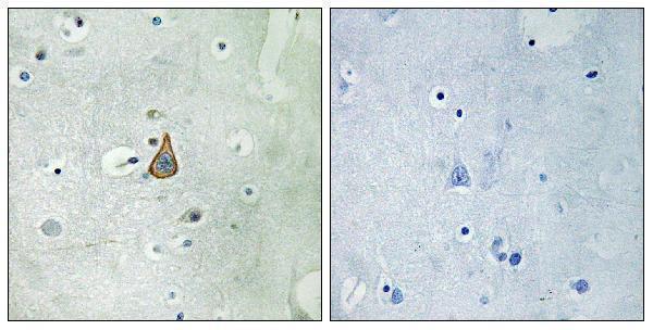
The catalog lists KCNC1 antibodies for IHC and IF in human, mouse, and rat (catalog: applications and reactivity); the supplied figure shows paraffin-embedded human brain IHC only (A06871-1 image caption).
A06871-1 lists IHC and IF for human, mouse, and rat, with an IHC figure captioned as paraffin-embedded human brain (catalog: applications, reactivity, image caption). A06871-2 lists IHC, IF, and ICC for human, mouse, and rat; no image caption is supplied (catalog: applications, reactivity, image alts).
Which to pick: For tissue IHC, choose A06871-1 when a paraffin-section example matters: its own caption shows human brain, but reports no fixative (A06871-1 image caption). For IF/ICC, choose A06871-2 because both applications are listed; A06871-1 lists IF but not ICC (catalog: applications). Both list human, mouse, and rat reactivity for cross-species planning; both have rabbit hosts, and neither has a reported clone (catalog: reactivity, host, clone).