This website uses cookies to ensure you get the best experience on our website.


- Table of Contents
Plan paraffin-section KCNJ2 IHC with tissue controls, antibody titration, and scoring. Compare the cytoplasmic tissue staining (HPA tissue IHC) with the channel’s annotated membrane location (UniProt).

Expected localisation, validated protocols, controls and antibodies — the at-a-glance facts below, then the full design guide.
| Expected localisation | Diffuse cytoplasm (HPA tissue IHC); cell membrane (UniProt) | |
| Staining pattern | Cytoplasmic staining in most tissues (HPA tissue IHC) | |
| Antigen retrieval | Citrate pH 6.0 HIER, 95–98 °C, 20 min (rule: cytoplasmic / membrane antigen) | |
| Positive control | Adrenal gland+4 more · see all | |
| Negative control | None in HPA (detected in all 45 tissues); use no-primary + isotype controls |
| Fixation | Keep formalin fixation consistent across sections (standard IHC practice; not target-specific); Matched tissue-IHC evidence does not establish the fixation claim. Validate the specimen-specific method before use. (selected-SKU IHC image A01850-1) | |
| Caveat | Staining has low consistency with RNA expression (HPA tissue IHC) | |
| Regulation | No expression regulator reported (UniProt) | |
| Isoform / epitope | No annotated isoforms; cytoplasmic and extracellular regions (UniProt) |
The catalog antibody’s IHC-P protocol is followed by published KCNJ2 IHC methods for osteosarcoma and lung sections (PMC9979522; PMC13205818).
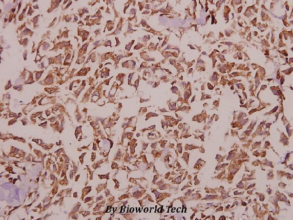
| Sample | Paraffin-embedded human breast carcinoma tissue; fixative not specified (datasheet A01850-1) |
| Fixation | Image fixative and duration unreported (datasheet A01850-1); verify before use. |
| Sectioning | 4–5 µm sections on charged slides (standard) |
| Deparaffinisation | Xylene, graded ethanol series to water (standard) |
| Antigen retrieval | Heat-induced epitope retrieval in citrate buffer, pH 6.0, 20 min at 95–98 °C (standard rule: cytoplasmic / membrane antigen) |
| Peroxidase block | 3% H2O2, 10 min, room temperature (standard) |
| Blocking | 10% normal serum of the secondary host, 30 min, room temperature (standard) |
| Primary antibody | Rabbit anti-KCNJ2, 1:50-1:200 (datasheet A01850-1) |
| Primary incubation | Overnight at 4 °C (standard) |
| Detection | HRP-polymer secondary, DAB chromogen 5–10 min (standard) |
| Counterstain | Hematoxylin, blue, dehydrate and mount (standard) |
| Expected result | KCNJ2-positive staining in glandular cells of adrenal gland (HPA tissue IHC: High). HPA tissue profile: Cytoplasmic expression in most tissues. No signal in the no-primary control. |
KCNJ2 is a cell membrane channel with cytoplasmic and extracellular regions (UniProt P63252 topology). In IHC, expect staining in cells such as cerebellar Purkinje cells, cortical neurons and bronchial respiratory epithelial cells (HPA: High). HPA also reports cytoplasmic expression in most tissues (HPA: tissue profile). Its IHC antibody is Approved, with low consistency between staining and RNA expression (HPA: reliability).
| Defined staining in Purkinje cells or bronchial respiratory epithelium, with a membrane-associated pattern. | These are useful positive-pattern examples (HPA: High in both cell types). Membrane staining fits KCNJ2 localisation (UniProt P63252: cell membrane). Judge the cell type and compartment together; HPA also describes cytoplasmic staining in most tissues (HPA: tissue profile). |
| Strong, isolated nuclear staining with little staining at the cell edge or in the cytoplasm. | A nuclear-only pattern conflicts with the recorded membrane localisation (UniProt P63252: cell membrane; HPA: subcellular summary, Membrane). Treat it as suspect staining and assess controls before assigning it to KCNJ2 (general IHC practice). |
| Strong staining in unexpected cells while the expected cells on the same section remain faint. | Review cell identification and antibody specificity; cross-reactivity or endogenous detection activity are possibilities, not established causes (general IHC practice). HPA names particular High-staining cell populations but also reports cytoplasmic staining in most tissues, so an unlisted cell is not automatically negative (HPA: tissue profile). |
| Broad, nearly uniform colour across unrelated structures or outside identifiable cells. | The result lacks the cell-specific contrast needed to interpret an IHC stain (general IHC practice). Diffuse background cannot establish KCNJ2 localisation, even though HPA reports cytoplasmic expression across most tissues (HPA: tissue profile). Compare with an appropriate detection control (general IHC practice). |
| No convincing signal in a section containing identifiable Purkinje cells or cortical neurons. | Both populations have High HPA tissue staining, so the result calls for a run-level check (HPA: High in cerebellar Purkinje cells and cerebral-cortex neurons). Absence on one slide does not establish biological absence; verify tissue identification, antibody conditions and detection controls (general IHC practice). |
| Compartment and topology | KCNJ2 has two transmembrane segments and substantial cytoplasmic regions (UniProt P63252 topology). A membrane-associated result fits the annotated location (UniProt P63252: cell membrane); HPA's predominantly cytoplasmic tissue IHC profile creates an interpretation caveat rather than proof that every cytoplasmic signal is specific (HPA: tissue profile). |
| Choice of tissue and cell population | HPA reports High staining in several defined populations, including Purkinje cells, cortical neurons, respiratory epithelial cells and colon endothelial cells (HPA: tissue IHC). Adipocytes are listed as Low, not negative (HPA: adipose tissue). Use the identified cell population when assessing a section; whole-tissue staining can conceal that distinction (general IHC practice). |
| Strength of the HPA IHC evidence | The listed antibody, HPA029109, has Approved IHC status; its record does not state Enhanced IHC status (HPA: antibody validation). HPA explicitly reports low consistency between staining and RNA expression (HPA: reliability description). Interpret a plausible pattern with that qualification, especially when using RNA abundance as a comparison. |
| Protein processing and epitope interpretation | The supplied UniProt record lists one chain spanning residues 1–427, with no signal peptide, propeptide or annotated isoforms (UniProt P63252: processing and isoforms). It does not identify this antibody's epitope, so topology alone cannot determine whether retrieval or permeabilisation will expose it in a paraffin section (UniProt P63252 topology; general IHC practice). |
| IF/ICC cross-check — what pattern is documented? | HPA summarises KCNJ2's subcellular location as Membrane, but supplies no ICC-IF image-bearing cell lines or main-location entry here (HPA: subcellular record). This supports a location expectation, not an image-based IF validation or an IF/ICC protocol choice. |
| Situation | Likely cause | Next action |
|---|---|---|
| A known High-staining cell population is blank. | A failed stain or detection step is possible; absence alone does not identify the cause (HPA: High in Purkinje cells and cortical neurons; general IHC practice). | Check that the expected cells are present, then review the run's antibody and detection controls and the documented IHC-P conditions (general IHC practice). |
| The slide shows nuclear-only staining. | The compartment conflicts with KCNJ2's membrane annotation and HPA's Membrane summary (UniProt P63252: cell membrane; HPA: subcellular record). | Compare the same run with a defined High-staining cell population and a detection control before interpreting the nuclear signal (HPA: tissue IHC; general IHC practice). |
| Colour is widespread and cell boundaries are hard to distinguish. | Background or excess detection signal may obscure cell-level interpretation (general IHC practice). HPA's broad cytoplasmic profile alone does not validate uniform slide-wide colour (HPA: tissue profile). | Assess detection-control staining and adjust general IHC blocking, washing or detection conditions as indicated by the controls (general IHC practice). |
| Unexpected cells stain more strongly than the named High-staining population. | Cell misidentification, cross-reactivity or endogenous detection activity are possible explanations (general IHC practice); HPA does not designate all other cells negative (HPA: tissue profile). | Recheck morphology and compare an appropriate detection control with the expected cells on the same section (general IHC practice). |
| Cytoplasmic staining appears without a crisp membrane outline. | HPA reports cytoplasmic expression in most tissues, while UniProt places KCNJ2 at the membrane (HPA: tissue profile; UniProt P63252: cell membrane). The records do not resolve every cytoplasmic signal's specificity. | Record the observed compartment precisely; weigh its cell distribution and controls before calling it KCNJ2-specific (general IHC practice). |
| Adipocytes show weak staining, or staining differs from an RNA-based expectation. | HPA lists adipocytes as Low and reports low consistency between antibody staining and RNA expression (HPA: adipose tissue; HPA: reliability description). | Treat Low as a comparison level rather than a zero-signal control, and report the IHC observation separately from RNA evidence (HPA: adipose tissue and reliability; general IHC practice). |


Comprehensive Human Protein Atlas IHC scoring per tissue (reliability: Approved — Low consistency between antibody staining and RNA expression data.). Rows are taken directly from the HPA tissue chart — click any row's HPA link to view the source.
| Tissue | Cell type | Level | Evidence | Source |
|---|---|---|---|---|
| Adrenal gland | Glandular cells | High | Protein (IHC) | HPA → |
| Breast | Glandular cells | High | Protein (IHC) | HPA → |
| Bronchus | Respiratory epithelial cells | High | Protein (IHC) | HPA → |
| Caudate | Neuronal cells | High | Protein (IHC) | HPA → |
| Cerebellum | Purkinje cells | High | Protein (IHC) | HPA → |
| Tissue | Cell type | Level | Evidence | Source |
|---|---|---|---|---|
| None in HPA: KCNJ2 is detected in all 45 scored tissues. Use a no-primary (secondary-only) and an isotype control instead. | ||||
Troubleshoot KCNJ2 staining in paraffin sections by checking retrieval, compartment, cell identity and controls before interpreting chromogenic signal.
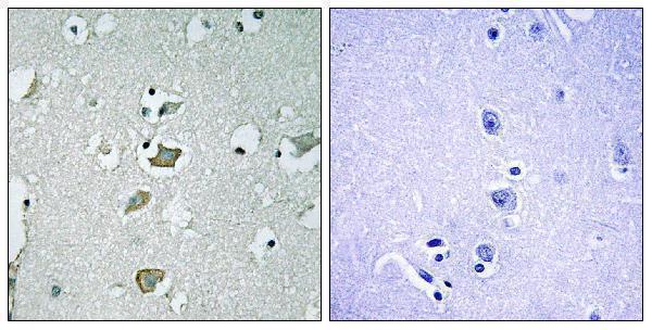
Two anti-KCNJ2 antibodies have IHC images from paraffin-embedded human breast carcinoma and brain tissue (catalog image captions); A30730 also lists IF/ICC applications (catalog applications).
A01850-1 shows IHC staining of paraffin-embedded human breast carcinoma at 1:50 and lists Human, Mouse, and Rat reactivity (A01850-1 image caption; catalog reactivity). A30730 shows IHC staining of paraffin-embedded human brain tissue with a peptide-blocked comparison and lists Human and Rat reactivity plus IF/ICC applications (A30730 image caption; catalog applications/reactivity).
Which to pick: For tissue IHC, choose A01850-1 for paraffin-embedded breast carcinoma or A30730 for paraffin-embedded brain tissue, matching each antibody’s own image (catalog image captions). For IF/ICC, choose A30730 because those applications are listed, although no IF image is supplied (A30730 catalog applications; IF image data). For mouse samples, A01850-1 lists Mouse reactivity; both IHC captions describe paraffin-embedded tissue without reporting the fixative (catalog reactivity; catalog image captions).