KCNQ1 / Potassium voltage-gated channel subfamily KQT member 1 · IHC design guide

Design Immunohistochemistry for KCNQ1

Plan chromogenic KCNQ1 IHC in paraffin sections using adrenal gland or stomach glandular cells as positive tissue controls (HPA tissue IHC). The guide covers the catalog antibody’s 2–5 μg/ml IHC range (datasheet A00310-1) and scoring of cytoplasmic and membranous staining (HPA tissue IHC).

Evidence assembled Oct 2026 · For research use; verify linked source records and product datasheet before use
Immunohistochemistry protocol sheet for KCNQ1 (IHC for KCNQ1): expected localisation Cytoplasmic and membranous in glandular cells (HPA tissue IHC), antibody A00310-1, validated IHC image, and IHC protocol steps
Printable KCNQ1 IHC protocol sheet — expected localisation Cytoplasmic and membranous in glandular cells (HPA tissue IHC), antibody A00310-1, controls and protocol steps. Open the full KCNQ1 IHC guide →

KCNQ1 Immunohistochemistry Experimental Design Guide

Expected localisation, validated protocols, controls and antibodies — the at-a-glance facts below, then the full design guide.

Must know before staining
Expected localisation Cytoplasmic and membranous in glandular cells (HPA tissue IHC)
Staining pattern Adrenal, thyroid and stomach: cytoplasmic and membranous glandular staining (HPA tissue IHC)
Antigen retrieval EDTA pH 8.0 HIER, heat-mediated (datasheet A00310-1)
Positive control ⓘ Adrenal gland+4 more · see all
Negative control ⓘ Adipose tissue+4 more · see all
Important caveats
Reasons your staining may differ from the expected pattern.
Fixation Matched tissue-IHC evidence does not establish the fixation claim. Validate the specimen-specific method before use. (selected-SKU IHC image A00310-1)
Caveat Epithelial polarization can shift KCNQ1 from ER to basolateral membrane (UniProt)
Regulation Estradiol promotes internalization (UniProt)
Isoform / epitope 2 isoforms; check whether the epitope is extracellular or cytoplasmic (UniProt)
Section 1

Recommended KCNQ1 IHC & IF Protocols

The catalog antibody’s IHC-P protocol (datasheet: A00310-1) is paired with two published KCNQ1 paraffin-section protocols (PMC3167289; PMC3935979).

Recommended immunohistochemistry (IHC-P) protocol parameters
SampleParaffin-embedded human liver cancer tissue; fixative not specified (datasheet A00310-1)
FixationImage fixative and duration unreported (datasheet A00310-1); verify before use.
Sectioning4–5 µm sections on charged slides (standard)
DeparaffinisationXylene, graded ethanol series to water (standard)
Antigen retrievalHeat retrieval: EDTA pH 8.0 (datasheet A00310-1); 20 min, 95–100 °C (standard)
Peroxidase block3% H2O2, 10 min, room temperature (standard)
Blocking10% goat serum (datasheet A00310-1)
Primary antibodyRabbit anti-KCNQ1, 2-5 μg/ml (datasheet A00310-1)
Primary incubationOvernight at 4 °C (datasheet A00310-1)
DetectionHRP-conjugated secondary, DAB chromogen (datasheet A00310-1)
CounterstainHematoxylin, blue, dehydrate and mount (standard)
Expected resultKCNQ1-positive staining in glandular cells of adrenal gland (HPA tissue IHC: High). HPA tissue profile: Cytoplasmic and membranous expression mainly in adrenal gland, thyroid gland and stomach. No signal in the no-primary control.
💡Decision noteStart with heat-mediated EDTA pH 8.0 retrieval (datasheet: A00310-1); the published protocols used Tris-EDTA pH 9.0 or citrate pH 6.0 (PMC3167289; PMC3935979).
Section 2

What Is the Expected KCNQ1 Staining Pattern?

KCNQ1 is a six-pass membrane protein with cytoplasmic regions (UniProt P51787 topology). In paraffin-section IHC, expect cytoplasmic and membranous staining in selected glandular cells and enterocytes (HPA tissue IHC). HPA rates its tissue pattern “Enhanced,” while reporting medium agreement with RNA data and pending retesting (HPA tissue IHC).

What am I looking at on my slide?
Membranous staining with some cytoplasmic signal in adrenal, stomach, thyroid, parathyroid, or seminal-vesicle glandular cells, or duodenal enterocytes.These cell types are reported as High by HPA (HPA tissue IHC). Cytoplasmic and membranous staining fits HPA’s tissue profile; UniProt also places KCNQ1 at the cell membrane and intracellular membranes (HPA tissue IHC; UniProt P51787 subcellular location).
Signal appears chiefly nuclear, with little plausible membrane or cytoplasmic staining in an otherwise positive cell population.Nuclear staining falls outside the supplied KCNQ1 locations (UniProt P51787 subcellular location; HPA tissue IHC). Treat it as an unexplained pattern and assess antibody and detection controls before assigning it to KCNQ1 (general IHC practice).
Strong staining appears in adipocytes, endometrial stromal cells, or prostate glandular cells.HPA reports these specific cell populations as Not detected (HPA tissue IHC). Consider cross-reactivity or endogenous detection activity, while retaining the cell-level distinction: UniProt reports abundant expression in prostate without specifying its IHC-positive cells (UniProt P51787 tissue specificity).
Chromogen is spread across stroma, lumina, or many unrelated cell types without clear cell boundaries.That distribution does not match HPA’s selected-cell tissue profile (HPA tissue IHC). Diffuse background can obscure genuine staining; examine reagent-only controls, blocking, washing, and development conditions as general IHC checks (general IHC practice).
A known-positive section has no convincing staining in its expected cells.For example, absent signal in duodenal enterocytes conflicts with their High HPA staining level (HPA tissue IHC). First check section integrity, antibody application, retrieval, and detection controls; this record supplies no KCNQ1-specific fixation-sensitivity finding (general IHC practice; supplied source scope).
💡Expected KCNQ1 appearanceCall a result positive when selected glandular cells or duodenal enterocytes show clear membranous and cytoplasmic staining, potentially strong in HPA High populations; isolated nuclear signal or widespread cell-independent color is suspect (HPA tissue IHC; UniProt P51787 subcellular location; general IHC practice).
How each factor affects the staining
Topology and epitope accessKCNQ1 has six transmembrane segments, extracellular loops, and cytoplasmic regions (UniProt P51787 topology). Interpret a staining failure against the antibody’s documented epitope, if available; the supplied record gives no antibody epitope or KCNQ1-specific retrieval requirement.
Compartment can varyUniProt lists cell membrane, endosome, vesicle membrane, and endoplasmic reticulum locations, with apical or basolateral membrane placement in some contexts (UniProt P51787 subcellular location). A cytoplasmic component alone does not identify which organelle contains the signal.
Protein formsUniProt lists two isoforms and a full-length chain spanning residues 1–676, with no signal peptide or propeptide annotated (UniProt P51787 isoforms and processing). The supplied evidence does not show which form any listed IHC antibody detects.
Strength of tissue evidenceHPA calls the tissue IHC pattern Enhanced but reports medium staining–RNA consistency and pending retesting (HPA tissue IHC). Its antibody records list IHC Enhanced for HPA048553; ICC Supported applies to HPA011306 and HPA071107, a different application (HPA antibodies).
Prostate evidence differs by methodUniProt lists abundant prostate expression, whereas HPA reports prostate glandular cells as Not detected by tissue IHC (UniProt P51787 tissue specificity; HPA tissue IHC). Avoid using that cell population as an assured positive IHC control.
Why is my staining missing, weak or wrong?
SituationLikely causeNext action
No staining in a selected positive tissue.A failed IHC step or poorly preserved section may be responsible; HPA reports High staining in duodenal enterocytes and stomach glandular cells (HPA tissue IHC; general IHC practice).Review tissue morphology, antibody application, retrieval, and detection controls on the same run (general IHC practice). Do not infer a KCNQ1-specific fixation effect from these sources.
Color appears in a reported negative cell population.Cross-reactivity or endogenous detection activity is possible (general IHC practice); HPA reports adipocytes and endometrial stromal cells as Not detected (HPA tissue IHC).Compare reagent-only and detection controls, then check whether color follows cells or background structures (general IHC practice). Interpret the named cell population, not the whole tissue.
Most cells show a faint, uniform haze.Nonspecific background or overdevelopment may overwhelm a selective pattern (general IHC practice); HPA describes chiefly cytoplasmic and membranous staining in selected tissues (HPA tissue IHC).Check blocking, washes, antibody concentration, and development time using appropriate run controls (general IHC practice); preserve a visible distinction between expected cells and background.
Staining is exclusively nuclear.The supplied KCNQ1 locations are membranous and intracellular, without a nuclear assignment (UniProt P51787 subcellular location; HPA tissue IHC).Treat the nuclear pattern as unconfirmed; compare a known-positive section and control stains before scoring it as KCNQ1 (general IHC practice).
Prostate glandular cells lack signal despite the UniProt tissue entry.UniProt reports abundant prostate expression, while HPA scores those glandular cells Not detected by IHC (UniProt P51787 tissue specificity; HPA tissue IHC).Use an HPA High cell population, such as duodenal enterocytes, to judge IHC performance; do not call prostate glandular cells a guaranteed positive control (HPA tissue IHC).
IF/ICC Q: Where should signal localize?A: HPA reports mainly endoplasmic reticulum localization, with additional plasma membrane and cytosol signal (HPA subcellular ICC-IF).Interpret that as ICC-IF evidence, distinct from HPA’s paraffin-section tissue IHC pattern; this section provides no IF/ICC protocol (HPA subcellular ICC-IF; HPA tissue IHC).

Sample controls for KCNQ1 IHC & IF

🧪Run duodenum first and expect staining in enterocytes (HPA: High in duodenal enterocytes). Use adipose tissue adipocytes as the negative tissue (HPA: Not detected in adipocytes); on the duodenal slide, non-enterocyte areas should provide a background comparison, but their KCNQ1 status is not established by the supplied HPA row.
Positive control tissue: Adrenal gland (Glandular cells, HPA High)
Negative control tissue: Adipose tissue (HPA Not detected)
ICC-IF cell lines (HPA subcellular resource): HPA ICC-IF images show KCNQ1 in OE19, SH-SY5Y, THP-1, U2OS, with annotated localisation: Endoplasmic reticulum (enhanced) (HPA subcellular).
Technical controls: Include a no-primary, secondary-only control and an isotype-matched rabbit IgG control; the selected IHC caption identifies a rabbit primary antibody but does not state its clonality (catalog antibody A00310-1 caption). Use a KCNQ1 knockout specimen as a biological specificity control where available, and quench endogenous peroxidase before HRP/DAB detection in duodenum (catalog antibody A00310-1 caption: HRP/DAB).
⚠️Feasibility: The selected paraffin-section caption reports heat retrieval in EDTA at pH 8.0, but its fixative is unreported; no target-specific fixation window or fixation effect is reported in the supplied evidence (catalog antibody A00310-1 caption). Paraffin IHC has a documented example here, while the supplied evidence does not establish that frozen sections or IF are easier; HPA reports ICC-IF localization mainly in the endoplasmic reticulum, with additional plasma membrane and cytosol signal (HPA subcellular). In duodenum, assess any brown background from endogenous peroxidase separately from enterocyte staining (HPA: High in duodenal enterocytes; catalog antibody A00310-1 caption: HRP/DAB).

HPA tissue IHC evidence for KCNQ1

Comprehensive Human Protein Atlas IHC scoring per tissue (reliability: Enhanced — Medium consistency between antibody staining and RNA expression data. Pending retesting.). Rows are taken directly from the HPA tissue chart — click any row's HPA link to view the source.

Positive expression · recommended positive controls

TissueCell typeLevelEvidenceSource
Adrenal gland Glandular cells High Protein (IHC) HPA →
Duodenum Enterocytes High Protein (IHC) HPA →
Parathyroid gland Glandular cells High Protein (IHC) HPA →
Seminal vesicle Glandular cells High Protein (IHC) HPA →
Stomach Glandular cells High Protein (IHC) HPA →

Undetected expression · recommended negative controls

TissueCell typeLevelEvidenceSource
Adipose tissue Adipocytes Not detected Protein (IHC) HPA →
Caudate Glial cells Not detected Protein (IHC) HPA →
Endometrium Cells in endometrial stroma Not detected Protein (IHC) HPA →
Lymph node Germinal center cells Not detected Protein (IHC) HPA →
Prostate Glandular cells Not detected Protein (IHC) HPA →
Section 3

Advanced KCNQ1 IHC Tips

Troubleshoot KCNQ1 staining in paraffin sections by checking retrieval, compartment, cell identity, controls and scoring before interpreting signal.

Which retrieval condition should I start with for weak KCNQ1 staining?
Start with heat-mediated antigen retrieval in EDTA at pH 8.0 for paraffin sections (datasheet A00310-1). The selected image used this retrieval before overnight incubation at 4°C with 2 μg/ml catalog antibody, so keep those conditions together as an initial comparison (datasheet A00310-1). If staining remains weak, compare retrieval durations on adjacent sections while keeping antibody concentration, detection and exposure to DAB constant (standard IHC practice). Check whether any gain occurs in glandular cells of stomach or thyroid gland, both reported as high, without a matching rise in a no-primary control (HPA tissue IHC; standard IHC practice).
Could fixation explain a weak or patchy KCNQ1 result?
Target-specific sensitivity to fixation is unknown because the selected paraffin-section caption does not state a fixative (datasheet A00310-1). Record the specimen’s actual fixative and fixation duration, then compare sections with matched processing and the same EDTA pH 8.0 retrieval (datasheet A00310-1; standard IHC practice). Examine morphology alongside signal: uneven preservation, damaged edges and poor nuclear detail can make a staining difference difficult to attribute to KCNQ1 (standard IHC practice). Include a consistent positive control, such as stomach glandular cells reported high by HPA, and compare it across runs before changing antibody concentration (HPA tissue IHC; standard IHC practice).
How should I assess cytoplasmic staining from a membrane channel?
Score membrane and intracellular patterns separately because KCNQ1 is reported at the cell membrane, endoplasmic reticulum and early endosomes (UniProt P51787 subcellular location). Cytoplasmic and membranous tissue staining is reported mainly in adrenal gland, thyroid gland and stomach, while cell imaging places KCNQ1 mainly in the endoplasmic reticulum (HPA tissue IHC; HPA subcellular). In polarized epithelium, inspect apical and basolateral borders independently because both membrane locations are annotated for KCNQ1 (UniProt P51787 subcellular location). Compare each pattern with the same compartment in a no-primary control, and record diffuse DAB separately from a discernible membrane or intracellular distribution (standard IHC practice).
Why might two KCNQ1 antibodies stain different compartments?
KCNQ1 has 2 annotated isoforms and 6 transmembrane segments, so establish which sequence and epitope each antibody recognizes before comparing stains (UniProt P51787 isoforms and topology). The annotated N terminus, residues 1–120, and C-terminal region, residues 349–676, are cytoplasmic; several short loops are extracellular (UniProt P51787 topology). Glycosylation at residue 289 and phosphorylation at residues 27, 407 and 409 are annotated, but their effects on this antibody’s IHC staining are unreported (UniProt P51787 processing; datasheet A00310-1). Compare antibodies on adjacent sections with the same retrieval and controls, and avoid assigning an isoform from staining pattern alone (standard IHC practice).
How can I use IF to investigate an ambiguous IHC pattern?
Use IF/ICC as a separate assay to test whether KCNQ1 overlaps a marker of the expected glandular or enterocyte population, then compare that cellular pattern with chromogenic IHC (HPA tissue IHC; standard IF practice). Choose fluorophores after checking tissue autofluorescence and include single-stain controls, since broad background can mimic intracellular overlap (standard IF practice). Select permeabilisation according to the antibody epitope: intracellular epitopes require access across membranes, whereas an extracellular epitope can be assessed with minimal or no permeabilisation (UniProt P51787 topology; standard IF practice). HPA cell imaging reports mainly endoplasmic reticulum localisation with additional plasma membrane and cytosolic signal, which can guide compartment checks without validating this catalog antibody for IF (HPA subcellular).
What should I adjust when DAB obscures KCNQ1 localisation?
First inspect a no-primary section and the tissue edges to distinguish nonspecific DAB deposition from a cell-associated KCNQ1 pattern (standard IHC practice). The selected paraffin-section image used 10% goat serum blocking, 2 μg/ml primary antibody overnight at 4°C, and a peroxidase-linked detection system with DAB (datasheet A00310-1). If background is excessive, titrate primary antibody and shorten chromogen development on adjacent sections while preserving the same retrieval and control tissues (standard IHC practice). Include an endogenous peroxidase block as a general chromogenic workflow step, then require the expected glandular-cell pattern to exceed background in a no-primary control (HPA tissue IHC; standard IHC practice).
How should I quantify KCNQ1 across heterogeneous tissue sections? ⚠ ANSWER MARKED FOR VERIFICATION
Define the cell population and compartment before scoring; HPA reports high staining in stomach glandular cells and duodenal enterocytes, with cytoplasmic and membranous patterns in its tissue profile (HPA tissue IHC). For a defined region, record the percentage of positive target cells and an H-score based on intensity categories, then report membrane and cytoplasmic scores separately (standard IHC practice). Normalize cell-based scores to the number of evaluable target cells, or normalize positive-cell counts to analyzed tissue area in mm² (standard IHC practice). Keep retrieval, DAB development and image thresholds consistent across specimens, and document excluded necrotic or damaged areas before comparison (standard IHC practice).
When is an apparent KCNQ1-positive area likely to be artefact?
A credible KCNQ1 result should align with cell identity and a plausible membrane or intracellular compartment, including the endoplasmic reticulum (HPA tissue IHC; HPA subcellular; UniProt P51787 subcellular location). Check suspicious staining in the wrong cell population against a no-primary section and a positive control such as stomach glandular cells, reported high by HPA (HPA tissue IHC; standard IHC practice). Edge-limited color, necrotic areas and signal reproduced without primary antibody favor processing, DAB or endogenous-enzyme artefact (standard IHC practice). Interpret weak or discordant areas cautiously because HPA rates tissue-IHC reliability as Enhanced while reporting medium staining–RNA consistency and pending retesting (HPA tissue IHC).
Boster reagents

Best KCNQ1 / Potassium voltage-gated channel subfamily KQT member 1 IHC Antibodies

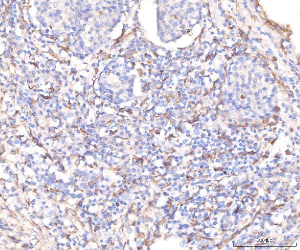
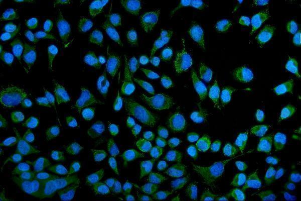
The catalog antibody has IHC images from human paraffin sections and an IF image from HeLa cells (datasheet: image captions). Human, mouse, and rat reactivity is listed (catalog: reactivity).

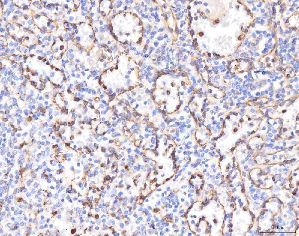
Real IHC data IHC analysis of KCNQ1 using anti-KCNQ1 antibody (A00310-1). KCNQ1 was detected in a paraffin-embedded section of human liver cancer tissue. Heat mediated antigen retrieval was performed in EDTA buffer (pH 8.0, epitope retrieval solution). The tissue section was blocked with 10% goat serum. The tissue section was then incubated with 2 μg/ml rabbit anti-KCNQ1 Antibody (A00310-1) overnight at 4°C. Peroxidase Conjugated Goat Anti-rabbit IgG was used as secondary antibody and incubated for 30 minutes at 37°C. The tissue section was developed using HRP Conjugated Rabbit IgG Super Vision Assay Kit (Catalog # SV0002) with DAB as the chromogen.
Anti-KCNQ1 Antibody ®
Cat # A00310-1

A00310-1 has IHC images from paraffin sections of human liver cancer, lung cancer, placenta, and breast cancer (datasheet: IHC image captions). Its IF image shows HeLa cells (datasheet: IF image caption); the catalog lists IHC, IF, and ICC applications and human, mouse, and rat reactivity (catalog: applications/reactivity).

Which to pick: Choose A00310-1 for paraffin-section IHC: its IHC captions show human tissue with EDTA retrieval at pH 8.0, and the fixative is unreported (datasheet: IHC image captions). The same SKU is listed for IF/ICC and has a HeLa-cell IF image (catalog: applications; datasheet: IF image caption). For mouse or rat work, it lists reactivity with both species, although the supplied IHC and IF images show human samples; clonality is unreported (catalog: reactivity/clone; datasheet: image captions).

Each figure is that product's own IHC / IF validation image from its datasheet.

References

  1. UniProt Consortium. UniProt entry P51787 (KCNQ1_HUMAN, Potassium voltage-gated channel subfamily KQT member 1).
  2. Human Protein Atlas. KCNQ1 tissue IHC expression (reliability: Enhanced).
  3. Human Protein Atlas. KCNQ1 subcellular location (ICC-IF): Mainly localized to the endoplasmic reticulum. In addition localized to the plasma membrane and cytosol..
  4. Human Protein Atlas. KCNQ1 antibody validation summary (3 antibodies).
  5. Prox1 maintains taste bud structure via inhibition of apoptosis. Cell and tissue research 2026 — PMC12872690.
  6. Differential expression of KCNQ1 K+ channel in tubular cells of frog kidney. European journal of histochemistry : EJH 2010 — PMC3167289.
  7. The role of KCNQ1 in mouse and human gastrointestinal cancers. Oncogene 2014 — PMC3935979.
  8. A Novel Role of Arrhythmia-Related Gene KCNQ1 Revealed by Multi-Omic Analysis: Theragnostic Value and Potential Mechanisms in Lung Adenocarcinoma. International journal of molecular sciences 2022 — PMC8874518.
  9. PubMed PMID:9312006 — UniProt-cited evidence.
  10. PubMed PMID:9305853 — UniProt-cited evidence.
  11. PubMed PMID:9799083 — UniProt-cited evidence.