This website uses cookies to ensure you get the best experience on our website.


- Table of Contents
Plan chromogenic KDELR3 IHC in paraffin sections using glandular cells in appendix and small intestine as positive references (HPA tissue IHC). Expect cytoplasmic, sometimes granular staining, but interpret it cautiously because tissue-IHC reliability is uncertain (HPA tissue IHC).

Expected localisation, validated protocols, controls and antibodies — the at-a-glance facts below, then the full design guide.
| Expected localisation | Cytoplasmic staining, sometimes granular (HPA tissue IHC) | |
| Staining pattern | Glandular cells: cytoplasmic, sometimes granular (HPA tissue IHC) | |
| Antigen retrieval | Citrate pH 6.0 HIER, 95–98 °C, 20 min (rule: cytoplasmic / membrane antigen) | |
| Positive control | Appendix+4 more · see all | |
| Negative control | Adipose tissue+4 more · see all |
| Fixation | Keep fixation consistent across sections (standard IHC practice; not target-specific); Matched tissue-IHC evidence does not establish the fixation claim. Validate the specimen-specific method before use. (selected-SKU IHC image A14682-1) | |
| Caveat | Very low staining–RNA concordance (HPA tissue IHC) | |
| Regulation | No regulation annotated (UniProt) | |
| Isoform / epitope | 2 isoforms; map epitopes to lumenal or cytoplasmic loops (UniProt) |
The catalog antibody’s IHC-P protocol appears alongside four published KDELR3 chromogenic IHC examples (PMC11681132; PMC11625429; PMC11451269; PMC6965108).
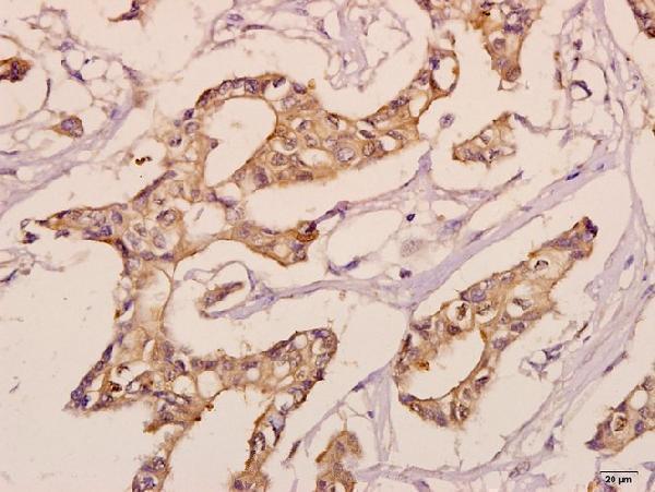
| Sample | Paraffin-embedded human breast carcinoma tissue; fixative not specified (datasheet A14682-1) |
| Fixation | Image fixative and duration unreported (datasheet A14682-1); verify before use. |
| Sectioning | 4–5 µm sections on charged slides (standard) |
| Deparaffinisation | Xylene, graded ethanol series to water (standard) |
| Antigen retrieval | Heat-induced epitope retrieval in citrate buffer, pH 6.0, 20 min at 95–98 °C (standard rule: cytoplasmic / membrane antigen) |
| Peroxidase block | 3% H2O2, 10 min, room temperature (standard) |
| Blocking | 10% normal serum of the secondary host, 30 min, room temperature (standard) |
| Primary antibody | Rabbit anti-KDELR3, 1:50-1:200 (datasheet A14682-1) |
| Primary incubation | Overnight at 4 °C (standard) |
| Detection | HRP-polymer secondary, DAB chromogen 5–10 min (standard) |
| Counterstain | Hematoxylin, blue, dehydrate and mount (standard) |
| Expected result | KDELR3-positive staining in glandular cells of appendix (HPA tissue IHC: High). HPA tissue profile: Cytoplasmic, sometimes granular expression in a few tissues. No signal in the no-primary control. |
KDELR3 is a seven-pass membrane receptor that cycles between the Golgi and endoplasmic reticulum with KDEL-bearing cargo (UniProt O43731 topology and localization). In paraffin-section IHC, expect cytoplasmic, sometimes granular staining, particularly in appendix and small-intestine glandular cells (HPA: High). Treat this as a provisional pattern: HPA rates tissue IHC reliability Uncertain because antibody staining and RNA expression show very low consistency (HPA: tissue IHC).
| Glandular cells show cytoplasmic, sometimes granular chromogen in appendix or small intestine (HPA: High in both). | This matches the reported tissue pattern and the ER/Golgi membrane location (HPA: tissue IHC; UniProt O43731 localization). Evaluate cell boundaries and morphology before scoring; HPA's Uncertain IHC rating prevents treating matching stain alone as proof of specificity (HPA: tissue IHC). |
| Most signal appears inside nuclei, along cell surfaces, or in extracellular material. | Those dominant compartments do not match the reported cytoplasmic IHC pattern or membrane-bound ER/Golgi localization (HPA: tissue IHC; UniProt O43731 localization). Review morphology and controls for artefact before interpreting the signal as KDELR3. |
| Unexpected strong stain appears in adipocytes or bronchial respiratory epithelium (HPA: Not detected in those cells). | Investigate antibody cross-reactivity or endogenous detection activity, especially if the stain is diffuse or lacks cytoplasmic structure. A mismatch is a warning, not a definitive exclusion: the HPA tissue IHC assessment is Uncertain (HPA: tissue IHC). |
| Chromogen covers many cell types and the surrounding section without clear cell-associated granules. | Poorly localized, widespread color is more consistent with background than the reported cytoplasmic, sometimes granular pattern (HPA: tissue IHC). Compare a no-primary control and assess blocking, washing, and chromogen development as general IHC checks. |
| Appendix or small-intestine glandular cells have no discernible stain despite preserved morphology (HPA: High in both). | Check that the assay can reveal signal before calling the sample KDELR3-negative. Review antibody identity, retrieval, detection, and the positive-control section as general IHC checks; HPA's Uncertain rating means even these reported high-staining tissues are imperfect controls (HPA: tissue IHC). |
| Cell compartment and topology (UniProt O43731) | KDELR3 has seven transmembrane segments and is assigned to ER, Golgi, and COPI-vesicle membranes (UniProt O43731 topology and localization). Cytoplasmic organelle-associated staining is plausible; topology alone does not specify how a particular antibody will stain a paraffin section. |
| Tissue-control choice (HPA: tissue IHC) | Appendix and small-intestine glandular cells are reported High; adipocytes and bronchial respiratory epithelial cells are Not detected (HPA: tissue IHC). Use these as comparison tissues or cell types while retaining the Uncertain IHC reliability caveat. |
| Antibody evidence (HPA: antibody validation) | HPA043477 has Uncertain IHC status, while HPA076405 has Supported ICC status and no listed IHC status (HPA: antibody validation). The ICC result does not validate either antibody's paraffin-section IHC pattern; keep application-specific evidence separate. |
| Isoforms and epitope (UniProt O43731) | Two isoforms are listed, and their coverage by any particular antibody cannot be inferred from this record (UniProt O43731 isoforms). If staining differs between samples, check the antibody's stated epitope and isoform coverage before assigning the difference to KDELR3 abundance. |
| IF/ICC Q&A: What pattern is supported? | An ER pattern is supported in ICC-IF, with images listed for MCF-7, SiHa, and U2OS (HPA: subcellular). That finding can inform localization interpretation; it does not establish an IF protocol or override the Uncertain tissue IHC assessment (HPA: tissue IHC). |
| Situation | Likely cause | Next action |
|---|---|---|
| No signal in appendix or small-intestine glandular cells (HPA: High). | The IHC assay may have failed, or the antibody may not reproduce the reported tissue pattern; HPA rates tissue IHC Uncertain (HPA: tissue IHC). | Confirm the expected cells are present, run a documented positive control, and check retrieval and detection against the antibody's IHC instructions. Treat retrieval review as general IHC practice, not a KDELR3-specific fixation claim. |
| Strong nuclear signal dominates the section. | Nuclear dominance conflicts with the cytoplasmic HPA pattern and ER/Golgi membrane assignment (HPA: tissue IHC; UniProt O43731 localization). | Compare the no-primary control and inspect staining at the cell level. Reassess antibody specificity and detection background before scoring nuclear color as KDELR3. |
| Adipocytes or bronchial respiratory epithelial cells stain strongly (HPA: Not detected). | The observed cell-type pattern may reflect nonspecific binding or endogenous detection activity; the HPA IHC profile is itself Uncertain (HPA: tissue IHC). | Include a no-primary control, review detection blocking, and compare the same run with appendix or small intestine. Investigate the mismatch without declaring the unexpected cells definitively KDELR3-negative. |
| Diffuse chromogen obscures glandular-cell boundaries. | Background from binding, inadequate washing, or detection development can mask the reported cytoplasmic, sometimes granular pattern (HPA: tissue IHC; general IHC practice). | Inspect the no-primary control; optimize blocking, washes, and development using standard IHC practice. Score only distinguishable cell-associated staining, and record any unresolved background. |
| Only a faint or patchy signal appears in a reported High tissue (HPA: appendix and small intestine). | Variation in assay performance or section quality may weaken visible staining; HPA's Uncertain rating also limits how firmly its High calls predict another run (HPA: tissue IHC). | Check preserved tissue morphology, reagent performance, and staining consistency across the section. Repeat with an appropriate control if needed; avoid converting weak signal into a categorical absence call. |
| IF/ICC shows an ER pattern while tissue IHC is weak or inconsistent (HPA: subcellular; HPA: tissue IHC). | The assays have different validation evidence: HPA reports Supported ICC localization for HPA076405 but Uncertain tissue IHC for HPA043477 (HPA: antibody validation). | Interpret each application with its own antibody and controls. Use the ER result as localization context, then evaluate paraffin-section IHC on its own cytoplasmic pattern and tissue controls. |
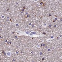


Comprehensive Human Protein Atlas IHC scoring per tissue (reliability: Uncertain — Very low consistency between antibody staining and RNA expression data.). Rows are taken directly from the HPA tissue chart — click any row's HPA link to view the source.
| Tissue | Cell type | Level | Evidence | Source |
|---|---|---|---|---|
| Appendix | Glandular cells | High | Protein (IHC) | HPA → |
| Small intestine | Glandular cells | High | Protein (IHC) | HPA → |
| Adrenal gland | Glandular cells | Medium | Protein (IHC) | HPA → |
| Caudate | Glial cells | Medium | Protein (IHC) | HPA → |
| Cerebellum | Cells in granular layer | Medium | Protein (IHC) | HPA → |
| Tissue | Cell type | Level | Evidence | Source |
|---|---|---|---|---|
| Adipose tissue | Adipocytes | Not detected | Protein (IHC) | HPA → |
| Bone marrow | Hematopoietic cells | Not detected | Protein (IHC) | HPA → |
| Breast | Adipocytes | Not detected | Protein (IHC) | HPA → |
| Bronchus | Respiratory epithelial cells | Not detected | Protein (IHC) | HPA → |
| Cervix | Glandular cells | Not detected | Protein (IHC) | HPA → |
Troubleshoot KDELR3 staining in paraffin sections by checking retrieval, controls, subcellular pattern, and scoring before interpreting tissue differences.
Anti-KDELR3 catalog entries list IHC for human, mouse, and rat and IF/ICC for human and mouse (catalog applications/reactivity). The supplied image documents paraffin-embedded human breast carcinoma IHC (A14682-1 image caption).
A14682-1 is the only card that renders; its IHC image shows paraffin-embedded human breast carcinoma at 1:50 (A14682-1 image caption). Its listed IHC reactivity includes human, mouse, and rat, while the supplied image documents human tissue only (A14682-1 applications/reactivity; A14682-1 image caption).
Which to pick: For paraffin-section IHC, choose A14682-1: its own image documents human breast carcinoma at 1:50; the fixative is unreported (A14682-1 image caption). For IF/ICC, A30677 lists both applications for human and mouse, with no IF image supplied (A30677 applications/reactivity/image alts). For cross-species IHC, A14682-1 has the broadest listed reactivity—human, mouse, and rat—although its image documents human tissue only (A14682-1 reactivity/image caption).