This website uses cookies to ensure you get the best experience on our website.


- Table of Contents
Plan paraffin section KDM4A IHC with nuclear localisation in mind (UniProt) while evaluating the reported cytoplasmic tissue staining (HPA tissue IHC). The catalog antibody A02850-1 has a documented IHC dilution of 2–5 μg/mL (datasheet A02850-1); score nuclear and cytoplasmic signal separately.

Expected localisation, validated protocols, controls and antibodies — the at-a-glance facts below, then the full design guide.
| Expected localisation | Cytoplasmic; nuclear in neurons and fallopian glands (HPA tissue IHC) | |
| Staining pattern | Cytoplasm across tissues; neuronal and fallopian gland nuclei (HPA tissue IHC) | |
| Antigen retrieval | EDTA pH 8.0 HIER, heat-mediated (datasheet A02850-1) | |
| Positive control | Caudate+4 more · see all | |
| Negative control | Adipose tissue+4 more · see all |
| Fixation | Keep fixation consistent across sections (standard IHC practice; not target-specific). Selected-image fixative and duration unreported (datasheet A02850-1); verify before use. | |
| Caveat | Reported tissue staining has uncertain reliability (HPA tissue IHC) | |
| Regulation | FBXO22 promotes KDM4A ubiquitination (UniProt) | |
| Isoform / epitope | 2 isoforms; epitope differences unspecified (UniProt) |
The catalog antibody’s IHC-P protocol is accompanied by published KDM4A methods for endometrial tissues, mouse xenografts, and paraffin specimens (PMC4741634; PMC8417295).
| Sample | Paraffin-embedded human ovarian serous carcinoma tissue; fixative not specified (datasheet A02850-1) |
| Fixation | Image fixative and duration unreported (datasheet A02850-1); verify before use. |
| Sectioning | 4–5 µm sections on charged slides (standard) |
| Deparaffinisation | Xylene, graded ethanol series to water (standard) |
| Antigen retrieval | Heat retrieval: EDTA pH 8.0 (datasheet A02850-1); 20 min, 95–100 °C (standard) |
| Peroxidase block | 3% H2O2, 10 min, room temperature (standard) |
| Blocking | 10% goat serum (datasheet A02850-1) |
| Primary antibody | Rabbit anti-KDM4A, 2-5 μg/ml (datasheet A02850-1) |
| Primary incubation | Overnight at 4 °C (datasheet A02850-1) |
| Detection | HRP-conjugated secondary, DAB chromogen (datasheet A02850-1) |
| Counterstain | Hematoxylin, blue, dehydrate and mount (standard) |
| Expected result | KDM4A-positive staining in neuronal cells of caudate (HPA tissue IHC: High). HPA tissue profile: Cytoplasmic expression in all tissues. Nuclear expression in neuronal cells and glandular cells of fallopian tube. No signal in the no-primary control. |
KDM4A is a nuclear histone demethylase with no transmembrane segment (UniProt O75164). In tissue IHC, HPA reports cytoplasmic staining across tissues, with nuclear staining in neuronal cells and fallopian tube glandular cells (HPA: tissue IHC). High staining is reported in caudate and cerebral cortex neurons and duodenal glandular cells (HPA: tissue IHC). Treat compartment calls cautiously: the tissue profile is rated Uncertain (HPA: reliability).
| High staining in caudate or cerebral cortex neurons, or duodenal glandular cells; cytoplasmic staining may accompany neuronal nuclear staining (HPA: tissue IHC). | This resembles the reported tissue pattern, but agreement alone does not establish antibody specificity. HPA rates the tissue IHC profile Uncertain because antibody staining and RNA expression have medium consistency and external verification is pending (HPA: reliability). |
| A predominantly nuclear result, or a wholly cytoplasmic result, seems inconsistent with the other source (UniProt O75164; HPA: tissue IHC). | UniProt assigns KDM4A to the nucleus, whereas HPA reports widespread cytoplasmic tissue staining and nuclear staining in selected cells. Review cell boundaries and controls before calling either compartment an artefact; these sources do not resolve the discrepancy (UniProt O75164; HPA: tissue IHC). |
| Strong staining appears in adipocytes, pancreatic exocrine glandular cells, or smooth muscle cells (HPA: Not detected in these cells). | These cells are poor positive comparators in HPA tissue IHC. Unexpected signal raises questions about cross-reactivity, endogenous chromogen activity, or scoring, but the Uncertain HPA profile does not make their staining proof of a false positive (HPA: tissue IHC; standard IHC practice). |
| Color covers tissue broadly without clear cell boundaries or an interpretable intracellular pattern (standard IHC practice). | Diffuse background can obscure both the reported cytoplasmic staining and the more restricted nuclear staining. Examine the negative detection control and background in surrounding tissue before assigning a KDM4A score (HPA: tissue IHC; standard IHC practice). |
| No signal appears in caudate neurons or duodenal glandular cells, where HPA reports High staining (HPA: tissue IHC). | Check whether tissue, detection, and controls worked before interpreting absence as biological. HPA's Uncertain reliability means a negative result in one section cannot by itself settle the expected KDM4A pattern (HPA: reliability; standard IHC practice). |
| Tissue and cell selection (HPA: tissue IHC) | Caudate neurons and duodenal glandular cells are reported High; adipocytes and pancreatic exocrine glandular cells are Not detected. Compare the named cell populations, since a whole-section score can hide their different staining levels (HPA: tissue IHC; standard IHC practice). |
| Compartment interpretation (UniProt O75164; HPA: tissue IHC) | UniProt lists a nuclear location. HPA tissue IHC reports cytoplasmic expression across tissues plus nuclear expression in neuronal and fallopian tube glandular cells. Record nuclear and cytoplasmic scores separately rather than forcing one consensus location (UniProt O75164; HPA: tissue IHC). |
| Strength of IHC evidence (HPA: reliability; HPA007610 validation) | The tissue profile and HPA007610 IHC validation are Uncertain; HPA says external verification is pending. A matching image is therefore supportive of a working assay, while independent specificity controls remain important for interpretation (HPA: reliability; HPA007610 validation; standard IHC practice). |
| Protein form and epitope coverage (UniProt O75164) | UniProt lists 2 isoforms, no signal peptide, and no transmembrane segment. The payload gives no antibody epitope, so it cannot establish which isoforms the IHC-validated antibody detects or predict an isoform-specific tissue pattern (UniProt O75164). |
| IF/ICC Q: Where should fluorescence localize? (HPA: subcellular ICC-IF) | A: HPA reports a supported main location in the nucleoli fibrillar center and an additional supported location in the nucleoplasm. That ICC-IF observation informs image interpretation but does not establish the compartment or intensity of chromogenic tissue IHC (HPA: subcellular ICC-IF; HPA: tissue IHC). |
| Situation | Likely cause | Next action |
|---|---|---|
| A reported High cell population is blank (HPA: tissue IHC). | Possible assay or section failure; a blank section alone cannot identify the cause (standard IHC practice). | Confirm the named cells are present, then review the positive control, antigen retrieval, and detection steps before scoring the result (HPA: tissue IHC; standard IHC practice). |
| Most of the section has diffuse brown color (standard IHC practice). | Background may reflect nonspecific binding or endogenous detection activity; appearance alone cannot distinguish them (standard IHC practice). | Inspect the negative detection control and blocking steps, then reassess whether staining is confined to identifiable cells and compartments (standard IHC practice). |
| Strong color appears in HPA Not detected cells (HPA: tissue IHC). | Cross-reactivity or endogenous detection activity is possible; HPA's tissue profile is itself Uncertain (HPA: reliability; standard IHC practice). | Compare the same run with a reported High population and negative detection control before assigning a KDM4A-positive score (HPA: tissue IHC; standard IHC practice). |
| Only cytoplasm stains, despite UniProt's nuclear assignment (UniProt O75164). | HPA reports cytoplasmic tissue staining, so location alone cannot identify an artefact; its IHC profile remains Uncertain (HPA: tissue IHC; HPA: reliability). | Record cytoplasmic and nuclear scores separately, verify morphology, and interpret the result with the assay controls (HPA: tissue IHC; standard IHC practice). |
| Only nuclei stain in a tissue expected to show cytoplasmic signal (HPA: tissue IHC). | UniProt's nuclear assignment supports nuclear KDM4A, but it does not explain the missing HPA cytoplasmic pattern (UniProt O75164; HPA: tissue IHC). | Check a reported High cell population in the same run and document the compartment discrepancy without assuming its cause (HPA: tissue IHC; standard IHC practice). |
| A proposed dilution or retrieval change is presented as KDM4A-specific (standard IHC practice). | The supplied UniProt and HPA records provide no target-specific retrieval or fixation sensitivity data (UniProt O75164; HPA: tissue IHC). | Treat such changes as assay optimization, compare controlled sections, and report the tested conditions without attributing an effect to KDM4A chemistry (standard IHC practice). |
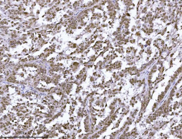
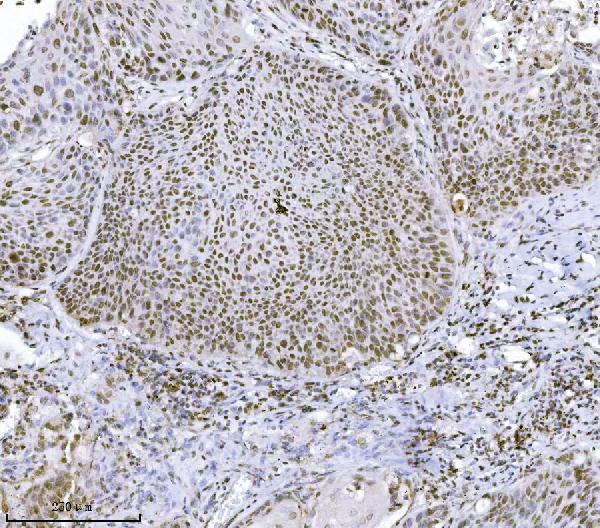
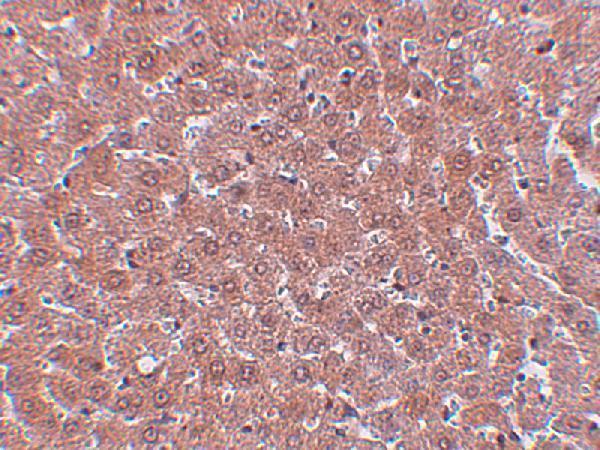
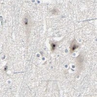


Comprehensive Human Protein Atlas IHC scoring per tissue (reliability: Uncertain — Medium consistency between antibody staining and RNA expression data. Pending external verification.). Rows are taken directly from the HPA tissue chart — click any row's HPA link to view the source.
| Tissue | Cell type | Level | Evidence | Source |
|---|---|---|---|---|
| Caudate | Neuronal cells | High | Protein (IHC) | HPA → |
| Cerebellum | Cells in molecular layer | High | Protein (IHC) | HPA → |
| Cerebral cortex | Neuronal cells | High | Protein (IHC) | HPA → |
| Duodenum | Glandular cells | High | Protein (IHC) | HPA → |
| Hippocampus | Neuronal cells | High | Protein (IHC) | HPA → |
| Tissue | Cell type | Level | Evidence | Source |
|---|---|---|---|---|
| Adipose tissue | Adipocytes | Not detected | Protein (IHC) | HPA → |
| Pancreas | Exocrine glandular cells | Not detected | Protein (IHC) | HPA → |
| Salivary gland | Glandular cells | Not detected | Protein (IHC) | HPA → |
| Smooth muscle | Smooth muscle cells | Not detected | Protein (IHC) | HPA → |
| Soft tissue | Peripheral nerve | Not detected | Protein (IHC) | HPA → |
Troubleshoot chromogenic IHC for KDM4A in paraffin sections using the catalog antibody’s tissue staining conditions and compartment evidence (datasheet A02850-1; UniProt O75164).
Two anti-KDM4A antibodies have real IHC images covering human and mouse paraffin sections and rat liver tissue (A02850-1 and A02850 image captions); no IF images are supplied (catalog: IF image alts).
A02850-1 has IHC images from human ovarian serous carcinoma and mouse colon paraffin sections (A02850-1 image captions). A02850 has a rat liver IHC image at 5 μg/mL (A02850 image caption).
Which to pick: For cross-species paraffin-section IHC in human and mouse samples, choose A02850-1: its own captions show both species using 2 μg/ml antibody after EDTA heat retrieval (A02850-1 image captions). For rat tissue IHC, choose A02850, whose own image shows rat liver at 5 μg/mL; its listed human and mouse reactivity does not establish IHC validation in those species (A02850 image caption; catalog: reactivity). Neither SKU has documented IF/ICC use or IF images, and the fixative is unreported in both IHC captions (catalog: applications and IF image alts; A02850-1 and A02850 image captions).