This website uses cookies to ensure you get the best experience on our website.


- Table of Contents
Plan chromogenic KDM8 IHC on paraffin sections with the catalog antibody at a starting concentration of 2.5 μg/mL (datasheet). Compare observed cytoplasmic staining with tissue IHC evidence while treating nuclear and chromosomal location as a molecular expectation (HPA tissue IHC; UniProt).

Expected localisation, validated protocols, controls and antibodies — the at-a-glance facts below, then the full design guide.
| Expected localisation | Cytoplasmic tissue staining (HPA tissue IHC); nuclear/chromosomal molecular location (UniProt) | |
| Staining pattern | General cytoplasmic staining across tissue cell types (HPA tissue IHC) | |
| Antigen retrieval | Tris-EDTA pH 9.0 HIER, 95–98 °C, 20 min (rule: nuclear antigen) | |
| Positive control | Adrenal gland+4 more · see all | |
| Negative control | Adipose tissue |
| Fixation | Keep fixation consistent across sections. (standard IHC practice; not target-specific) | |
| Caveat | Staining has low consistency with RNA expression (HPA tissue IHC) | |
| Regulation | Highly expressed in breast cancer cells (UniProt) | |
| Isoform / epitope | 3 isoforms; check antibody epitope coverage (UniProt) |
Compare the catalog antibody’s IHC-P protocol with reported KDM8 staining of prostate tissue microarrays and mouse pancreatic tumors (PMC6755995; PMC12622147; PMC12407890).
| Sample | Tissue sections; selected-image fixative not specified (standard IHC workflow) |
| Fixation | Image fixative and duration unreported (datasheet A08840); verify before use. |
| Sectioning | 4–5 µm sections on charged slides (standard) |
| Deparaffinisation | Xylene, graded ethanol series to water (standard) |
| Antigen retrieval | Heat-induced epitope retrieval in Tris-EDTA buffer, pH 9.0, 20 min at 95–98 °C (standard rule: nuclear antigen) |
| Peroxidase block | 3% H2O2, 10 min, room temperature (standard) |
| Blocking | 10% normal serum of the secondary host, 30 min, room temperature (standard) |
| Primary antibody | Rabbit anti-KDM8, 2.5 μg/mL (datasheet A08840) |
| Primary incubation | Overnight at 4 °C (standard) |
| Detection | HRP-polymer secondary, DAB chromogen 5–10 min (standard) |
| Counterstain | Hematoxylin, blue, dehydrate and mount (standard) |
| Expected result | KDM8-positive staining in glandular cells of adrenal gland (HPA tissue IHC: High). HPA tissue profile: General cytoplasmic expression. No signal in the no-primary control. |
KDM8 is annotated in the nucleus and on chromosomes (UniProt Q8N371), while tissue IHC shows general cytoplasmic staining (HPA tissue IHC) and ICC-IF places it mainly in the nucleoplasm, with additional cytosolic signal (HPA ICC-IF). High IHC staining is reported in several glandular, epithelial, neuronal and muscle cell populations (HPA tissue IHC). Interpret the compartment carefully: HPA rates its tissue IHC Approved but reports low consistency with RNA expression (HPA tissue IHC).
| Cytoplasmic staining in adrenal, salivary or stomach glandular cells, or bronchial respiratory epithelial cells. | This matches cell populations reported as High in tissue IHC (HPA tissue IHC). Record the stained cell type and intensity alongside the compartment: the tissue profile is generally cytoplasmic (HPA tissue IHC), although UniProt places KDM8 in the nucleus and on chromosomes (UniProt Q8N371). |
| Strong staining confined to a compartment that disagrees with both the reported tissue IHC and nucleoplasmic ICC-IF patterns. | Treat an isolated, unexpected compartment as a possible artefact until controls support it (general IHC practice). Nuclear signal alone is not automatically wrong: nucleoplasm is the main ICC-IF location (HPA ICC-IF), and nucleus and chromosome are UniProt locations (UniProt Q8N371). |
| Prominent staining in a cell population reported as undetected, such as adipocytes. | Adipocytes are reported as Not detected (HPA tissue IHC). Check whether the signal belongs to adipocytes or adjacent cells; if it persists in the wrong cell type, consider cross-reactivity or endogenous detection activity (general IHC practice). An unexpected result is not, by itself, proof of either cause. |
| Diffuse color across cells, extracellular areas and the slide, with little distinction between stained and unstained structures. | This is difficult to score as cell-specific KDM8 staining (general IHC practice). Check the no-primary control and assess blocking, washes and chromogen development (general IHC practice); the HPA tissue pattern does not establish a target-specific cause for diffuse background (HPA tissue IHC). |
| No visible staining in a section expected to contain a reported High cell population. | First check that the relevant cells are present: cardiomyocytes, cerebral cortical neuronal cells and splenic white-pulp cells are among the High populations (HPA tissue IHC). An absent signal calls for a run-control check before concluding biological absence (general IHC practice), especially given the reported low IHC–RNA consistency (HPA tissue IHC). |
| Compartment differs by assay | Tissue IHC is described as generally cytoplasmic (HPA tissue IHC); ICC-IF is mainly nucleoplasmic with additional cytosol (HPA ICC-IF), and UniProt lists nucleus and chromosome (UniProt Q8N371). Report the method when describing location. |
| Cell type matters more than a whole-tissue label | High staining is assigned to specified cells within tissues, including cerebellar granular-layer cells and splenic white-pulp cells (HPA tissue IHC). Sample composition therefore matters when selecting a comparison field (general IHC practice). |
| Strength of tissue evidence | The tissue IHC reliability label is Approved, with low consistency between antibody staining and RNA expression (HPA tissue IHC). The listed High and Not detected calls describe observed staining; they should be weighed with slide controls rather than treated as independent proof of specificity. |
| Antibody validation scope | HPA026545 is Approved for IHC and Supported for ICC (HPA antibodies). The supplied record does not assign it Enhanced IHC validation (HPA antibodies); do not infer independent reproduction of this tissue pattern from the listed status. |
| Isoforms and epitope uncertainty | UniProt lists three KDM8 isoforms and a JmjC domain at residues 271–416 (UniProt Q8N371). The supplied evidence gives no antibody epitope or isoform coverage, so a difference in staining cannot be assigned to a particular isoform. |
| Protein topology and processing | KDM8 has no transmembrane segment, signal peptide, propeptide or listed glycosylation site, and its annotated chain spans residues 1–416 (UniProt Q8N371). These annotations do not establish how fixation or antigen retrieval affects IHC signal. |
| Situation | Likely cause | Next action |
|---|---|---|
| No color appears in a reported High cell population. | The expected cells may be missing from the section, or the staining run may have failed (general IHC practice); HPA reports High staining only for specified cell types (HPA tissue IHC). | Confirm the cell population morphologically, then review the run control, primary-antibody step, detection reagents and development (general IHC practice). Do not use tissue RNA enrichment alone to decide whether the stain succeeded: IHC–RNA consistency is low (HPA tissue IHC). |
| Color appears broadly, including outside recognizable cells. | Non-specific reagent binding, incomplete washes or overdevelopment can produce diffuse chromogenic background (general IHC practice). | Compare with a no-primary control; review blocking, wash steps and development time (general IHC practice). Score KDM8 only where cell boundaries and a reproducible compartment can be assessed (general IHC practice). |
| A high-intensity signal appears in adipocytes. | Adipocytes are reported as Not detected (HPA tissue IHC); an unexpected signal could reflect misidentified adjacent cells, cross-reactivity or endogenous detection activity (general IHC practice). | Verify cell identity on the counterstained section and compare a no-primary control (general IHC practice). If an enzyme-based chromogen is used, check the corresponding endogenous-activity control (general IHC practice). |
| Only nuclei stain in a tissue IHC section. | Nuclear staining agrees with UniProt location and the main ICC-IF location (UniProt Q8N371; HPA ICC-IF), but differs from HPA's general cytoplasmic tissue IHC profile (HPA tissue IHC). | Document the compartment and cell type, compare control sections and avoid calling the result either confirmed or artifactual from location alone (general IHC practice). |
| Cytoplasmic color is present, but the expected High cells are hard to distinguish. | The High calls refer to particular populations, such as glandular cells in adrenal gland, salivary gland and stomach (HPA tissue IHC); surrounding cells can complicate visual attribution (general IHC practice). | Use morphology and the counterstain to identify the reported population, then score its staining separately from neighboring cells and background (general IHC practice). |
| IF/ICC Q: Is cytosolic KDM8 signal compatible with the reference pattern? | A: Yes. HPA reports an additional cytosolic location alongside its approved main nucleoplasmic location (HPA ICC-IF); UniProt lists nucleus and chromosome (UniProt Q8N371). | Describe nucleoplasmic and cytosolic signal separately and compare appropriate imaging controls (general IF practice). Do not transfer the tissue IHC intensity calls directly to IF/ICC images, because those calls come from tissue IHC (HPA tissue IHC). |


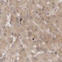


Comprehensive Human Protein Atlas IHC scoring per tissue (reliability: Approved — Low consistency between antibody staining and RNA expression data.). Rows are taken directly from the HPA tissue chart — click any row's HPA link to view the source.
| Tissue | Cell type | Level | Evidence | Source |
|---|---|---|---|---|
| Adrenal gland | Glandular cells | High | Protein (IHC) | HPA → |
| Bronchus | Respiratory epithelial cells | High | Protein (IHC) | HPA → |
| Cerebellum | Cells in granular layer | High | Protein (IHC) | HPA → |
| Cerebral cortex | Neuronal cells | High | Protein (IHC) | HPA → |
| Heart muscle | Cardiomyocytes | High | Protein (IHC) | HPA → |
| Tissue | Cell type | Level | Evidence | Source |
|---|---|---|---|---|
| Adipose tissue | Adipocytes | Not detected | Protein (IHC) | HPA → |
Use compartment-aware controls to troubleshoot KDM8 staining in paraffin-section chromogenic IHC; assess IF/ICC separately.
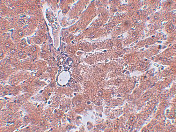
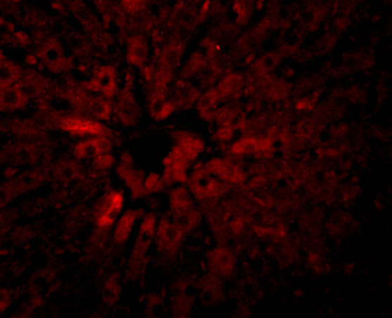
A08840 has real IHC data from rat liver tissue and IF data from rat liver cells (IHC and IF image captions); human, mouse and rat reactivity is listed (catalog reactivity).
A08840 is listed for IHC-P and shown staining rat liver tissue at 2.5 μg/mL (catalog applications; IHC image caption). A08840 is also listed for IF and shown in rat liver cells at 20 μg/mL (catalog applications; IF image caption).
Which to pick: Choose A08840 for paraffin-section tissue IHC based on its IHC-P listing and rat liver tissue image; the fixative is unreported (catalog applications; IHC image caption). For IF/ICC, A08840 has an IF image in rat liver cells, while ICC-specific validation is unreported (catalog applications; IF image caption). A08840 is listed as reactive with human, mouse and rat, but the supplied IHC and IF images show rat samples only; its host is rabbit and clonality is unreported (catalog reactivity and host; IHC and IF image captions).