This website uses cookies to ensure you get the best experience on our website.


- Table of Contents
Plan chromogenic IHC for KHDRBS1 in paraffin sections using 2–5 μg/ml of catalog antibody A01717-1 as a starting range (datasheet: A01717-1). Expect widespread nuclear staining (HPA tissue IHC) and compare signal with appropriate background controls (standard IHC practice).

Expected localisation, validated protocols, controls and antibodies — the at-a-glance facts below, then the full design guide.
| Expected localisation | Predominantly nuclear tissue staining (HPA tissue IHC) | |
| Staining pattern | Nuclear staining across diverse tissue cell types (HPA tissue IHC) | |
| Antigen retrieval | EDTA pH 8.0 HIER, heat-mediated (datasheet A01717-1) | |
| Positive control | Adrenal gland+4 more · see all | |
| Negative control | None in HPA (detected in all 45 tissues); use no-primary + isotype controls |
| Fixation | Keep fixation conditions consistent across sections. (standard IHC practice; not target-specific) | |
| Caveat | No tissue-negative comparator reported (HPA tissue IHC) | |
| Regulation | Isoform 1 lower in brain, muscle, liver (UniProt) | |
| Isoform / epitope | 3 isoforms; no processing; epitope coverage unknown (UniProt) |
Start with the catalog antibody’s EDTA pH 8.0 retrieval protocol (datasheet: A01717-1); the published IHC methods below provide tissue-specific conditions (PMC10277899; PMC7698510; PMC5626414).
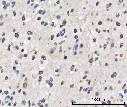
| Sample | Paraffin-embedded human glioblastoma tissue; fixative not specified (datasheet A01717-1) |
| Fixation | Image fixative and duration unreported (datasheet A01717-1); verify before use. |
| Sectioning | 4–5 µm sections on charged slides (standard) |
| Deparaffinisation | Xylene, graded ethanol series to water (standard) |
| Antigen retrieval | Heat retrieval: EDTA pH 8.0 (datasheet A01717-1); 20 min, 95–100 °C (standard) |
| Peroxidase block | 3% H2O2, 10 min, room temperature (standard) |
| Blocking | 10% goat serum (datasheet A01717-1) |
| Primary antibody | Rabbit anti-KHDRBS1, 2-5 μg/ml (datasheet A01717-1) |
| Primary incubation | Overnight at 4 °C (datasheet A01717-1) |
| Detection | HRP-conjugated secondary, DAB chromogen (datasheet A01717-1) |
| Counterstain | Hematoxylin, blue, dehydrate and mount (standard) |
| Expected result | KHDRBS1-positive staining in glandular cells of adrenal gland (HPA tissue IHC: High). HPA tissue profile: Ubiquitous nuclear expression. No signal in the no-primary control. |
KHDRBS1 should appear predominantly in nuclei across many cell types: HPA describes ubiquitous nuclear tissue staining with Supported reliability and high consistency with RNA expression (HPA tissue IHC). UniProt also places KHDRBS1 partly in cytoplasm and at membranes, although it has no transmembrane segment (UniProt Q07666). Use the nuclear pattern as the primary IHC-P interpretation (HPA tissue IHC; UniProt Q07666).
| Clear nuclear chromogen in glandular cells, respiratory epithelium, neurons, granular-layer cells, or endothelium. | This fits the reported pattern when the corresponding tissue is present: HPA rates staining High in adrenal gland, appendix, breast and cervix glandular cells; bronchus respiratory epithelial cells; caudate neurons; cerebellar granular-layer cells; and cerebral-cortex endothelial cells (HPA tissue IHC). Compare nuclei with neighboring tissue structures before scoring intensity. |
| Strong cytoplasmic or membrane staining with little nuclear signal. | This conflicts with the predominant nuclear pattern (HPA tissue IHC; UniProt Q07666). Some cytoplasmic localisation is reported, so cytoplasmic signal alone is not proof of an artefact (UniProt Q07666). Reassess compartment boundaries and controls; a membrane-only pattern needs independent validation because KHDRBS1 has no transmembrane segment (UniProt Q07666). |
| Signal is concentrated in an unexpected cell population while expected nuclei remain weak. | Consider cross-reactivity or endogenous chromogenic activity, particularly if the staining ignores nuclear boundaries (general IHC practice). HPA reports low tissue specificity and supplies no negative tissue list here, so an unlisted cell type cannot be declared KHDRBS1-negative from this record (HPA tissue IHC). Check morphology and detection controls before assigning specificity. |
| Diffuse chromogen obscures nuclei or spreads across the section. | The reported nuclear pattern cannot be scored reliably under diffuse background (HPA tissue IHC). Excess primary or detection reagent, inadequate blocking, or incomplete washing can produce background in IHC (general IHC practice). A diffuse deposit does not establish cytoplasmic KHDRBS1, even though partial cytoplasmic localisation is reported (UniProt Q07666). |
| No nuclear signal in a section expected to stain strongly. | High staining is reported in the specified cells of the listed tissues, but the result depends on those cells being represented in the section (HPA tissue IHC). First verify tissue identity and preservation of recognizable nuclei; then check the IHC-P workflow and detection controls (general IHC practice). A single blank section does not establish biological absence. |
| Tissue and cell context | HPA calls KHDRBS1 ubiquitous and RNA expression low in tissue specificity, yet its supplied High examples name particular cells within each tissue (HPA tissue IHC). Score the cells actually present; do not treat unlisted tissues or cell populations as negative controls. |
| Compartment and topology | The nucleus is the main expectation; partial cytoplasmic localisation is possible (UniProt Q07666; HPA tissue IHC). UniProt lists membrane localisation but no transmembrane segment, so a crisp membrane-only IHC pattern should be checked rather than accepted from topology alone (UniProt Q07666). |
| Isoforms and epitope coverage | UniProt lists 3 isoforms and reports lower isoform 1 expression in brain, skeletal muscle and liver, with intensified isoform 3 in skeletal muscle and liver (UniProt Q07666). Antibody epitope and isoform coverage are not supplied; these facts cannot predict this assay's staining intensity. |
| Antibody evidence | HPA lists IHC Supported status for HPA051280, HPA056813 and CAB005355; its tissue profile is also Supported (HPA antibodies; HPA tissue IHC). Those ratings support the reported pattern but do not establish specificity for an unspecified catalog antibody or every staining condition. |
| IF/ICC Q&A: where should fluorescence appear? | Primarily in the nucleoplasm: HPA calls it the enhanced main location in ICC-IF images (HPA subcellular ICC-IF). This is a localisation comparison for the separate IF/ICC guide, not an IHC-P fluorescence protocol or proof that every IHC antibody performs in IF. |
| Situation | Likely cause | Next action |
|---|---|---|
| Expected positive tissue is blank. | The expected High cell population may be absent from the section (HPA tissue IHC), or an IHC detection step may have failed (general IHC practice). | Locate the HPA-listed cell population on a matched section; verify a working positive control and the chromogenic detection sequence before interpreting absence (HPA tissue IHC; general IHC practice). |
| Nuclei are weak while the section is generally stained. | Weak nuclear contrast limits comparison with HPA's ubiquitous nuclear profile (HPA tissue IHC). Retrieval, primary dilution, incubation, and counterstain balance can affect ordinary IHC readability (general IHC practice); KHDRBS1-specific fixation sensitivity is unreported here. | Review the catalog antibody's IHC-P instructions and optimize one general workflow variable at a time with a positive control (general IHC practice). Do not infer a KHDRBS1-specific retrieval requirement from HPA or UniProt. |
| Cytoplasm dominates and nuclear staining is missing. | Partial cytoplasmic localisation is reported, but a cytoplasm-dominant result departs from the main nuclear pattern (UniProt Q07666; HPA tissue IHC). | Confirm nuclear outlines and compare with the expected positive control. If the pattern persists, seek antibody-specific validation before scoring it as KHDRBS1 (general IHC practice). |
| Colour appears in cells or areas unrelated to the expected nuclear pattern. | Cross-reactivity or endogenous activity from the chosen chromogenic detection system may contribute (general IHC practice). HPA provides no negative cell or tissue list for this record (HPA tissue IHC). | Run an appropriate primary-omission detection control and the detection system's activity block where applicable; compare the remaining colour with cell morphology (general IHC practice). |
| A widespread haze hides nuclear boundaries. | Excess reagent, insufficient blocking, or inadequate washing can raise IHC background (general IHC practice). The haze cannot be assigned to KHDRBS1 localisation from appearance alone. | Check primary concentration against its IHC-P instructions, improve washing and blocking as needed, and confirm that nuclei become individually readable (general IHC practice). |
| Different listed tissues show different overall intensity. | HPA reports High staining in selected cells, not identical staining in every cell or section (HPA tissue IHC). UniProt reports tissue-dependent isoform expression, but assay epitope coverage is unknown (UniProt Q07666). | Compare like cell populations under the same staining and scoring conditions; record nuclear localisation separately from intensity (HPA tissue IHC; general IHC practice). |


Comprehensive Human Protein Atlas IHC scoring per tissue (reliability: Supported — High consistency between antibody staining and RNA expression data.). Rows are taken directly from the HPA tissue chart — click any row's HPA link to view the source.
| Tissue | Cell type | Level | Evidence | Source |
|---|---|---|---|---|
| Adrenal gland | Glandular cells | High | Protein (IHC) | HPA → |
| Appendix | Glandular cells | High | Protein (IHC) | HPA → |
| Breast | Glandular cells | High | Protein (IHC) | HPA → |
| Bronchus | Respiratory epithelial cells | High | Protein (IHC) | HPA → |
| Caudate | Neuronal cells | High | Protein (IHC) | HPA → |
| Tissue | Cell type | Level | Evidence | Source |
|---|---|---|---|---|
| None in HPA: KHDRBS1 is detected in all 45 scored tissues. Use a no-primary (secondary-only) and an isotype control instead. | ||||
Troubleshoot KHDRBS1 staining in paraffin sections by checking retrieval, nuclear localisation, controls and cell-specific scoring (datasheet A01717-1; HPA tissue IHC).
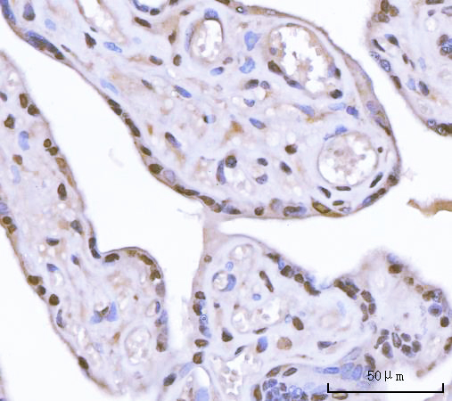
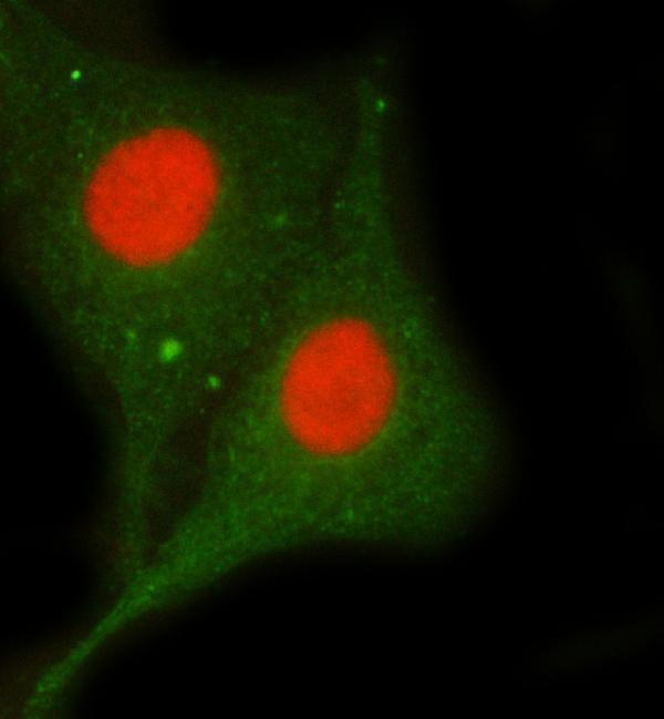
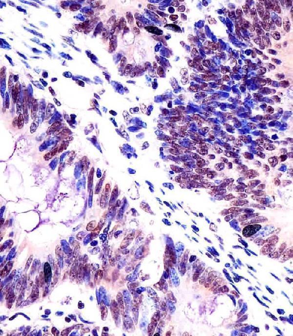
KHDRBS1 IHC images cover human glioblastoma and colorectal carcinoma paraffin sections (catalog IHC captions). IF/ICC images cover A549 cells, human intestinal cancer sections, and mouse and rat brain sections (A01717-1 IF captions).
A01717-1 will render with its human glioblastoma paraffin-section IHC image; its catalog also lists IF/ICC and Human, Mouse, and Rat reactivity (A01717-1 IHC caption; catalog applications and reactivity). M01717 will render with its formaldehyde-fixed, paraffin-embedded human colorectal carcinoma IHC image; its catalog lists IHC-P and Human and Mouse reactivity (M01717 IHC caption; catalog applications and reactivity).
Which to pick: For tissue IHC, choose M01717 when its paraffin-section paraffin preparation and citrate pH 6 retrieval match your sections; A01717-1 documents EDTA pH 8 retrieval in paraffin sections, but its IHC caption does not report a fixative (M01717 IHC caption; A01717-1 IHC caption). For IF/ICC, choose A01717-1: its catalog lists both applications and provides cell and tissue IF images, while M01717 lists IHC-P without IF/ICC (A01717-1 catalog applications and IF captions; M01717 catalog applications). For work across species, A01717-1 lists Human, Mouse, and Rat reactivity and shows mouse and rat brain IF; its IHC image documents human tissue (A01717-1 catalog reactivity, IF captions, and IHC caption). The selected A01717-1 tissue-IHC caption documents paraffin sections, but does not specify the fixative (selected-SKU IHC image A01717-1).