KIF20A / Kinesin-like protein KIF20A · IHC design guide

Design Immunohistochemistry for KIF20A

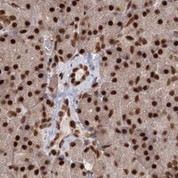
Plan chromogenic KIF20A IHC in paraffin sections with an antibody dilution of 1:50–1:200 (datasheet). Expect nuclear staining in varying fractions of cells (HPA tissue IHC); bone marrow hematopoietic cells show high staining (HPA tissue IHC).

Evidence assembled Oct 2026 · For research use; verify linked source records and product datasheet before use
Immunohistochemistry protocol sheet for KIF20A (IHC for KIF20A): expected localisation Nuclear tissue staining (HPA tissue IHC); Golgi and spindle localisation (UniProt), antibody A05142-1, validated IHC image, and IHC protocol steps
Printable KIF20A IHC protocol sheet — expected localisation Nuclear tissue staining (HPA tissue IHC); Golgi and spindle localisation (UniProt), antibody A05142-1, controls and protocol steps. Open the full KIF20A IHC guide →

KIF20A Immunohistochemistry Experimental Design Guide

Expected localisation, validated protocols, controls and antibodies — the at-a-glance facts below, then the full design guide.

Must know before staining
Expected localisation Nuclear tissue staining (HPA tissue IHC); Golgi and spindle localisation (UniProt)
Staining pattern Nuclear staining in varying fractions of cells (HPA tissue IHC)
Antigen retrieval Citrate pH 6.0 HIER, 95–98 °C, 20 min (rule: cytoplasmic / membrane antigen)
Positive control ⓘ Bone marrow+4 more · see all
Negative control ⓘ Heart muscle+2 more · see all
Important caveats
Reasons your staining may differ from the expected pattern.
Fixation Keep fixation consistent across paraffin sections. (standard IHC practice; not target-specific)
Caveat Staining has low consistency with RNA data (HPA tissue IHC)
Regulation Expression regulation is unannotated (UniProt)
Isoform / epitope 2 isoforms; antibody epitope coverage is unknown (UniProt; catalog)
Section 1

Recommended KIF20A IHC & IF Protocols

The catalog antibody’s IHC-P protocol (datasheet) is accompanied by published KIF20A protocols for ovarian, nasopharyngeal and prostate cancer tissue (PMC6140728; PMC5230771; PMC6745134).

Recommended immunohistochemistry (IHC-P) protocol parameters
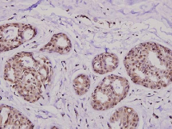
SampleParaffin-embedded human breast carcinoma tissue; fixative not specified (datasheet A05142-1)
FixationImage fixative and duration unreported (datasheet A05142-1); verify before use.
Sectioning4–5 µm sections on charged slides (standard)
DeparaffinisationXylene, graded ethanol series to water (standard)
Antigen retrievalHeat-induced epitope retrieval in citrate buffer, pH 6.0, 20 min at 95–98 °C (standard rule: cytoplasmic / membrane antigen)
Peroxidase block3% H2O2, 10 min, room temperature (standard)
Blocking10% normal serum of the secondary host, 30 min, room temperature (standard)
Primary antibodyRabbit anti-KIF20A, 1:50-1:200 (datasheet A05142-1)
Primary incubationOvernight at 4 °C (standard)
DetectionHRP-polymer secondary, DAB chromogen 5–10 min (standard)
CounterstainHematoxylin, blue, dehydrate and mount (standard)
Expected resultKIF20A-positive staining in hematopoietic cells of bone marrow (HPA tissue IHC: High). HPA tissue profile: Nuclear expression in varying fractions of cells in most tissues. No signal in the no-primary control.
💡Decision noteStart with citrate pH 6.0 HIER at 95–98 °C for 20 min (page antigen retrieval). The ovarian and nasopharyngeal protocols use microwave EDTA retrieval (PMC6140728; PMC5230771).
Section 2

What Is the Expected KIF20A Staining Pattern?

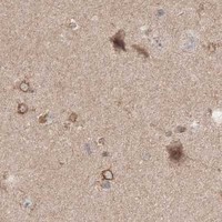
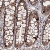
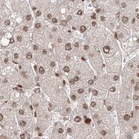
In paraffin-section IHC, expect KIF20A staining in varying fractions of nuclei across many tissues, including strong staining in selected hematopoietic, germinal-center and seminiferous-duct cells (HPA: tissue IHC). Mitotic cells may show spindle-associated signal (UniProt O95235: spindle-midzone localization; HPA: mitotic spindle supported by ICC-IF). KIF20A has no transmembrane segment (UniProt O95235: topology). HPA rates tissue IHC Approved but reports low agreement between staining and RNA expression (HPA: reliability).

What am I looking at on my slide?
Distinct nuclear staining in a subset of cells, with strong signal in germinal-center cells or bone-marrow hematopoietic cells (HPA: tissue IHC).This fits the reported paraffin-section pattern: nuclear expression varies among cells and tissues (HPA: tissue IHC). Score the positive cell fraction and intensity within the named cell population, rather than treating every unstained neighbor as a failed assay (standard IHC practice).
Signal appears exclusively at cell borders or as broad extracellular deposits, without a recognizable nuclear or mitotic-cell pattern.That distribution does not match HPA tissue IHC or the reported intracellular locations (HPA: tissue IHC; UniProt O95235: localization). Treat it as suspect; inspect morphology, detection controls and the staining run before assigning it to KIF20A (standard IHC practice).
Strong chromogen appears in muscle cells chosen as a negative comparator (HPA: cardiomyocytes, skeletal myocytes and smooth-muscle cells not detected).This conflicts with the sampled HPA tissue pattern, but alone does not identify its cause (HPA: tissue IHC). Consider antibody cross-reactivity or endogenous detection activity, and compare with an appropriate reagent-omission control (standard IHC practice).
A diffuse haze covers nuclei and surrounding tissue, making individual positive cells hard to distinguish.The slide cannot reliably show the reported cell-restricted nuclear pattern (HPA: tissue IHC). Background may reflect nonspecific antibody binding, inadequate blocking or chromogenic detection activity; use controls to locate the source (standard IHC practice).
No discernible signal appears in a known-positive comparator such as lymph-node germinal-center cells (HPA: High).A blank comparator makes negative calls elsewhere unreliable (standard IHC practice). Check tissue identity, antibody and detection controls, then review the assay conditions; HPA's Approved rating does not guarantee signal in every run (HPA: reliability).
💡Expected KIF20A appearanceCall positive when discrete staining marks nuclei in a subset of the expected cells, potentially strong in HPA High populations; broad extracellular or cell-border staining is suspect (HPA: tissue IHC; UniProt O95235: intracellular localization).
How each factor affects the staining
Cell population and tissue context (HPA: tissue IHC)High staining is reported in bone-marrow hematopoietic cells, lymph-node germinal-center cells, melanocytes, seminiferous-duct cells and urothelial cells; several other sampled populations are Medium or Low (HPA: tissue IHC). Compare like cell types when judging intensity.
Cell-cycle-dependent location (UniProt O95235: function and localization)KIF20A participates in cytokinesis and localizes to the spindle midzone during anaphase and telophase (UniProt O95235). A mitotic spindle pattern is supported in ICC-IF, while HPA describes variable nuclear staining in tissue IHC; interpret each assay in its own context (HPA: subcellular; HPA: tissue IHC).
Evidence strength (HPA: reliability and antibody validation)Tissue IHC is rated Approved despite low consistency with RNA expression (HPA: reliability). HPA036909 and HPA036910 are IHC Approved; HPA036910 is ICC Enhanced (HPA: antibodies). These ratings support using the images as references, with controls for any new specimen.
Topology, isoforms and epitope uncertainty (UniProt O95235)The protein has no transmembrane segment and has two listed isoforms (UniProt O95235). The supplied record does not map the IHC antibody epitope to either isoform, so staining cannot distinguish them. It also supplies no target-specific fixation-sensitivity result.
Chromogenic detection background (standard IHC practice)Endogenous tissue activity or nonspecific detection reagents can produce chromogen unrelated to the target (standard IHC practice). Read staining against cell morphology and a suitable detection control, especially when signal lies outside the HPA-reported cell populations (HPA: tissue IHC).
Why is my staining missing, weak or wrong?
SituationLikely causeNext action
Expected positive cells stain weakly or not at all (HPA: High populations).The assay may have insufficient primary or detection signal; a blank control tissue also prevents a confident negative interpretation (standard IHC practice).Confirm the comparator tissue and cell population, then review the catalog antibody's IHC-P conditions, antigen retrieval, detection reagents and run controls (standard IHC practice). No KIF20A-specific retrieval condition is established here.
Nearly every cell shows equally strong nuclear staining, obscuring the expected variation (HPA: tissue IHC).Excessive assay signal or nonspecific binding may overwhelm differences between cells (standard IHC practice).Compare a negative tissue region and reagent-omission control; adjust primary-antibody or detection conditions within the validated IHC workflow if background persists (standard IHC practice).
Chromogen collects at tissue edges or forms diffuse deposits.Uneven reagent coverage, washing or detection background can create patterns unrelated to cell location (standard IHC practice).Review section integrity, reagent coverage, wash steps and a detection control; score only intact cells with interpretable morphology (standard IHC practice).
Strong staining appears in cardiomyocytes or other HPA-negative muscle cells (HPA: tissue IHC).Cross-reactivity or endogenous chromogenic activity is possible; the unexpected result alone cannot separate them (standard IHC practice).Compare primary-antibody omission and detection controls, check cell identity on the counterstain, and avoid calling the pattern KIF20A until resolved (standard IHC practice).
A specimen has fewer positive cells than the HPA reference.HPA reports nuclear staining in varying fractions of cells, and its tissue IHC has low agreement with RNA expression (HPA: profile and reliability).Identify the same cell population, report positive fraction alongside intensity, and check a same-run positive comparator before interpreting the difference (standard IHC practice).
IF/ICC Q: Which compartments should an image be checked against?HPA supports mitotic-spindle localization; nucleoplasm, cleavage furrow and cytokinetic bridge are marked uncertain (HPA: subcellular ICC-IF).Compare cell-cycle stage and compartment with those HPA annotations and UniProt's anaphase/telophase spindle-midzone note (HPA: subcellular ICC-IF; UniProt O95235). Use the separate IF/ICC guide for assay design.

Sample controls for KIF20A IHC & IF

🧪Run lymph node first and look for staining in germinal center cells (HPA: High in germinal center cells). Use heart muscle cardiomyocytes as the negative tissue (HPA: Not detected); on the lymph node slide, cells outside germinal centers should show little or no specific chromogen to serve as an internal background comparator, although their KIF20A status is not established by the supplied HPA rows.
Positive control tissue: Bone marrow (Hematopoietic cells, HPA High)
Negative control tissue: Heart muscle (HPA Not detected)
ICC-IF cell lines (HPA subcellular resource): HPA ICC-IF images show KIF20A in A-431, U-251MG, U2OS, hTERT-RPE1 (serum starved), Sperm, U2OS, siRNA 2 (10x), U2OS, siRNA 2 (40x), U2OS, scrambled (10x), U2OS, scrambled (40x), with annotated localisation: Nucleoplasm (uncertain), Cytokinetic bridge (uncertain), Cleavage furrow (uncertain), Mitotic spindle (supported) (HPA subcellular).
Technical controls: Include a no-primary (secondary-only) slide, an isotype control matched to the primary antibody’s host species and immunoglobulin class or polyclonal IgG as applicable, and KIF20A knockout material as a biological negative (standard IHC practice). Block endogenous peroxidase and check background in the lymph node section before interpreting chromogenic staining (standard chromogenic IHC practice).
⚠️Feasibility: Paraffin-section IHC has been shown for the selected antibody in human breast carcinoma at 1:100, but its fixative is unreported (selected-SKU tissue-IHC caption). No target-specific fixation window, fixation effect, or antigen-retrieval dependency is reported in the supplied evidence; optimize retrieval empirically (supplied target/application evidence; standard IHC practice). IF/ICC images are available, but the evidence does not establish that IF or frozen sections are easier than paraffin IHC (HPA: subcellular ICC-IF images); endogenous peroxidase can complicate interpretation in lymph node sections (standard chromogenic IHC practice).

HPA tissue IHC evidence for KIF20A

Comprehensive Human Protein Atlas IHC scoring per tissue (reliability: Approved — Low consistency between antibody staining and RNA expression data.). Rows are taken directly from the HPA tissue chart — click any row's HPA link to view the source.

Positive expression · recommended positive controls

TissueCell typeLevelEvidenceSource
Bone marrow Hematopoietic cells High Protein (IHC) HPA →
Lymph node Germinal center cells High Protein (IHC) HPA →
Skin Melanocytes High Protein (IHC) HPA →
Testis Cells in seminiferous ducts High Protein (IHC) HPA →
Urinary bladder Urothelial cells High Protein (IHC) HPA →

Undetected expression · recommended negative controls

TissueCell typeLevelEvidenceSource
Heart muscle Cardiomyocytes Not detected Protein (IHC) HPA →
Skeletal muscle Myocytes Not detected Protein (IHC) HPA →
Smooth muscle Smooth muscle cells Not detected Protein (IHC) HPA →
Section 3

Advanced KIF20A IHC Tips

Use compartment, cell type, and matched section controls to troubleshoot KIF20A staining in paraffin section chromogenic IHC.

How should I adjust retrieval when KIF20A staining is weak or uneven?
Start with citrate pH 6.0 heat induced retrieval at 95–98 °C for 20 min (page retrieval rule). Cool sections consistently, then compare a short retrieval time series on adjacent sections while keeping antibody dilution and chromogen development fixed (standard IHC practice). Use the catalog antibody’s paraffin section image at 1:100 as evidence that tissue staining is feasible, without assuming its unreported fixation or retrieval conditions (caption: A05142-1). If signal remains weak, trial another buffer or pH only as a fallback, with a known positive section and a no primary control beside each condition (standard IHC practice). Judge improvement by preserved morphology and discrete staining, rather than maximum brown intensity alone (standard IHC practice).
Could fixation explain loss of KIF20A staining in my sections?
Target specific KIF20A sensitivity to fixative type or duration is unknown from the supplied evidence; the paraffin section caption does not state a fixative (caption: A05142-1). Record the actual fixation conditions for each specimen, then compare sections processed together with the same citrate pH 6.0 retrieval for 20 min (page retrieval rule; standard IHC practice). If fixation histories differ, avoid attributing an intensity difference to KIF20A biology until a matched processing comparison is available (standard IHC practice). Include a positive control section in every run and check whether morphology and an independent tissue control also deteriorate (standard IHC practice). Do not infer fixation sensitivity from phosphorylation sites or reported tissue staining patterns (UniProt O95235; HPA: tissue IHC).
Which staining compartments are plausible for KIF20A in tissue IHC?
Assess cytoplasmic and mitotic structures separately: KIF20A is annotated at the Golgi and spindle, including the spindle midzone during anaphase and telophase (UniProt O95235). The supported mitotic spindle location and uncertain nucleoplasm, cleavage furrow, and cytokinetic bridge locations add context, but come from cell imaging (HPA: subcellular). HPA tissue IHC instead reports nuclear staining in varying fractions of cells, with low consistency against RNA expression (HPA: tissue IHC). In paraffin sections, annotate nuclear, cytoplasmic, and mitotic staining independently, using morphology to identify dividing cells (standard IHC practice). Treat diffuse staining across every compartment as a reason to review controls and detection conditions before assigning a biological location (standard IHC practice).
Could isoforms or modification sites alter what this antibody stains?
KIF20A has 2 annotated isoforms and a kinesin motor domain spanning residues 64–507 (UniProt O95235). Multiple modified residues are annotated, including PLK1 associated phosphorylation at residue 528 (UniProt O95235). The supplied catalog caption gives no epitope map, so neither isoform recognition nor modification sensitivity can be assigned to this antibody (caption: A05142-1). Request or consult the antibody’s immunogen coordinates before interpreting an unexpected tissue distribution, and compare an independently mapped antibody on adjacent sections if available (standard IHC practice). Keep retrieval, detection, and scoring constant during that comparison so differences can be evaluated without conflating processing variables (standard IHC practice).
How can IF help assess a puzzling chromogenic KIF20A pattern?
Use IF as a separate localization check when chromogenic IHC cannot resolve a suspected mitotic structure (UniProt O95235: spindle midzone localization; standard microscopy practice). For a bone marrow comparison, multiplex KIF20A with a hematopoietic cell marker and a nuclear counterstain; hematopoietic cells show high tissue staining in the supplied reference (HPA: bone marrow). Choose a fluorophore in a channel with low measured tissue autofluorescence and include single stain controls before judging overlap (standard IF practice). KIF20A has no transmembrane segment and is annotated at intracellular sites, so use permeabilisation appropriate for an intracellular epitope in IF specimens (UniProt O95235). Do not transfer an IF result directly into the paraffin IHC score without checking matched cell types and compartments (standard microscopy practice).
What should I check when brown staining obscures specific KIF20A signal?
Run a no primary control to identify signal from the detection system, and inspect pigment or precipitate before changing antibody concentration (standard chromogenic IHC practice). Block endogenous peroxidase before peroxidase based DAB detection and use a blocking step suited to the specimen and secondary reagent (standard chromogenic IHC practice). Compare clean and affected regions within the same section, including tissue edges and damaged areas, at identical illumination and development time (standard IHC practice). If background persists, titrate the catalog antibody around the caption’s 1:100 image dilution without treating that image as an optimized protocol for your tissue (caption: A05142-1). Accept a change only when plausible cellular staining improves relative to its paired no primary control (standard IHC practice).
How should I quantify KIF20A across sections with different cell composition? ⚠ ANSWER MARKED FOR VERIFICATION
Define the cell population and compartment before scoring, because the supplied references describe variable nuclear tissue staining and mitotic localization (HPA: tissue IHC; UniProt O95235). For comparable epithelial regions, record percent positive cells and an H-score using consistent intensity categories; keep nuclear and cytoplasmic scores separate (standard IHC scoring practice). For sparse mitotic staining, count positive cells per mm² of viable analyzed tissue and record the total cells examined (standard IHC scoring practice). Normalize comparisons to the relevant cell population or viable area, rather than the whole section when its composition varies (standard IHC scoring practice). Set thresholds with matched positive and no primary controls, then apply the same scoring rules to every section (standard IHC practice).
When is a KIF20A positive IHC result biologically convincing?
A convincing result has reproducible cellular staining above controls in a plausible compartment, especially identifiable mitotic structures consistent with spindle midzone localization (UniProt O95235; standard IHC practice). Check cell identity: high staining is reported in bone marrow hematopoietic cells and lymph node germinal center cells, while cardiomyocytes are reported as not detected (HPA: tissue IHC). Nuclear staining alone needs caution because the tissue IHC profile has low consistency with RNA expression and the subcellular nucleoplasm assignment is uncertain (HPA: tissue IHC; HPA: subcellular). Exclude edge accentuation, necrotic areas, and signal retained in the no primary control before calling a region positive (standard IHC practice). Document compartment, cell type, and control behavior alongside the score so the interpretation can be reviewed (standard IHC practice).
Boster reagents

Best KIF20A / Kinesin-like protein KIF20A IHC Antibodies

A05142-1 has an IHC image from human paraffin-embedded breast carcinoma (catalog image caption); IF is listed as an application, but no IF image is supplied (catalog payload).

Real IHC data Immunohistochemistry (IHC) analyzes of KIF20A (I524) pAb in paraffin-embedded human breast carcinoma tissue at 1:100.
Anti-KIF20A (I524) Antibody
Cat # A05142-1

A05142-1 is listed for human IHC and IF (catalog applications and reactivity). Its IHC image shows staining in paraffin-embedded human breast carcinoma at 1:100 (catalog image caption); no IF image is supplied (catalog payload).

Which to pick: For tissue IHC, choose A05142-1: its own image documents paraffin-embedded human breast carcinoma (catalog image caption), and the fixative is unreported (catalog image caption). For IF/ICC, A05142-1 lists IF, but the payload provides no IF image or ICC validation (catalog applications and image list). No cross-species option is documented: A05142-1 is rabbit polyclonal (catalog host and image caption) and lists human reactivity only (catalog reactivity).

Each figure is that product's own IHC / IF validation image from its datasheet.

References

  1. UniProt Consortium. UniProt entry O95235 (KI20A_HUMAN, Kinesin-like protein KIF20A).
  2. Human Protein Atlas. KIF20A tissue IHC expression (reliability: Approved).
  3. Human Protein Atlas. KIF20A subcellular location (ICC-IF): Localized to the nucleoplasm, cleavage furrow, cytokinetic bridge and mitotic spindle..
  4. Human Protein Atlas. KIF20A antibody validation summary (2 antibodies).
  5. Overexpression of kinesin family member 20A is associated with unfavorable clinical outcome and tumor progression in epithelial ovarian cancer. Cancer management and research 2018 — PMC6140728.
  6. Overexpression of Kinesin Family Member 20A Correlates with Disease Progression and Poor Prognosis in Human Nasopharyngeal Cancer: A Retrospective Analysis of 105 Patients. PloS one 2017 — PMC5230771.
  7. Aberrant KIF20A Expression Is Associated with Adverse Clinical Outcome and Promotes Tumor Progression in Prostate Cancer. Disease markers 2019 — PMC6745134.
  8. Identification of Differentially Expressed Genes (DEGs) Relevant to Prognosis of Ovarian Cancer by Use of Integrated Bioinformatics Analysis and Validation by Immunohistochemistry Assay. Medical science monitor : international medical journal of experimental and clinical research 2019 — PMC6941780.
  9. PubMed PMID:10233894 — UniProt-cited evidence.
  10. PubMed PMID:10806357 — UniProt-cited evidence.
  11. PubMed PMID:14702039 — UniProt-cited evidence.