KIF22 / Kinesin-like protein KIF22 · IHC design guide

Design Immunohistochemistry for KIF22

Plan chromogenic KIF22 IHC around the nuclear and cytoplasmic tissue pattern (HPA tissue IHC). Use high-staining kidney distal tubules as a positive tissue reference and assess staining by cell type (HPA tissue IHC).

Evidence assembled Oct 2026 · For research use; verify linked source records and product datasheet before use
Immunohistochemistry protocol sheet for KIF22 (IHC for KIF22): expected localisation Nuclear and cytoplasmic tissue staining (HPA tissue IHC), antibody M05570, validated IHC image, and IHC protocol steps
Printable KIF22 IHC protocol sheet — expected localisation Nuclear and cytoplasmic tissue staining (HPA tissue IHC), antibody M05570, controls and protocol steps. Open the full KIF22 IHC guide →

KIF22 Immunohistochemistry Experimental Design Guide

Expected localisation, validated protocols, controls and antibodies — the at-a-glance facts below, then the full design guide.

Must know before staining
Expected localisation Nuclear and cytoplasmic tissue staining (HPA tissue IHC)
Staining pattern General nuclear and cytoplasmic staining across tissues (HPA tissue IHC)
Antigen retrieval Citrate pH 6 HIER, heat-mediated (datasheet M05570)
Positive control ⓘ Adrenal gland+4 more · see all
Negative control ⓘ Adipose tissue+4 more · see all
Important caveats
Reasons your staining may differ from the expected pattern.
Fixation Formaldehyde-fixed paraffin sections were used (datasheet M05570). (selected-SKU IHC image M05570)
Caveat Cell-type variation: distal tubules high; adipocytes undetected (HPA tissue IHC)
Regulation Intensity regulation unreported (UniProt)
Isoform / epitope 2 isoforms; confirm antibody epitope coverage (UniProt)
Section 1

Recommended KIF22 IHC & IF Protocols

Compare the catalog antibody’s IHC-P protocol with four published KIF22 immunohistochemistry protocols (PMC7176455; PMC8522959; PMC6996685; PMC6247746).

Recommended immunohistochemistry (IHC-P) protocol parameters
SampleFormaldehyde-fixed, paraffin-embedded human skin tissue (datasheet M05570)
FixationImage formalin-fixed; duration unreported (datasheet M05570); verify before use.
Sectioning4–5 µm sections on charged slides (standard)
DeparaffinisationXylene, graded ethanol series to water (standard)
Antigen retrievalHeat retrieval: Citrate pH 6 (datasheet M05570); 20 min, 95–100 °C (standard)
Peroxidase block3% H2O2, 10 min, room temperature (standard)
Blocking10% normal serum of the secondary host, 30 min, room temperature (standard)
Primary antibodyRabbit anti-KIF22, 1:25 (datasheet M05570)
Primary incubation1 hours at 37°C (datasheet M05570)
DetectionHRP-polymer secondary, DAB chromogen 5–10 min (standard)
CounterstainHematoxylin, blue, dehydrate and mount (standard)
Expected resultKIF22-positive staining in glandular cells of adrenal gland (HPA tissue IHC: High). HPA tissue profile: General cytoplasmic and nuclear expression. No signal in the no-primary control.
💡Decision noteStart with heat-mediated citrate pH 6 retrieval (datasheet M05570); published citrate retrieval conditions provide tissue-specific starting points (PMC8522959; PMC6996685; PMC6247746).
Section 2

What Is the Expected KIF22 Staining Pattern?

KIF22 staining in paraffin IHC should be assessed in nuclei and cytoplasm, with the expressing cell type identified before scoring (HPA: general cytoplasmic and nuclear expression; UniProt Q14807: nucleus and cytoplasm). High staining is reported in kidney distal tubules, several glandular cell populations, and testis pachytene spermatocytes (HPA: High). The tissue IHC profile has Enhanced reliability, reflecting consistency between antibody staining and RNA expression (HPA: Enhanced). KIF22 has no transmembrane segment (UniProt Q14807: topology).

What am I looking at on my slide?
Nuclear and cytoplasmic signal appears in kidney distal tubules or duodenal glandular cells, with little staining in adjacent tissue.This matches reported high staining in those cell populations (HPA: High in kidney distal tubules and duodenal glandular cells). Score the identified cells and compartments; a whole-section average can obscure a cell-specific result (general IHC practice).
The dominant signal outlines cell membranes or fills extracellular spaces, without convincing nuclear or cytoplasmic staining.Membrane or extracellular staining is discordant with the reported compartments and the absence of a transmembrane segment (HPA: general cytoplasmic and nuclear expression; UniProt Q14807: topology). Treat it as a possible artefact and check controls before assigning it to KIF22 (general IHC practice).
Strong staining appears in adipocytes or cardiomyocytes, while the intended positive cells also stain.These cell types are reported as not detected, so their staining warrants a specificity check (HPA: Not detected in adipocytes and cardiomyocytes). Cross-reactivity or endogenous chromogenic detection activity are possible technical explanations, not conclusions established by the tissue profile (general IHC practice).
Color spreads across negative-control tissue, stromal areas, and slide background without clear cellular boundaries.This pattern does not provide a reliable cell-level KIF22 call (general IHC practice). Compare the no-primary control and inspect blocking, washing, antibody concentration, and detection conditions; diffuse cytoplasmic staining alone can be genuine because cytoplasmic expression is reported (HPA: general cytoplasmic and nuclear expression).
No signal is visible in kidney distal tubules or testis pachytene spermatocytes.Both are reported as high-staining populations, so a blank result needs technical review before it is interpreted biologically (HPA: High in kidney distal tubules and pachytene spermatocytes). Confirm that the expected cells are present, then review the IHC-P antibody and detection workflow (general IHC practice).
💡Expected KIF22 appearanceA credible chromogenic positive shows cell-associated nuclear and/or cytoplasmic staining, potentially strong in kidney distal tubules or testis pachytene spermatocytes; isolated membrane outlines or staining in reported non-detecting cell types is suspect (HPA: general cytoplasmic and nuclear expression; HPA: High in distal tubules and pachytene spermatocytes; HPA: Not detected in adipocytes and cardiomyocytes; UniProt Q14807: topology).
How each factor affects the staining
Tissue and cell selectionUse a reported high-staining cell population as a positive reference and identify that population on the section (HPA: High in adrenal and duodenal glandular cells, kidney distal tubules, and testis pachytene spermatocytes). Reported non-detection applies to the named cells, not necessarily every cell in their tissues (HPA: Not detected in adipocytes, cardiomyocytes, and other listed populations).
Compartment and antibody evidenceExpect a nuclear and cytoplasmic tissue pattern (HPA: general cytoplasmic and nuclear expression; UniProt Q14807: subcellular location). HPA075670 has Enhanced IHC validation; HPA048213 has Supported ICC validation without an IHC status in this payload, so ICC evidence alone does not establish its paraffin IHC performance (HPA: antibody validation).
IF/ICC Q&A: must paraffin IHC show nuclear speckles?No speckle requirement is established for paraffin IHC: nuclear speckles are the supported main location in the separate ICC-IF record, while tissue IHC reports a general nuclear and cytoplasmic pattern (HPA: subcellular ICC-IF; HPA: tissue IHC profile). Evaluate IHC against its tissue profile and antibody validation (HPA: Enhanced tissue IHC).
KIF22 molecular formKIF22 is a 665-residue protein with two listed isoforms, no signal peptide, no propeptide, and no transmembrane segment (UniProt Q14807: length, isoforms, processing, topology). These annotations support an intracellular interpretation but do not identify the epitope recognized by an antibody or predict which isoform a given IHC stain detects.
Antigen retrieval and epitope accessRetrieval conditions can affect staining in paraffin IHC, so use the IHC-validated antibody's documented conditions as the starting point and assess controls if results fail (general IHC practice). The supplied UniProt and HPA records give no KIF22-specific retrieval requirement or fixation-sensitivity finding; neither can be inferred from a tissue staining level (HPA: tissue IHC profile; UniProt Q14807: record).
Why is my staining missing, weak or wrong?
SituationLikely causeNext action
A reported high-staining cell population is present but remains blank.Possible assay failure, an unsuitable antibody condition, or failed detection; the blank section alone cannot distinguish these causes (general IHC practice).Confirm cell identity and section quality, run a reported positive reference, and check the IHC-P antibody instructions, retrieval, primary incubation, and detection steps (HPA: High in kidney distal tubules; general IHC practice).
Color appears in the no-primary control or diffusely across the section.Background may arise from detection chemistry or insufficient blocking and washing (general IHC practice); this observation does not establish KIF22 expression.Check the no-primary control, endogenous activity control where relevant, blocking, washes, and detection exposure before scoring cells (general IHC practice). Compare any remaining cellular staining with the reported nuclear and cytoplasmic pattern (HPA: tissue IHC profile).
Strong membrane rims dominate the purported positive cells.The distribution conflicts with intracellular KIF22 localization and its lack of a transmembrane segment (UniProt Q14807: subcellular location and topology; HPA: tissue IHC profile).Review controls and staining morphology, then verify whether convincing nuclear or cytoplasmic signal remains in a reported positive cell population (HPA: general cytoplasmic and nuclear expression; HPA: High in kidney distal tubules).
Adipocytes, cardiomyocytes, or lung alveolar cells stain prominently.Those named cells are reported as not detected; cross-reactivity or endogenous detection activity is possible, but the image alone cannot identify which (HPA: Not detected in these cell types; general IHC practice).Compare no-primary and positive controls, assess whether staining follows the reported compartments, and verify the primary antibody's IHC validation (HPA: general cytoplasmic and nuclear expression; HPA: HPA075670 IHC Enhanced; general IHC practice).
Nuclear staining is visible but lacks discrete speckles.The speckle observation comes from ICC-IF, whereas the paraffin tissue profile describes general nuclear and cytoplasmic expression (HPA: subcellular ICC-IF; HPA: tissue IHC profile).Score paraffin IHC by its cell type and nuclear or cytoplasmic distribution; do not reject an otherwise concordant chromogenic result solely because speckles are unresolved (HPA: tissue IHC profile; general IHC practice).
A weak signal is seen in a low-staining reference tissue.A low reported level offers limited contrast for judging assay performance (HPA: Low in salivary glandular cells and urinary bladder urothelial cells). Weak signal by itself cannot establish a failed stain.Check a reported high-staining population in the same run before changing conditions, and score the named cells rather than the tissue as a whole (HPA: High in kidney distal tubules and duodenal glandular cells; general IHC practice).

Sample controls for KIF22 IHC & IF

🧪Run kidney first: distal tubules should stain because HPA rates them High (HPA: kidney distal tubules, High). Use adipose tissue as the negative comparator because adipocytes are Not detected (HPA: adipose adipocytes, Not detected); on the kidney slide, identify internal negative cells by intact morphology and chromogen no stronger than local background, without assuming that every cell outside distal tubules lacks KIF22.
Positive control tissue: Adrenal gland (Glandular cells, HPA High)
Negative control tissue: Adipose tissue (HPA Not detected)
ICC-IF cell lines (HPA subcellular resource): HPA ICC-IF images show KIF22 in A-431, MCF-7, U2OS, NIH 3T3, with annotated localisation: Nuclear speckles (supported) (HPA subcellular).
Technical controls: Include a no-primary (secondary-only) slide, a host-species- and isotype-matched control IgG, and KIF22 knockout tissue or a validated peptide-block control. If using a biotinylated secondary, check endogenous biotin in kidney (caption: biotinylated secondary; standard IHC practice); quench endogenous peroxidase if using chromogenic HRP detection (standard IHC practice).
⚠️Feasibility: A KIF22-specific fixation window or fixation effect is unreported, and the exact M05570 tissue-IHC caption leaves the fixative unreported (caption: fixative unreported). Start with heat-mediated citrate retrieval at pH 6, as used in the paraffin-section caption, but its necessity for KIF22 is unreported (caption: citrate retrieval, pH 6). Neither frozen sections nor IF are established as easier by the supplied evidence; kidney can show endogenous biotin signal with biotin-based detection (standard IHC practice), while the reported nuclear-speckle IF localization provides a separate localization check (HPA: nuclear speckles, supported).

HPA tissue IHC evidence for KIF22

Comprehensive Human Protein Atlas IHC scoring per tissue (reliability: Enhanced — High consistency between antibody staining and RNA expression data.). Rows are taken directly from the HPA tissue chart — click any row's HPA link to view the source.

Positive expression · recommended positive controls

TissueCell typeLevelEvidenceSource
Adrenal gland Glandular cells High Protein (IHC) HPA →
Duodenum Glandular cells High Protein (IHC) HPA →
Kidney Distal tubules High Protein (IHC) HPA →
Rectum Glandular cells High Protein (IHC) HPA →
Small intestine Glandular cells High Protein (IHC) HPA →

Undetected expression · recommended negative controls

TissueCell typeLevelEvidenceSource
Adipose tissue Adipocytes Not detected Protein (IHC) HPA →
Cerebellum Cells in granular layer Not detected Protein (IHC) HPA →
Heart muscle Cardiomyocytes Not detected Protein (IHC) HPA →
Lung Alveolar cells Not detected Protein (IHC) HPA →
Ovary Ovarian stroma cells Not detected Protein (IHC) HPA →
Section 3

Advanced KIF22 IHC Tips

Troubleshoot KIF22 staining in chromogenic paraffin IHC by checking retrieval, compartment, controls, and cell specific scoring.

What should I change when KIF22 staining is weak after antigen retrieval?
Start with heat-mediated citrate retrieval at pH 6 for KIF22 paraffin IHC (datasheet M05570). The selected skin-section image used this retrieval with primary antibody at 1:25 for 1 hour at 37°C, providing a concrete starting condition for that antibody and specimen (M05570 tissue-IHC caption). If signal is weak, verify complete deparaffinisation, retrieval temperature, and consistent heating before extending retrieval, because those steps affect access to fixed epitopes (standard IHC practice). If the citrate condition remains weak, compare a second retrieval buffer on matched sections while holding detection and exposure constant (standard IHC practice). Assess nuclear and cytoplasmic staining separately during that comparison (HPA: general cytoplasmic and nuclear expression).
How can I troubleshoot variable KIF22 staining between paraffin blocks?
KIF22-specific sensitivity to fixative type or fixation duration is unknown from the supplied evidence (provided evidence: no comparative KIF22 fixation data). Record the fixative, time in fixative, section age, and processing history for each block, then compare matched sections under identical retrieval and detection conditions (standard IHC practice). Include a positive control with high KIF22 staining, such as kidney distal tubules, to detect a run-wide failure (HPA: High in kidney distal tubules). Uneven signal within a block can warrant checking section thickness, deparaffinisation, and heat distribution before changing antibody concentration (standard IHC practice). Do not attribute a fixation effect to KIF22 without a controlled comparison across specimens (provided evidence: no comparative KIF22 fixation data).
Should KIF22 appear in nuclei, cytoplasm, or both in IHC?
Score nuclear and cytoplasmic KIF22 staining separately, since tissue IHC reports a general expression pattern in both compartments (HPA: general cytoplasmic and nuclear expression). Nuclear speckles are supported by subcellular IF evidence, but a chromogenic paraffin section may not resolve that fine pattern reliably (HPA: nuclear speckles supported; standard IHC practice). KIF22 also associates with the cytoskeleton and functions in chromosome movement during cell division, so inspect cell state when interpreting a compartment shift (UniProt Q14807: localisation and function). Compare the pattern within the expected cell population, such as kidney distal tubules, against adjacent controls on the same run (HPA: High in kidney distal tubules; standard IHC practice). Diffuse staining alone is insufficient to establish specificity (standard IHC practice).
Could isoforms or epitope masking explain discordant KIF22 staining?
KIF22 has 2 reported isoforms, so establish which sequence the antibody recognises before treating a negative cell population as protein absence (UniProt Q14807: isoforms 1 and 2; standard IHC practice). Its kinesin motor spans residues 43–368, while reported phosphoserines include positions 412, 427, 452, 543, and 562 (UniProt Q14807: domain and modified residues). The supplied evidence does not map the catalog antibody's epitope or show isoform-specific reactivity (provided evidence: no epitope map or isoform validation). Compare retrieval conditions on matched sections and, if available, an independently validated antibody against a defined region (standard IHC practice). Interpret discordance only after checking compartment, tissue controls, and detection background (standard IHC practice).
How can IF help check an ambiguous KIF22 IHC pattern?
Use IF as a separate check of an ambiguous IHC result, with a validated marker for the expected cell type in the same specimen (standard IF practice). For example, glandular cells in adrenal gland are a high KIF22 IHC population, making cell identity essential when comparing patterns (HPA: High in adrenal gland glandular cells). Choose fluorophores and imaging channels after measuring tissue autofluorescence, and include single-stain and no-primary controls for bleed-through and background (standard IF practice). Because KIF22 has no transmembrane segment and is reported in nuclear and cytoskeletal compartments, optimise permeabilisation for intracellular epitope access (UniProt Q14807: topology and localisation; standard IF practice). Nuclear speckles provide a supported IF localisation to examine without requiring that resolution in chromogenic IHC (HPA: nuclear speckles supported).
What causes diffuse brown background in KIF22 paraffin IHC?
First inspect a no-primary section to separate detection-system background from antibody-associated staining (standard IHC practice). Block endogenous peroxidase before chromogen development and control DAB development time, since either can produce misleading brown signal in chromogenic IHC (standard IHC practice). The selected M05570 image used a biotinylated secondary antibody, so a matched detection-only control can help reveal background from that detection route (M05570 tissue-IHC caption; standard IHC practice). Titrate primary antibody around the image's 1:25 condition and confirm that washing and blocking are consistent across slides (M05570 tissue-IHC caption; standard IHC practice). Require staining in plausible nuclear or cytoplasmic compartments rather than accepting uniform tissue haze as KIF22 (HPA: general cytoplasmic and nuclear expression; standard IHC practice).
How should I quantify KIF22 staining across tissue sections? ⚠ ANSWER MARKED FOR VERIFICATION
Define the cell population and compartment before scoring, because KIF22 tissue staining is reported in both nucleus and cytoplasm (HPA: general cytoplasmic and nuclear expression). For each compartment, record the percentage of viable positive cells and staining intensity on a consistent 0–3 scale; an H-score combines intensity weighted percentages on a 0–300 scale (standard IHC practice). If cell abundance varies, report positive-cell density per mm² of viable tissue alongside the H-score (standard IHC practice). Normalise comparisons to the same cell type or tissue area, rather than whole-slide area containing different amounts of stroma or necrosis (standard IHC practice). Keep retrieval, detection, counterstain, and image analysis thresholds constant across groups (standard IHC practice).
How do I distinguish true KIF22 signal from staining artefacts?
A convincing result follows plausible cell types and nuclear or cytoplasmic compartments, then reproduces in a control section (HPA: general cytoplasmic and nuclear expression; standard IHC practice). Kidney distal tubules provide a high staining reference, whereas adipocytes are reported as not detected; compare like cell populations and processing conditions (HPA: High in kidney distal tubules; HPA: Not detected in adipocytes; standard IHC practice). Check whether apparent positivity concentrates at section edges, folds, or necrotic areas, where staining artefacts can accumulate (standard IHC practice). Investigate strong signal confined to an implausible compartment or present in a no-primary control before assigning it to KIF22 (UniProt Q14807: nuclear and cytoskeletal localisation; standard IHC practice). Endogenous peroxidase activity can also mimic DAB positivity unless blocked and controlled (standard IHC practice).
Boster reagents

Best KIF22 / Kinesin-like protein KIF22 IHC Antibodies

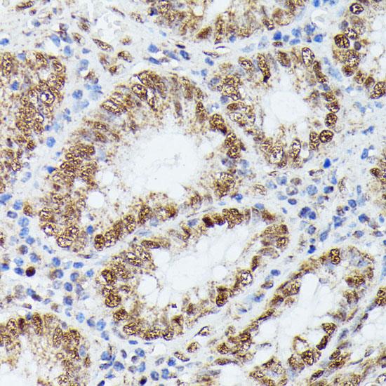
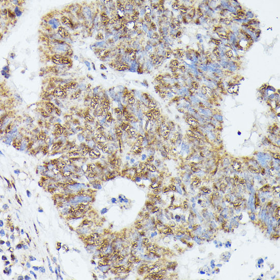
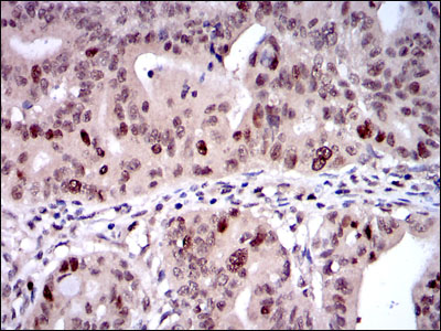
Anti-KIF22 antibodies have IHC images from human skin, kidney, colon, colon carcinoma and rectum cancer tissue (catalog IHC captions), plus MCF-7 IF data (M05570 IF caption).

Real IHC data M05570 staining KIF22 in human skin tissue sections by Immunohistochemistry (IHC-P -paraformaldehyde-fixed, paraffin-embedded sections). Tissue was fixed with formaldehyde and blocked with 3% BSA for 0. 5 hour at room temperature; antigen retrieval was by heat mediation with a citrate buffer (pH6). Samples were incubated with primary antibody (1/25) for 1 hours at 37°C. A undiluted biotinylated goat polyvalent antibody was used as the secondary antibody.
Anti-KIF22 Antibody (Center)
Cat # M05570
Real IHC data Immunohistochemistry of paraffin-embedded human colon using KIF22 antibody at dilution of 1:100 .Perform microwave antigen retrieval with 10 mM PBS buffer pH 7.2 before commencing with IHC staining protocol.
Anti-Kinesin-like protein KIF22 KIF22 Antibody
Cat # A05570-1
Real IHC data Immunohistochemistry analysis of paraffin-embedded rectum cancer tissues with DAB staining using KID Monoclonal Antibody.
Anti-KID KIF22 Monoclonal Antibody
Cat # A05570

M05570 shows human skin and kidney IHC-P and MCF-7 IF (M05570 image captions); A05570-1 shows paraffin-embedded human colon and colon carcinoma IHC (A05570-1 IHC captions). A05570 shows paraffin-embedded rectum cancer tissue by DAB IHC (A05570 IHC caption).

Which to pick: For human paraffin IHC, M05570 is a rabbit polyclonal with formaldehyde-fixed skin and kidney images, citrate pH 6 retrieval and a 1:25 primary dilution (M05570 catalog and IHC captions); A05570-1 shows colon and colon carcinoma with microwave PBS pH 7.2 retrieval and an unreported fixative (A05570-1 IHC captions), while human-reactive mouse monoclonal A05570 shows rectum cancer DAB staining with an unreported fixative (A05570 catalog and IHC caption). For IF, M05570 has a paraformaldehyde-fixed, permeabilized MCF-7 image (M05570 IF caption); for ICC/IF, A05570-1 lists both applications (A05570-1 catalog). For human and mouse specimens, choose rabbit polyclonal A05570-1 because both species and IHC/ICC/IF are listed (A05570-1 catalog).

Each figure is that product's own IHC / IF validation image from its datasheet.

References

  1. UniProt Consortium. UniProt entry Q14807 (KIF22_HUMAN, Kinesin-like protein KIF22).
  2. Human Protein Atlas. KIF22 tissue IHC expression (reliability: Enhanced).
  3. Human Protein Atlas. KIF22 subcellular location (ICC-IF): Mainly localized to the nuclear speckles..
  4. Human Protein Atlas. KIF22 antibody validation summary (2 antibodies).
  5. Effect of KIF22 on promoting proliferation and migration of gastric cancer cells via MAPK-ERK pathways. Chinese medical journal 2020 — PMC7176455.
  6. KIF22 promotes bladder cancer progression by activating the expression of CDCA3. International journal of molecular medicine 2021 — PMC8522959.
  7. Suppression of KIF22 Inhibits Cell Proliferation and Xenograft Tumor Growth in Tongue Squamous Cell Carcinoma. BioMed research international 2020 — PMC6996685.
  8. High Expression of KIF22/Kinesin-Like DNA Binding Protein (Kid) as a Poor Prognostic Factor in Prostate Cancer Patients. Medical science monitor : international medical journal of experimental and clinical research 2018 — PMC6247746.
  9. PubMed PMID:8599929 — UniProt-cited evidence.
  10. PubMed PMID:9790757 — UniProt-cited evidence.
  11. PubMed PMID:14702039 — UniProt-cited evidence.