KLF15 / Krueppel-like factor 15 · IHC design guide

Design Immunohistochemistry for KLF15

Plan paraffin section KLF15 IHC with the catalog antibody at 2–5 μg/ml (datasheet A03661-1). Assess granular cytoplasmic staining in renal tubules (HPA tissue IHC), while considering the annotated nuclear location (UniProt) and uncertain tissue IHC reliability (HPA tissue IHC).

Evidence assembled Oct 2026 · For research use; verify linked source records and product datasheet before use
Immunohistochemistry protocol sheet for KLF15 (IHC for KLF15): expected localisation Granular cytoplasm (HPA tissue IHC); nucleus (UniProt), antibody A03661-1, validated IHC image, and IHC protocol steps
Printable KLF15 IHC protocol sheet — expected localisation Granular cytoplasm (HPA tissue IHC); nucleus (UniProt), antibody A03661-1, controls and protocol steps. Open the full KLF15 IHC guide →

KLF15 Immunohistochemistry Experimental Design Guide

Expected localisation, validated protocols, controls and antibodies — the at-a-glance facts below, then the full design guide.

Must know before staining
Expected localisation Granular cytoplasm (HPA tissue IHC); nucleus (UniProt)
Staining pattern Granular cytoplasm, most abundant in renal tubules (HPA tissue IHC)
Antigen retrieval EDTA pH 8.0 HIER, heat-mediated (datasheet A03661-1)
Positive control ⓘ Cerebellum+4 more · see all
Negative control ⓘ Adipose tissue+4 more · see all
Important caveats
Reasons your staining may differ from the expected pattern.
Fixation Keep fixation consistent across sections (standard IHC practice; not target-specific)
Caveat Very low agreement between staining and RNA expression (HPA tissue IHC)
Regulation Reduced in cardiac and glomerular disease (UniProt)
Isoform / epitope One unprocessed chain; no annotated isoforms (UniProt)
Section 1

Recommended KLF15 IHC & IF Protocols

The catalog antibody protocol uses EDTA retrieval (datasheet A03661-1). Two published KLF15 IHC protocols provide cardiac and endometrial examples (PMC6029153; PMC11056958).

Recommended immunohistochemistry (IHC-P) protocol parameters
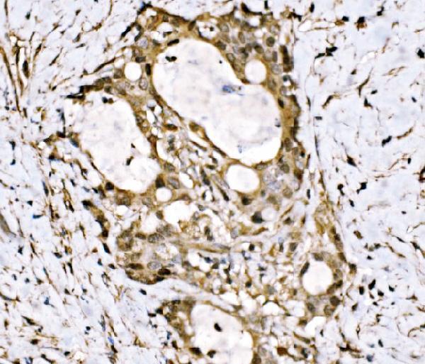
SampleParaffin-embedded human gall bladder adenosquamous carcinoma tissue; fixative not specified (datasheet A03661-1)
FixationImage fixative and duration unreported (datasheet A03661-1); verify before use.
Sectioning4–5 µm sections on charged slides (standard)
DeparaffinisationXylene, graded ethanol series to water (standard)
Antigen retrievalHeat retrieval: EDTA pH 8.0 (datasheet A03661-1); 20 min, 95–100 °C (standard)
Peroxidase block3% H2O2, 10 min, room temperature (standard)
Blocking10% goat serum (datasheet A03661-1)
Primary antibodyRabbit anti-KLF15, 2-5 μg/ml (datasheet A03661-1)
Primary incubationOvernight at 4 °C (datasheet A03661-1)
DetectionStreptavidin-biotin complex (SABC), DAB chromogen (datasheet A03661-1)
CounterstainHematoxylin, blue, dehydrate and mount (standard)
Expected resultKLF15-positive staining in cells in granular layer of cerebellum (HPA tissue IHC: Medium). HPA tissue profile: Granular cytoplasmic expression in several tissues most abundant in renal tubules. No signal in the no-primary control.
💡Decision noteStart with heat-mediated EDTA retrieval at pH 8.0 (datasheet A03661-1); the endometrial protocol reports retrieval without specifying its conditions (PMC11056958 methods).
Section 2

What Is the Expected KLF15 Staining Pattern?

KLF15 is a nuclear transcriptional regulator with no transmembrane segment (UniProt Q9UIH9). Renal tubular cells and cardiomyocytes show medium IHC staining (HPA tissue IHC). HPA describes the tissue signal as granular cytoplasmic, while supported ICC-IF localization is mainly nucleoplasmic and in nuclear speckles (HPA tissue IHC; HPA subcellular). Interpret any IHC pattern cautiously: HPA rates tissue staining Uncertain because antibody staining has very low consistency with RNA expression (HPA tissue IHC).

What am I looking at on my slide?
Nuclear staining in renal tubular cells, with a readable counterstain.This fits KLF15's nuclear localization and the supported ICC-IF nucleoplasmic pattern (UniProt Q9UIH9; HPA subcellular). Renal tubular cells are a plausible positive population, but HPA reports medium, granular cytoplasmic IHC staining there and rates tissue IHC Uncertain (HPA tissue IHC). Nuclear IHC alone therefore does not establish antibody specificity.
Predominantly granular cytoplasmic staining, with little nuclear enrichment.This resembles HPA's reported tissue IHC profile, especially in renal tubules (HPA tissue IHC). It conflicts with UniProt nuclear localization and supported ICC-IF nucleoplasmic localization (UniProt Q9UIH9; HPA subcellular). Treat the compartment mismatch as a possible staining artefact and seek independent validation before assigning it to KLF15; HPA's tissue IHC reliability is Uncertain (HPA tissue IHC).
Strong staining in bone marrow hematopoietic cells.That cell population is reported as not detected by HPA IHC, and UniProt reports no expression in bone marrow (HPA tissue IHC; UniProt Q9UIH9). Consider antibody cross-reactivity or endogenous detection activity (general IHC practice). Review compartment, background and controls before scoring these cells as KLF15 positive.
Diffuse color across cells and tissue, obscuring boundaries.A diffuse field cannot resolve the nuclear localization expected from UniProt and supported by ICC-IF (UniProt Q9UIH9; HPA subcellular). Nonspecific binding or detection background can produce this appearance (general IHC practice). Reassess the slide after background controls and detection conditions have been checked; do not use diffuse color as evidence of tissue distribution.
No staining in renal tubular cells.Renal tubular cells have medium staining in HPA tissue IHC, so an absent signal warrants a run-level check (HPA tissue IHC). It does not by itself prove KLF15 absence: HPA rates tissue IHC Uncertain, and a failed staining run can also yield no signal (HPA tissue IHC; general IHC practice). Compare positive tissue and detection controls before interpreting the specimen.
💡Expected KLF15 appearanceA provisional positive result is cell-resolved staining in renal tubular cells, with nuclear signal consistent with KLF15 localization and at most medium intensity as reported for those cells; diffuse color or unsupported staining in negative populations is suspect, and HPA tissue IHC remains Uncertain (UniProt Q9UIH9; HPA tissue IHC).
How each factor affects the staining
Cell and compartment selectionKLF15 is nuclear (UniProt Q9UIH9), while HPA reports medium staining in renal tubular cells and cardiomyocytes (HPA tissue IHC). Score the cell population and compartment separately because HPA's granular cytoplasmic IHC profile conflicts with its supported nuclear ICC-IF result (HPA tissue IHC; HPA subcellular).
Tissue evidenceUniProt reports high expression in liver, skeletal muscle and kidney (UniProt Q9UIH9). HPA calls liver RNA tissue enhanced but reports cholangiocytes as not detected by IHC (HPA tissue IHC). A liver result cannot be inferred from that RNA category, and a cholangiocyte negative should not override a separately assessed cell population.
Antibody and assay validationHPA028866 is Uncertain for IHC; HPA003286 is Supported for ICC, with no IHC status listed for it (HPA antibodies). HPA also rates the overall tissue IHC profile Uncertain because staining and RNA expression agree poorly (HPA tissue IHC). Keep assay-specific validation attached to the antibody used.
IF/ICC Q&A: Where should signal localize?Mainly to nucleoplasm and nuclear speckles; HPA lists these as supported, while its additional vesicle localization is uncertain (HPA subcellular). HPA lists ICC-IF images from SH-SY5Y and SK-MEL-30 (HPA subcellular). These observations help assess compartment agreement but do not validate an IHC-P staining pattern.
Why is my staining missing, weak or wrong?
SituationLikely causeNext action
Renal tubules show no signal.The run may have weak detection, or the antibody may not perform in this preparation (general IHC practice); HPA tissue IHC is Uncertain (HPA tissue IHC).Check positive tissue, reagent and detection controls, then review the validated IHC-P workflow for the antibody used (general IHC practice).
Granular cytoplasm dominates the slide.HPA reports granular cytoplasmic IHC staining, although KLF15 is nuclear and HPA's ICC-IF nuclear locations are supported (HPA tissue IHC; UniProt Q9UIH9; HPA subcellular).Record the compartment mismatch; compare controls and seek independent antibody or orthogonal evidence before calling it KLF15 (general IHC practice).
Bone marrow hematopoietic cells stain.HPA reports them as not detected, and UniProt reports no bone marrow expression (HPA tissue IHC; UniProt Q9UIH9). Cross-reactivity or endogenous detection activity is possible (general IHC practice).Inspect no-primary and detection controls, then reassess whether the signal is specific (general IHC practice).
Color appears broadly outside identifiable cells.Nonspecific reagent binding or detection background can obscure cell-level staining (general IHC practice).Check background controls and review blocking, antibody concentration and detection conditions for the run (general IHC practice).
The counterstain hides weak nuclear color.Heavy counterstaining can make a nuclear chromogen signal hard to resolve (general IHC practice).Review counterstain intensity and inspect a suitably counterstained section before assigning nuclear localization (general IHC practice).
ICC-IF looks nuclear but IHC looks cytoplasmic.HPA supports nucleoplasm and nuclear speckles in ICC-IF but describes granular cytoplasmic tissue IHC with Uncertain reliability (HPA subcellular; HPA tissue IHC).Document the assay-specific discrepancy; use tissue controls and independent evidence before transferring the ICC-IF interpretation to IHC-P (general IHC practice).

Sample controls for KLF15 IHC & IF

🧪Run kidney first and score nuclear staining in tubular cells (HPA: Medium in kidney tubular cells; UniProt: nucleus). Use bone marrow hematopoietic cells as the negative tissue (HPA: Not detected in bone marrow hematopoietic cells); any internal negative cells on the kidney slide should show counterstain without convincing nuclear chromogen, but HPA does not identify a validated negative kidney cell type.
Positive control tissue: Cerebellum (Cells in granular layer, HPA Medium)
Negative control tissue: Adipose tissue (HPA Not detected)
ICC-IF cell lines (HPA subcellular resource): HPA ICC-IF images show KLF15 in SH-SY5Y, SK-MEL-30, with annotated localisation: Nucleoplasm (supported), Nuclear speckles (supported) (HPA subcellular).
Technical controls: Include no-primary (secondary-only) and concentration-matched rabbit IgG isotype controls, plus KLF15 knockout tissue or a validated immunizing-peptide block (caption: rabbit primary at 2 μg/mL). Block endogenous peroxidase and check for endogenous biotin in kidney because the reported detection uses a biotinylated secondary, streptavidin–biotin complex, and DAB (caption: kidney tissue not specified; caption: biotin-based DAB detection).
⚠️Feasibility: No target-specific fixation window or fixation effect is reported in the supplied evidence; the selected A03661-1 paraffin-section caption does not state a fixative (caption: fixative not stated). The reported IHC staining used heat retrieval in EDTA at pH 8.0, so use that as the initial retrieval condition and verify nuclear signal against controls (caption: EDTA pH 8.0; UniProt: nucleus). HPA reports nucleoplasm and nuclear speckles in ICC-IF images, but the supplied evidence does not establish that frozen sections or IF are easier than paraffin IHC (HPA: supported nucleoplasm and nuclear speckles).

HPA tissue IHC evidence for KLF15

Comprehensive Human Protein Atlas IHC scoring per tissue (reliability: Uncertain — Very low consistency between antibody staining and RNA expression data.). Rows are taken directly from the HPA tissue chart — click any row's HPA link to view the source.

Positive expression · recommended positive controls

TissueCell typeLevelEvidenceSource
Cerebellum Cells in granular layer Medium Protein (IHC) HPA →
Colon Glandular cells Medium Protein (IHC) HPA →
Duodenum Glandular cells Medium Protein (IHC) HPA →
Heart muscle Cardiomyocytes Medium Protein (IHC) HPA →
Kidney Cells in tubules Medium Protein (IHC) HPA →

Undetected expression · recommended negative controls

TissueCell typeLevelEvidenceSource
Adipose tissue Adipocytes Not detected Protein (IHC) HPA →
Bone marrow Hematopoietic cells Not detected Protein (IHC) HPA →
Breast Adipocytes Not detected Protein (IHC) HPA →
Fallopian tube Glandular cells Not detected Protein (IHC) HPA →
Liver Cholangiocytes Not detected Protein (IHC) HPA →
Section 3

Advanced KLF15 IHC Tips

Troubleshoot KLF15 staining in paraffin sections by checking retrieval, compartment, cell type, and controls before interpreting chromogenic signal.

Which retrieval condition should I start with for KLF15 in paraffin sections?
Start with heat-mediated retrieval in EDTA at pH 8.0 for the catalog antibody (datasheet A03661-1). Keep heating and cooling conditions consistent across slides, then compare a retrieval-treated section with an untreated section from the same specimen (standard IHC practice). If staining is weak, adjust heating time in small increments before testing another buffer, while watching for tissue damage (standard IHC practice). The documented paraffin-section staining used this EDTA condition with 2 μg/ml primary antibody overnight at 4°C (caption A03661-1). Judge any improvement by specific cellular staining and tissue preservation, not overall DAB intensity alone (standard IHC practice).
How should I troubleshoot weak KLF15 staining when fixation history is uncertain?
The selected paraffin-section caption does not state a fixative, so target-specific KLF15 sensitivity to fixation is unknown (caption A03661-1). Record the specimen's actual fixation and processing history, and compare sections processed together before changing antibody concentration (standard IHC practice). Begin with EDTA retrieval at pH 8.0 and 2 μg/ml primary antibody overnight at 4°C, as documented for this antibody (caption A03661-1). If a preserved control section stains but the specimen does not, examine tissue preservation, retrieval consistency, and detection controls before assigning a biological negative (standard IHC practice). Do not infer fixation sensitivity from the reported tissue staining pattern (HPA: tissue IHC reliability Uncertain).
Should nuclear or cytoplasmic DAB staining count as KLF15 positive?
Prioritise staining that resolves within nuclei: KLF15 is annotated as nuclear (UniProt Q9UIH9), and nucleoplasm and nuclear speckles are supported IF locations (HPA: subcellular). Assess nuclei against a counterstain at high magnification, keeping faint diffuse DAB separate from clearly localised signal (standard IHC practice). HPA tissue IHC instead describes granular cytoplasmic staining, particularly in renal tubules, but rates agreement with RNA expression as very low (HPA: tissue IHC reliability Uncertain). Treat isolated cytoplasmic granules as provisional until they reproduce with an independent antibody or another target-specific control (standard IHC practice). Record nuclear and cytoplasmic staining separately rather than combining them into one positive score (standard IHC practice).
Could an isoform or inaccessible epitope explain discordant KLF15 staining?
The supplied record annotates a single 1–416 chain and 0 isoforms, but gives no antibody epitope position (UniProt Q9UIH9; caption A03661-1). Thus, an isoform-specific explanation cannot be established from these data (UniProt Q9UIH9). KLF15 has no annotated transmembrane segment, signal peptide, glycosylation sites, or modified residues in this record; none identifies the catalog antibody's accessible epitope (UniProt Q9UIH9). Compare retrieval conditions on adjacent sections only after applying the documented EDTA pH 8.0 condition (datasheet A03661-1; standard IHC practice). If results remain discordant, obtain the immunogen or epitope information and validate staining with an independent antibody (standard IHC practice).
How can IF help check a disputed KLF15 IHC pattern?
Use IF as a separate validation experiment, pairing KLF15 with a marker identifying the expected cell type in the specimen (standard IF practice). KLF15 is nuclear (UniProt Q9UIH9), with supported nucleoplasmic and nuclear-speckle IF localisation, while vesicular localisation is uncertain (HPA: subcellular). Include a nuclear counterstain and permeabilise fixed cells sufficiently to admit antibody to the nuclear epitope; the supplied data give no epitope position or IF fixation condition (standard IF practice; caption A03661-1). Choose fluorophores away from the specimen's strongest autofluorescence and inspect unstained and single-label controls (standard IF practice). Compare cell identity and compartment with IHC, without transferring the paraffin-section retrieval conditions to IF (caption A03661-1; standard IF practice).
What should I check when KLF15 DAB staining is widespread or granular?
Inspect a no-primary control and assess whether DAB persists in the same structures; persistent colour implicates the detection system or tissue background (standard IHC practice). The documented workflow uses a biotinylated secondary, streptavidin–biotin complex, and DAB, so include an appropriate detection control when interpreting granular deposits (caption A03661-1; standard IHC practice). Apply a peroxidase block as a general DAB workflow step, and evaluate endogenous biotin or nonspecific secondary binding if background remains (standard IHC practice). The caption reports 10% goat serum blocking and 2 μg/ml primary antibody overnight at 4°C (caption A03661-1). Compare background with nuclear localisation expected for KLF15 before calling diffuse or granular cytoplasmic signal positive (UniProt Q9UIH9; standard IHC practice).
How should I score KLF15 IHC when staining varies by compartment and cell type? ⚠ ANSWER MARKED FOR VERIFICATION
Define the eligible cell population before scoring, then report the percentage of cells with convincing nuclear DAB and an intensity-based H-score if intensity is reproducible (standard IHC practice; UniProt Q9UIH9: nuclear). Use the conventional 0–300 H-score scale, calculated from percentages at intensity grades 0–3 (standard IHC practice). Normalise positive counts to the number of eligible, intact cells, or report positive-cell density per mm² of viable analysed tissue (standard IHC practice). Score cytoplasmic staining separately because HPA describes granular cytoplasmic tissue staining with uncertain reliability (HPA: tissue IHC reliability Uncertain). Apply the same threshold, counterstain, and region-selection rules across compared sections (standard IHC practice).
When is a KLF15-positive section convincing rather than an artefact?
A convincing result places reproducible signal in the expected cells and predominantly in nuclei, consistent with KLF15's annotated location (UniProt Q9UIH9; standard IHC practice). Kidney tubule cells and cardiomyocytes have medium HPA tissue staining, but the tissue IHC dataset has uncertain reliability, so those patterns alone do not establish specificity (HPA: tissue IHC). Check whether staining follows section edges, damaged or necrotic areas, or structures positive in the no-primary control (standard IHC practice). Exclude endogenous peroxidase or detection-related DAB deposits with appropriate controls before assigning positivity (standard IHC practice). Interpret isolated granular cytoplasmic signal cautiously because it conflicts with the nuclear annotation and comes from uncertain tissue IHC evidence (UniProt Q9UIH9; HPA: tissue IHC).
Boster reagents

Best KLF15 / Krueppel-like factor 15 IHC Antibodies

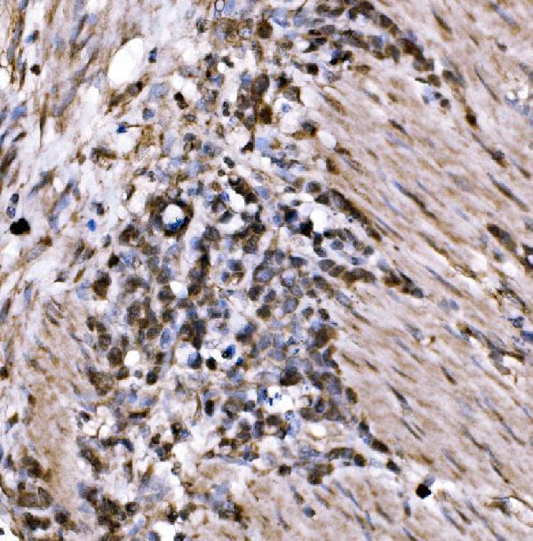
The IHC-validated anti-KLF15 antibody has real paraffin-section images from human gall bladder adenosquamous carcinoma, human gastric cancer, and mouse and rat brain (A03661-1 IHC image captions).

Real IHC data IHC analysis of KLF15 using anti-KLF15 antibody (A03661-1). KLF15 was detected in a paraffin-embedded section of human gall bladder adenosquamous carcinoma tissue. Heat mediated antigen retrieval was performed in EDTA buffer (pH 8.0, epitope retrieval solution). The tissue section was blocked with 10% goat serum. The tissue section was then incubated with 2 μg/ml rabbit anti-KLF15 Antibody (A03661-1) overnight at 4°C. Biotinylated goat anti-rabbit IgG was used as secondary antibody and incubated for 30 minutes at 37°C. The tissue section was developed using Strepavidin-Biotin-Complex (SABC) (Catalog # SA1022) with DAB as the chromogen.
Anti-KLF15 Antibody ®
Cat # A03661-1

A03661-1 is listed for IHC in human, mouse, and rat samples (A03661-1 applications and reactivity). Its IHC captions document paraffin sections of human gall bladder adenosquamous carcinoma, human gastric cancer, mouse brain, and rat brain (A03661-1 IHC image captions).

Which to pick: For tissue IHC, choose A03661-1 for paraffin sections; its captions describe EDTA retrieval at pH 8.0 and 2 μg/ml primary antibody, but do not report the fixative (A03661-1 IHC image captions). There is no IF/ICC-validated option in this catalog: A03661-1 lists IHC but not IF/ICC, and has no IF images (A03661-1 applications and IF image alts). For cross-species IHC, A03661-1 covers human, mouse, and rat; its host is rabbit and its clone is unreported (A03661-1 reactivity, host, and clone).

Each figure is that product's own IHC / IF validation image from its datasheet.

References

  1. UniProt Consortium. UniProt entry Q9UIH9 (KLF15_HUMAN, Krueppel-like factor 15).
  2. Human Protein Atlas. KLF15 tissue IHC expression (reliability: Uncertain).
  3. Human Protein Atlas. KLF15 subcellular location (ICC-IF): Mainly localized to the nucleoplasm and nuclear speckles. In addition localized to vesicles..
  4. Human Protein Atlas. KLF15 antibody validation summary (2 antibodies).
  5. Left ventricular hypertrophy in experimental chronic kidney disease is associated with reduced expression of cardiac Kruppel-like factor 15. BMC nephrology 2018 — PMC6029153.
  6. Clinicopathological significance of Kruppel-like factor 15 and epithelial-to-mesenchymal transition related factors in bladder cancer. Scientific reports 2025 — PMC12592540.
  7. Loss of KLF15 impairs endometrial receptivity by inhibiting EMT in endometriosis. The Journal of endocrinology 2024 — PMC11056958.
  8. Depletion of Kruppel-like factor 15 sensitized gliomas to temozolomide cytotoxicity through O (6)-methylguanine-DNA methyl-transferase. Biochemistry and biophysics reports 2021 — PMC8242961.
  9. PubMed PMID:10982849 — UniProt-cited evidence.
  10. PubMed PMID:15489334 — UniProt-cited evidence.
  11. PubMed PMID:17438289 — UniProt-cited evidence.