This website uses cookies to ensure you get the best experience on our website.


- Table of Contents
Plan KLK4 chromogenic IHC in paraffin sections using granular staining in prostate glandular cells as a reference (HPA tissue IHC). This guide covers fixation, retrieval, controls and scoring while accounting for KLK4 secretion (UniProt).

Expected localisation, validated protocols, controls and antibodies — the at-a-glance facts below, then the full design guide.
| Expected localisation | Secreted; tissue staining compartment unspecified (UniProt; HPA tissue IHC) | |
| Staining pattern | Granular staining in prostate glandular cells (HPA tissue IHC) | |
| Antigen retrieval | Citrate pH 6.0 HIER, 95–98 °C, 20 min (rule: cytoplasmic / membrane antigen) | |
| Positive control | Prostate | |
| Negative control | Adipose tissue+4 more · see all |
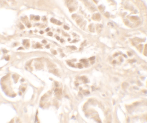
| Fixation | Keep formalin fixation consistent across paraffin sections (standard IHC practice; not target-specific); Matched tissue-IHC evidence does not establish the fixation claim. Validate the specimen-specific method before use. (selected-SKU IHC image A03430) | |
| Caveat | Secreted KLK4 may stain beyond producing cells (UniProt; HPA tissue IHC) | |
| Regulation | Prostate-enriched RNA expression (HPA tissue RNA) | |
| Isoform / epitope | 2 isoforms; cleaved regions affect epitope choice (UniProt) |
The catalog antibody’s IHC-P protocol is accompanied by three published KLK4 IHC methods (PMC13009279; PMC13201332; PMC7255463).
| Sample | Tissue sections; selected-image fixative not specified (standard IHC workflow) |
| Fixation | Image fixative and duration unreported (datasheet A03430); verify before use. |
| Sectioning | 4–5 µm sections on charged slides (standard) |
| Deparaffinisation | Xylene, graded ethanol series to water (standard) |
| Antigen retrieval | Heat-induced epitope retrieval in citrate buffer, pH 6.0, 20 min at 95–98 °C (standard rule: cytoplasmic / membrane antigen) |
| Peroxidase block | 3% H2O2, 10 min, room temperature (standard) |
| Blocking | 10% normal serum of the secondary host, 30 min, room temperature (standard) |
| Primary antibody | Rabbit anti-KLK4, 1:100–1:500 starting range (standard) |
| Primary incubation | Overnight at 4 °C (standard) |
| Detection | HRP-polymer secondary, DAB chromogen 5–10 min (standard) |
| Counterstain | Hematoxylin, blue, dehydrate and mount (standard) |
| Expected result | KLK4-positive staining in glandular cells of prostate (HPA tissue IHC: High). HPA tissue profile: Granular expression in prostate. No signal in the no-primary control. |
KLK4 is a secreted protein with no transmembrane segment (UniProt Q9Y5K2 topology). In paraffin sections, look for granular staining in prostate glandular cells, where staining is reported as High (HPA: prostate tissue IHC). HPA rates the tissue pattern Enhanced, citing consistency between antibody staining and RNA expression; secretion means the protein signal need not coincide exactly with the cells expressing its RNA (HPA: tissue reliability).
| Granular signal in prostate glandular cells. | This matches the reported positive pattern and High staining level (HPA: prostate tissue IHC). Judge it against the tissue architecture and negative controls; the secreted annotation alone does not specify every stained subcellular compartment (UniProt Q9Y5K2; HPA: subcellular summary). |
| Predominantly nuclear staining in prostate glandular cells. | A nuclear dominant pattern does not match the reported granular tissue pattern or secreted annotation (HPA: prostate tissue IHC; UniProt Q9Y5K2 topology). Treat it as suspect until the assay controls and an independently validated staining pattern support it. |
| Strong staining in cells reported as negative, such as adrenal glandular cells. | HPA reports KLK4 as Not detected in those cells (HPA: adrenal gland tissue IHC). Unexpected signal may reflect antibody cross-reactivity or endogenous chromogenic detection activity; assess the staining with appropriate controls before assigning it to KLK4. |
| Uniform haze across cells and surrounding tissue. | This lacks the reported granular, glandular pattern (HPA: prostate tissue IHC). Diffuse background can arise from nonspecific binding, incomplete blocking, or detection reagent background (general IHC practice). Its presence limits interpretation of a weak apparent positive signal. |
| No staining in prostate glandular cells. | This conflicts with the reported High positive tissue result (HPA: prostate tissue IHC). Check control performance and the actual antibody's IHC-P conditions before treating the section as biologically negative; HPA supplies no KLK4 specific fixation sensitivity here. |
| Secreted protein and processing | KLK4 has a signal peptide at residues 1–26, a propeptide at 27–30, and a mature chain at 31–254 (UniProt Q9Y5K2). Interpret staining with secretion in mind; these annotations do not establish where a particular antibody's epitope lies. |
| Prostate reference tissue | HPA reports granular expression and High glandular cell staining in prostate (HPA: prostate tissue IHC). Use that observed pattern as the positive benchmark; HPA notes that secreted protein location and RNA location may differ (HPA: tissue reliability). |
| Negative tissue comparison | HPA reports Not detected in adrenal glandular cells and appendix glandular cells, among other listed examples (HPA: negative tissue IHC). These are comparison patterns, not proof that every cell in every specimen must remain unstained. |
| Strength and limits of validation | The tissue profile is rated Enhanced and HPA051839 has Enhanced IHC validation (HPA: tissue reliability; HPA: antibody validation). Neither entry supplies antigen retrieval settings, an epitope map, or a KLK4 specific fixation effect. |
| IF/ICC: what pattern is established? | HPA summarizes KLK4 as secreted but provides no main subcellular location, ICC-IF images, or ICC validation for HPA051839 (HPA: subcellular summary; HPA: antibody validation). The IHC glandular pattern cannot by itself establish an IF/ICC staining pattern. |
| Situation | Likely cause | Next action |
|---|---|---|
| Prostate positive control is blank. | A failed IHC step or unsuitable antibody conditions are possibilities (general IHC practice); the result conflicts with High prostate glandular staining (HPA: prostate tissue IHC). | Confirm section integrity and detection controls, then check the actual IHC-validated antibody's recommended retrieval, dilution, and detection conditions. No KLK4 specific retrieval or dilution is supplied here. |
| Prostate signal is weak or hard to distinguish from background. | Poor signal separation can obscure the reported granular glandular pattern (HPA: prostate tissue IHC; general IHC practice). HPA does not establish a KLK4 specific fixation cause. | Compare with a clean negative control and review the antibody's IHC-P conditions. Adjust general assay variables one at a time, documenting whether glandular granules become distinguishable. |
| The slide shows mainly nuclear staining. | The compartment is inconsistent with KLK4's secreted annotation and HPA's granular prostate pattern (UniProt Q9Y5K2 topology; HPA: prostate tissue IHC). | Inspect matched controls and antibody specificity evidence. Do not score nuclear dominant signal as the expected KLK4 pattern solely because it appears in prostate tissue. |
| Adrenal or appendix glandular cells stain strongly. | Both are listed as Not detected (HPA: negative tissue IHC). Cross-reactivity or endogenous chromogenic activity may contribute (general IHC practice). | Review a no primary antibody control and the relevant endogenous activity block; compare staining distribution with the prostate positive control before interpreting unexpected signal. |
| Color develops broadly outside glandular cells. | Widespread color may reflect detection background or nonspecific binding (general IHC practice), rather than the granular prostate glandular pattern (HPA: prostate tissue IHC). | Check no primary and reagent controls, blocking and wash steps, and chromogen development. Repeat scoring only where cell boundaries and granular signal remain interpretable. |
| An IF/ICC result is used to justify the IHC pattern. | HPA lists no ICC-IF images or ICC validation for the listed antibody (HPA: subcellular summary; HPA: antibody validation). | Keep the IHC interpretation anchored to the HPA prostate tissue pattern. Evaluate any IF/ICC experiment under its separate guide and controls; do not infer an IF protocol from the IHC evidence. |


Comprehensive Human Protein Atlas IHC scoring per tissue (reliability: Enhanced — High consistency between antibody staining and RNA expression data. At least one protein variant secreted, tissue location of RNA and protein might differ and correlation is complex.). Rows are taken directly from the HPA tissue chart — click any row's HPA link to view the source.
| Tissue | Cell type | Level | Evidence | Source |
|---|---|---|---|---|
| Prostate | Glandular cells | High | Protein (IHC) | HPA → |
| Tissue | Cell type | Level | Evidence | Source |
|---|---|---|---|---|
| Adipose tissue | Adipocytes | Not detected | Protein (IHC) | HPA → |
| Adrenal gland | Glandular cells | Not detected | Protein (IHC) | HPA → |
| Appendix | Glandular cells | Not detected | Protein (IHC) | HPA → |
| Bone marrow | Hematopoietic cells | Not detected | Protein (IHC) | HPA → |
| Breast | Adipocytes | Not detected | Protein (IHC) | HPA → |
Troubleshoot KLK4 staining in paraffin sections by checking retrieval, controls, cellular distribution and scoring before interpreting chromogenic signal.
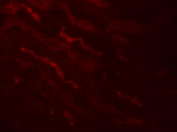
A03430 has human IHC-P and IF validation, with images from human kidney tissue (catalog applications; IHC and IF image captions).
A03430 is listed for human IHC-P and IF (catalog applications; reactivity: Human). Its IHC image shows human kidney tissue at 5 μg/mL, and its IF image shows human kidney tissue at 20 μg/mL (catalog image captions).
Which to pick: Choose A03430 for human paraffin-section IHC (catalog: IHC-P; reactivity: Human); its kidney IHC caption reports 5 μg/mL but does not report the fixative (catalog IHC image caption). A03430 also has human IF validation, but ICC is not separately listed (catalog applications; reactivity: Human). No cross-species option is supported by this catalog payload (catalog reactivity: Human only).