This website uses cookies to ensure you get the best experience on our website.


- Table of Contents
Plan chromogenic KMT2A IHC in paraffin sections around the observed nuclear tissue pattern (HPA tissue IHC). This guide covers fixation consistency, staining assessment, and catalog IHC dilution ranges of 1:100–1:300 for A00402 and 1:200–1:1000 for M00402 (datasheets).

Expected localisation, validated protocols, controls and antibodies — the at-a-glance facts below, then the full design guide.
| Expected localisation | Nuclear staining across tissues (HPA tissue IHC) | |
| Staining pattern | Nuclei across tissue cell types (HPA tissue IHC) | |
| Antigen retrieval | Tris-EDTA pH 9.0 HIER, 95–98 °C, 20 min (rule: nuclear antigen) | |
| Positive control | Placenta+4 more · see all | |
| Negative control | None in HPA (detected in all 45 tissues); use no-primary + isotype controls |
| Fixation | Keep formalin fixation consistent across paraffin sections. (standard IHC practice; not target-specific) | |
| Caveat | Staining and RNA show medium consistency (HPA tissue IHC) | |
| Regulation | Low tissue specificity (HPA tissue RNA) | |
| Isoform / epitope | 3 isoforms; N320/C180 cleavage makes epitope position relevant (UniProt) |
Compare the catalog antibody’s IHC-P protocol with 4 published KMT2A IHC methods (PMC11782969; PMC8133758; PMC5199165; PMC7288919).
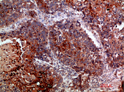
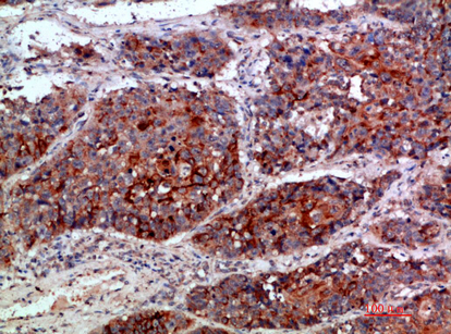
| Sample | Paraffin-embedded human lung-cancer tissue; fixative not specified (datasheet A00402) |
| Fixation | Image fixative and duration unreported (datasheet A00402); verify before use. |
| Sectioning | 4–5 µm sections on charged slides (standard) |
| Deparaffinisation | Xylene, graded ethanol series to water (standard) |
| Antigen retrieval | Heat-induced epitope retrieval in Tris-EDTA buffer, pH 9.0, 20 min at 95–98 °C (standard rule: nuclear antigen) |
| Peroxidase block | 3% H2O2, 10 min, room temperature (standard) |
| Blocking | 10% normal serum of the secondary host, 30 min, room temperature (standard) |
| Primary antibody | Rabbit anti-KMT2A, 1:100-1:300 (datasheet A00402) |
| Primary incubation | Overnight at 4 °C (standard) |
| Detection | HRP-polymer secondary, DAB chromogen 5–10 min (standard) |
| Counterstain | Hematoxylin, blue, dehydrate and mount (standard) |
| Expected result | KMT2A-positive staining in endothelial cells of placenta (HPA tissue IHC: High). HPA tissue profile: Ubiquitous nuclear expression. No signal in the no-primary control. |
KMT2A should appear predominantly in nuclei across many cell types in paraffin sections (UniProt Q03164: nucleus; HPA: ubiquitous nuclear expression). Strong reference staining occurs in placental endothelial cells and testicular pachytene spermatocytes (HPA: High in both). KMT2A has no transmembrane segment (UniProt Q03164: topology). HPA rates the tissue IHC evidence Enhanced, while reporting medium consistency between antibody staining and RNA expression (HPA: reliability).
| Nuclear chromogen in placental endothelial cells or testicular pachytene spermatocytes, with identifiable tissue morphology. | This matches the expected compartment and two High reference cell populations (UniProt Q03164: nucleus; HPA: High in both). Compare nuclei within the same section and confirm that the counterstain still permits cell identification (general IHC practice). |
| Strong staining is confined to cytoplasm, with little or no nuclear signal. | Treat a cytoplasm-only IHC pattern as suspect because the established tissue profile is nuclear (HPA: ubiquitous nuclear expression). Review localization in a known-positive section and check the detection controls before assigning biological meaning (general IHC practice). |
| Intense staining appears in cells expected to show low signal, or in cells whose identity is uncertain. | HPA reports low staining in caudate glial cells, parathyroid glandular cells, liver cholangiocytes and adipocytes (HPA: Low in each). Verify cell identity and compare a no-primary control; unexpected chromogen can reflect cross-reactivity or endogenous detection activity (general IHC practice). |
| Brown color spreads across tissue structures or covers nuclei and surrounding areas uniformly. | A diffuse deposit cannot reliably establish nuclear localization (general IHC practice). Compare a no-primary control and inspect blocking, washing and detection conditions; widespread color may be assay background rather than KMT2A staining (general IHC practice). |
| No nuclear signal appears in a known-positive reference section. | Absence in placental endothelial cells or testicular pachytene spermatocytes conflicts with their High HPA staining (HPA: High in both). Check tissue integrity, antigen retrieval, primary antibody use and detection performance before interpreting a test section as negative (general IHC practice). |
| Reference cell population | Placental endothelial cells and testicular pachytene spermatocytes are High; bone marrow hematopoietic cells and bronchial respiratory epithelial cells are Medium (HPA: tissue IHC). Choose the comparison cell type before judging intensity. |
| Low staining is not absence | HPA lists low staining in glial cells of the caudate, cholangiocytes and adipocytes (HPA: tissue IHC). A weak result in these cells is a poor stand-alone failure criterion; compare a High reference population. |
| Protein processing and antibody epitope | KMT2A yields N320 and C180 cleavage products (UniProt Q03164: processing). Which products an antibody detects depends on its epitope; the supplied HPA validation entries do not specify an epitope, so do not infer fragment-specific staining. |
| Antibody validation | Two listed antibodies have Enhanced IHC status, CAB017794 and CAB024270 (HPA: antibodies). Enhanced means agreement from independent antibodies or orthogonal data (HPA: validation definition); it does not resolve every unexpected cell-level signal. |
| IF/ICC Q&A: Where should signal appear? | A: Mainly in the nucleoplasm (HPA: supported ICC-IF location). HPA also lists cytosol as an uncertain additional location (HPA: subcellular); this does not establish a cytoplasm-only IHC pattern. |
| Situation | Likely cause | Next action |
|---|---|---|
| Known-positive section lacks nuclear staining. | A failed staining run is possible when cells reported High are blank (HPA: placental endothelial cells; testicular pachytene spermatocytes). | Check section quality, antigen retrieval settings, primary antibody application and detection reagents against the run controls (general IHC practice). |
| Test section looks negative beside a strongly stained reference. | The tested cell population may normally stain less strongly; HPA reports both Medium and Low populations (HPA: tissue IHC). | Identify the cells being scored, retain the High reference for run acceptance, and interpret weak staining relative to the appropriate population (HPA: tissue IHC; general IHC practice). |
| Signal is mainly cytoplasmic. | The pattern conflicts with the established nuclear tissue profile (HPA: ubiquitous nuclear expression; UniProt Q03164: nucleus). | Recheck morphology and a known-positive nuclear control; assess no-primary background before accepting the staining as specific (general IHC practice). |
| Diffuse chromogen obscures nuclei. | Excess background or endogenous detection activity may obscure localization (general IHC practice). | Inspect a no-primary control, then review blocking, washing and chromogen development conditions; score only cells with interpretable nuclei (general IHC practice). |
| Unexpectedly strong staining appears in a low-staining population. | Cell misidentification, cross-reactivity or endogenous detection activity are possibilities (HPA: Low populations; general IHC practice). | Confirm cell morphology and examine a no-primary control; compare the pattern with a High reference section before assigning positivity (HPA: High populations; general IHC practice). |
| Results differ between antibodies. | The listed antibodies have different application validation records, and the supplied entries do not establish matching epitopes (HPA: antibodies). | Check each antibody’s IHC validation and documented epitope, if available; compare nuclear localization and the same reference cell populations (HPA: tissue IHC; general IHC practice). |


Comprehensive Human Protein Atlas IHC scoring per tissue (reliability: Enhanced — Medium consistency between antibody staining and RNA expression data.). Rows are taken directly from the HPA tissue chart — click any row's HPA link to view the source.
| Tissue | Cell type | Level | Evidence | Source |
|---|---|---|---|---|
| Placenta | Endothelial cells | High | Protein (IHC) | HPA → |
| Testis | Pachytene spermatocytes | High | Protein (IHC) | HPA → |
| Adrenal gland | Glandular cells | Medium | Protein (IHC) | HPA → |
| Appendix | Glandular cells | Medium | Protein (IHC) | HPA → |
| Bone marrow | Hematopoietic cells | Medium | Protein (IHC) | HPA → |
| Tissue | Cell type | Level | Evidence | Source |
|---|---|---|---|---|
| None in HPA: KMT2A is detected in all 45 scored tissues. Use a no-primary (secondary-only) and an isotype control instead. | ||||
Troubleshoot KMT2A staining in paraffin sections by checking nuclear localisation, retrieval, antibody epitope, detection controls, and scoring consistency (UniProt Q03164; HPA tissue IHC).
Two anti-KMT2A antibodies have paraffin-section IHC images (catalog IHC captions); both list IF, but no IF images are supplied (catalog applications; catalog IF image lists).
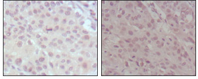
A00402 lists IHC and IF for human, mouse and rat; its IHC captions show paraffin-embedded human lung cancer and brain (catalog applications; catalog reactivity; A00402 IHC captions). M00402 lists IHC and IF for human samples; its IHC caption shows paraffin-embedded human lung and esophagus cancers (catalog applications; catalog reactivity; M00402 IHC caption).
Which to pick: For human tissue IHC, either SKU has a paraffin-section example: A00402 in lung cancer and brain, and M00402 in lung and esophagus cancers (A00402 IHC captions; M00402 IHC caption). For IF/ICC, both list IF, but neither has an IF image in the payload; A00402 is a rabbit antibody and M00402 is a mouse monoclonal, so choose based on the planned detection system (catalog applications; catalog IF image lists; catalog host and clone fields). For mouse or rat samples, choose A00402 because its reactivity list includes both, whereas M00402 lists human only; the captions report paraffin embedding but do not report the fixative (catalog reactivity; A00402 IHC captions; M00402 IHC caption).