This website uses cookies to ensure you get the best experience on our website.


- Table of Contents
Plan chromogenic KMT2E IHC in paraffin sections using hematopoietic cells in bone marrow as a medium-staining reference (HPA tissue IHC). The catalog antibody lists an IHC dilution range of 1:50–1:200 (datasheet); assess cytoplasmic staining in immune-cell subsets (HPA tissue IHC).

Expected localisation, validated protocols, controls and antibodies — the at-a-glance facts below, then the full design guide.
| Expected localisation | Cytoplasm in immune-cell subsets (HPA tissue IHC) | |
| Staining pattern | Subsets of immune cells show cytoplasmic staining (HPA tissue IHC) | |
| Antigen retrieval | Tris-EDTA pH 9.0 HIER, 95–98 °C, 20 min (rule: nuclear antigen) | |
| Positive control | Bone marrow | |
| Negative control | Adipose tissue+4 more · see all |
| Fixation | Keep paraffin-section fixation consistent (standard IHC practice; not target-specific); Matched tissue-IHC evidence does not establish the fixation claim. Validate the specimen-specific method before use. (selected-SKU IHC image A08526) | |
| Caveat | Marrow peroxidase may mimic positive staining (HPA tissue IHC; standard IHC practice) | |
| Regulation | OGT and USP7 stabilize isoform 3 (UniProt) | |
| Isoform / epitope | 8 isoforms; check epitope coverage; no TM sidedness (UniProt) |
Compare the catalog antibody’s IHC-P protocol (datasheet) with the published esophageal cancer tumor-section protocol (PMC12527620).
| Sample | Paraffin-embedded rat ovary tissue; fixative not specified (datasheet A08526) |
| Fixation | Image fixative and duration unreported (datasheet A08526); verify before use. |
| Sectioning | 4–5 µm sections on charged slides (standard) |
| Deparaffinisation | Xylene, graded ethanol series to water (standard) |
| Antigen retrieval | Heat-induced epitope retrieval in Tris-EDTA buffer, pH 9.0, 20 min at 95–98 °C (standard rule: nuclear antigen) |
| Peroxidase block | 3% H2O2, 10 min, room temperature (standard) |
| Blocking | 10% normal serum of the secondary host, 30 min, room temperature (standard) |
| Primary antibody | Rabbit anti-KMT2E, 1:50-1:200 (datasheet A08526) |
| Primary incubation | Overnight at 4 °C (standard) |
| Detection | HRP-polymer secondary, DAB chromogen 5–10 min (standard) |
| Counterstain | Hematoxylin, blue, dehydrate and mount (standard) |
| Expected result | KMT2E-positive staining in hematopoietic cells of bone marrow (HPA tissue IHC: Medium). HPA tissue profile: Cytoplasmic expression in subsets of immune cells in several tissues. No signal in the no-primary control. |
KMT2E can localize to the nucleoplasm, cytoplasm and other compartments (UniProt Q8IZD2 subcellular location); it has no transmembrane segment (UniProt Q8IZD2 topology). In tissue IHC, expect cytoplasmic staining in subsets of immune cells, including medium staining in bone marrow hematopoietic cells (HPA tissue IHC). HPA rates the tissue pattern Approved, with external verification pending (HPA tissue IHC reliability).
| Cytoplasmic signal in some bone marrow hematopoietic cells, with unstained neighbors. | This fits the reported medium staining in hematopoietic cells and subset pattern in immune cells (HPA tissue IHC). Score the stained cells and compartment; a whole-section average would obscure the subset pattern (general IHC practice). |
| Predominant nuclear staining, without the expected immune-cell cytoplasmic pattern. | Check the antibody and slide controls before rejecting it: nucleoplasmic localization is supported by ICC-IF (HPA subcellular), while the reported tissue IHC pattern is cytoplasmic (HPA tissue IHC). Uniform nucleolar staining is especially discordant with the reported absence from nucleoli (UniProt Q8IZD2 subcellular location). |
| Strong staining in cells reported as unstained, such as adipocytes or adrenal glandular cells. | These cell types were not detected in the supplied HPA tissue IHC observations (HPA tissue IHC). If staining persists in controls, consider cross-reactivity or endogenous detection activity as general IHC possibilities (general IHC practice); one discrepant slide cannot identify the cause. |
| Color spreads across stroma or slide areas without clear cell boundaries. | Treat diffuse staining as background until controls support a cellular signal (general IHC practice). It does not establish KMT2E expression or a subcellular location; compare tissue morphology, antibody-omission controls and the reported immune-cell subset pattern (HPA tissue IHC; general IHC practice). |
| No detectable signal in bone marrow hematopoietic cells. | The result conflicts with the supplied medium IHC observation (HPA tissue IHC). Check section quality and assay controls, then review the catalog antibody's IHC-P conditions (general IHC practice). Absence of staining alone does not establish absence of protein (general IHC practice). |
| Tissue-pattern confidence | HPA calls the tissue IHC pattern Approved but says staining involves cells or structures not annotated and awaits external verification (HPA tissue IHC reliability); interpret a new tissue result with its controls (general IHC practice). |
| Cell-specific expression | HPA reports cytoplasmic staining in immune-cell subsets and medium staining in bone marrow hematopoietic cells (HPA tissue IHC). Its low tissue RNA specificity does not predict uniform staining of every cell in a section (HPA tissue IHC RNA specificity; general IHC practice). |
| Compartment and cell cycle | UniProt places KMT2E on interphase chromosomes and at mitotic centrosomes, as well as in nuclear and cytoplasmic compartments (UniProt Q8IZD2 subcellular location). A small atypical cell should be judged against morphology and the surrounding pattern (general IHC practice). |
| Isoforms and membrane interpretation | UniProt lists 8 isoforms and reports tumor-associated expression of NKp44L, while the canonical record has no transmembrane segment (UniProt Q8IZD2 isoforms, tissue specificity and topology). Do not assign a membrane-only stain to a particular isoform without isoform-specific evidence (general IHC practice). |
| Antibody evidence | The supplied antibody records give IHC Approved for HPA056125 and ICC Supported for HPA042866 (HPA antibodies). Those ratings apply to different antibodies and applications; they do not show that the catalog antibody reproduces either pattern (HPA antibodies). |
| IF/ICC Q: Should its pattern match tissue IHC? | A: ICC-IF mainly shows nucleoplasm, with nuclear bodies and cytosol also reported (HPA subcellular); tissue IHC reports cytoplasm in immune-cell subsets (HPA tissue IHC). Compare each result with its own application evidence. |
| Situation | Likely cause | Next action |
|---|---|---|
| Bone marrow positive control is blank. | The slide conflicts with reported medium hematopoietic-cell staining (HPA tissue IHC); the failed step is unknown. | Confirm that hematopoietic cells are present, inspect the detection control, and compare retrieval and antibody use with the catalog antibody's IHC-P instructions (general IHC practice). Target-specific retrieval sensitivity is unreported in the supplied evidence. |
| Most cells stain equally, obscuring immune-cell subsets. | Uniform staining conflicts with the reported subset pattern (HPA tissue IHC); background or nonspecific binding is possible (general IHC practice). | Compare an antibody-omission control, inspect cell boundaries, and review blocking, washes and detection conditions (general IHC practice). Score only interpretable cellular staining (general IHC practice). |
| Reported unstained cell types appear positive. | The observation differs from HPA's unstained adipocytes or adrenal glandular cells (HPA tissue IHC); cross-reactivity or endogenous detection activity is possible (general IHC practice). | Run appropriate detection controls and compare staining across cell types on the same section (general IHC practice). Treat the discrepancy as unresolved until controlled evidence supports it. |
| Staining appears exclusively nucleolar. | UniProt reports KMT2E absent from the nucleolus (UniProt Q8IZD2 subcellular location). | Verify nuclear boundaries with the counterstain and check background and antibody controls (general IHC practice). Reassess compartment scoring before calling the signal KMT2E. |
| Nuclear ICC-IF and cytoplasmic tissue IHC appear inconsistent. | The supplied applications report different dominant patterns and use different antibody-validation records (HPA subcellular; HPA tissue IHC; HPA antibodies). | Interpret each image within its application and antibody record (HPA antibodies; general IHC practice). Use the separate IF/ICC guide for IF design. |
| A darker section seems to show more KMT2E-positive cells. | Detection intensity or diffuse background can alter apparent scoring (general IHC practice); HPA reports expression in subsets rather than all immune cells (HPA tissue IHC). | Review cell morphology and control staining, then score the fraction and intensity of clearly stained cells consistently across sections (general IHC practice). |
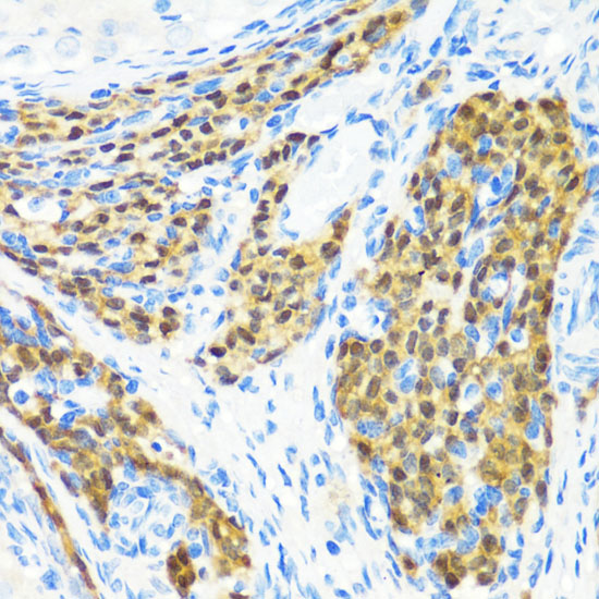
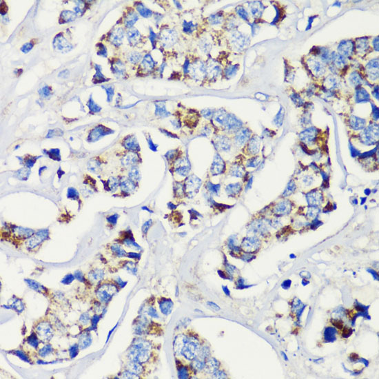


Comprehensive Human Protein Atlas IHC scoring per tissue (reliability: Approved — Antibody staining in cells/structures not annotated, view images. Pending external verification.). Rows are taken directly from the HPA tissue chart — click any row's HPA link to view the source.
| Tissue | Cell type | Level | Evidence | Source |
|---|---|---|---|---|
| Bone marrow | Hematopoietic cells | Medium | Protein (IHC) | HPA → |
| Tissue | Cell type | Level | Evidence | Source |
|---|---|---|---|---|
| Adipose tissue | Adipocytes | Not detected | Protein (IHC) | HPA → |
| Adrenal gland | Glandular cells | Not detected | Protein (IHC) | HPA → |
| Appendix | Glandular cells | Not detected | Protein (IHC) | HPA → |
| Breast | Adipocytes | Not detected | Protein (IHC) | HPA → |
| Bronchus | Respiratory epithelial cells | Not detected | Protein (IHC) | HPA → |
Troubleshoot KMT2E staining in paraffin sections by checking retrieval, cell compartment, controls and scoring before interpreting tissue differences.
A08526 has pictured paraffin-section IHC in rat ovary, human breast cancer and mouse lung (catalog IHC captions); IF/ICC is listed, but no IF image is supplied (catalog applications; IF images).
A08526 will render with its rat ovary paraffin-section IHC figure at 1:200 (catalog IHC caption). Its other captions show paraffin-section IHC in human breast cancer and mouse lung; IF/ICC is listed for Human, Mouse and Rat without an IF figure (catalog IHC captions; applications; reactivity; IF images).
Which to pick: For tissue IHC, choose A08526: its captions document paraffin-section staining after microwave retrieval in 10 mM PBS, pH 7.2, at 1:200 (catalog IHC captions). For IF/ICC, A08526 is the listed option with a 1:50–1:200 range, although no IF image is supplied (catalog applications; IF dilution; IF images). For cross-species work, A08526 lists Human, Mouse and Rat reactivity (catalog reactivity); its IHC captions do not report a fixative, and its clone field is blank (catalog IHC captions; clone).