KNL1 / Outer kinetochore KNL1 complex subunit KNL1 · IHC design guide

Design Immunohistochemistry for KNL1

This guide plans chromogenic IHC for KNL1 in paraffin sections, using the strong nuclear staining of testis pachytene spermatocytes as a reference (HPA tissue IHC). It covers fixation, controls and scoring, with a catalog antibody starting dilution of 1:50–1:100 (datasheet: A32138).

Evidence assembled Oct 2026 · For research use; verify linked source records and product datasheet before use
Immunohistochemistry protocol sheet for KNL1 (IHC for KNL1): expected localisation Nuclear staining in several tissues, strongest in testis (HPA tissue IHC), antibody A32138, validated IHC image, and IHC protocol steps
Printable KNL1 IHC protocol sheet — expected localisation Nuclear staining in several tissues, strongest in testis (HPA tissue IHC), antibody A32138, controls and protocol steps. Open the full KNL1 IHC guide →

KNL1 Immunohistochemistry Experimental Design Guide

Expected localisation, validated protocols, controls and antibodies — the at-a-glance facts below, then the full design guide.

Must know before staining
Expected localisation Nuclear staining in several tissues, strongest in testis (HPA tissue IHC)
Staining pattern Strong nuclear staining in testis pachytene spermatocytes (HPA tissue IHC)
Antigen retrieval Tris-EDTA pH 9.0 HIER, 95–98 °C, 20 min (rule: nuclear antigen)
Positive control ⓘ Testis+3 more · see all
Negative control ⓘ Adipose tissue+4 more · see all
Important caveats
Reasons your staining may differ from the expected pattern.
Fixation Keep fixation consistent across sections (standard IHC practice; not target-specific)
Caveat Cell-cycle phase can change KNL1 abundance and location (UniProt)
Regulation Rises through late anaphase; falls by early G1 (UniProt)
Isoform / epitope Four isoforms; epitope coverage is unknown (UniProt)
Section 1

Recommended KNL1 IHC & IF Protocols

The catalog antibody’s IHC-P protocol is accompanied by published KNL1 protocols for uterine corpus endometrial carcinoma (PMC9913269) and pancreatic adenocarcinoma (PMC10625436).

Recommended immunohistochemistry (IHC-P) protocol parameters
SampleParaffin-embedded rat testis tissue; fixative not specified (datasheet A32138)
FixationImage fixative and duration unreported (datasheet A32138); verify before use.
Sectioning4–5 µm sections on charged slides (standard)
DeparaffinisationXylene, graded ethanol series to water (standard)
Antigen retrievalHeat-induced epitope retrieval in Tris-EDTA buffer, pH 9.0, 20 min at 95–98 °C (standard rule: nuclear antigen)
Peroxidase block3% H2O2, 10 min, room temperature (standard)
Blocking10% normal serum of the secondary host, 30 min, room temperature (standard)
Primary antibodyRabbit anti-KNL1, 1:50-1:100 (datasheet A32138)
Primary incubationOvernight at 4 °C (standard)
DetectionHRP-polymer secondary, DAB chromogen 5–10 min (standard)
CounterstainHematoxylin, blue, dehydrate and mount (standard)
Expected resultKNL1-positive staining in pachytene spermatocytes of testis (HPA tissue IHC: High). HPA tissue profile: Nuclear expression in several tissues, most abundant in testis. No signal in the no-primary control.
💡Decision noteStart with Tris-EDTA pH 9.0 HIER at 95–98 °C for 20 min (page retrieval rule); the pancreatic study does not report retrieval (PMC10625436).
Section 2

What Is the Expected KNL1 Staining Pattern?

KNL1 is an outer kinetochore protein with nuclear and cytoplasmic localisation and no transmembrane segment (UniProt Q8NG31). In paraffin-section IHC, expect predominantly nuclear staining, strongest in testicular pachytene spermatocytes, with medium staining reported in selected bone marrow, placental and tonsillar cells (HPA tissue IHC: Enhanced reliability). HPA describes medium consistency between staining and RNA expression, so interpret the pattern alongside cell identity and controls (HPA tissue IHC).

What am I looking at on my slide?
Strong nuclear staining in pachytene spermatocytes; some nearby cells are faint or unstained (HPA tissue IHC: High in pachytene spermatocytes).This fits the principal tissue IHC pattern. KNL1 is weak in interphase nuclei and increases from prophase through late anaphase, so uniform staining of every cell is unnecessary (UniProt Q8NG31).
Predominantly cytoplasmic staining across a paraffin section, without the expected nuclear pattern (HPA tissue IHC: nuclear expression).Review localisation and controls for artefact (general IHC practice). Do not call every cytoplasmic signal false: UniProt reports cytoplasmic localisation during meiotic prophase I (UniProt Q8NG31).
Prominent signal in cells HPA lists as undetected, such as adipocytes, alongside weak staining of expected cells (HPA tissue IHC: adipocytes Not detected).Consider cross-reactivity or endogenous detection activity; the mismatch alone does not identify its cause (general IHC practice). Check a no-primary control and compare cell types on the same section.
Diffuse brown colour obscures cell boundaries or appears in tissue spaces and the no-primary control (general IHC practice).Interpret the slide as background-limited. Review blocking, washes and chromogen development before scoring nuclear KNL1; diffuse colour cannot establish its subcellular location (general IHC practice).
No detectable signal in identifiable pachytene spermatocytes of a testis control (HPA tissue IHC: High in pachytene spermatocytes).First check section quality, retrieval, primary antibody and detection controls (general IHC practice). A negative test section is hard to interpret until the known-positive control stains as expected.
💡Expected KNL1 appearanceCall a section positive when pachytene spermatocytes show clear, predominantly nuclear chromogenic signal, with High staining reported by HPA; widespread colour in HPA-undetected cells or a no-primary control raises concern for a false positive (HPA tissue IHC; general IHC practice).
How each factor affects the staining
Cell-cycle stage (UniProt Q8NG31 localisation)KNL1 is weak in interphase nuclei, rises from prophase to late anaphase, then falls through telophase and cytokinesis into early G1. Compare like cell populations when judging a faint result (UniProt Q8NG31).
Tissue and cell selection (HPA tissue IHC)Testis pachytene spermatocytes are High; bone marrow hematopoietic cells, placental Hofbauer cells and tonsil germinal-center cells are Medium. Choose identifiable target cells for comparison (HPA tissue IHC).
Antibody evidence (HPA antibodies; HPA tissue IHC)HPA026624 has Enhanced IHC validation; HPA049647 has no listed IHC status. The overall tissue profile has Enhanced reliability but only medium consistency with RNA expression (HPA antibodies; HPA tissue IHC).
Isoforms and epitope coverage (UniProt Q8NG31)UniProt lists 4 KNL1 isoforms. The supplied evidence does not map an IHC antibody epitope, so equal detection of all isoforms cannot be assumed (UniProt Q8NG31).
IF/ICC Q: What localisation is reported? (HPA subcellular)A: Mainly nucleoplasm, with additional nuclear bodies, is supported in ICC-IF (HPA subcellular). This is a separate imaging observation; judge paraffin IHC against the HPA tissue pattern.
Why is my staining missing, weak or wrong?
SituationLikely causeNext action
Testis control is blank, including identifiable pachytene spermatocytes (HPA tissue IHC: High).A failed staining step or unsuitable section is possible; one blank slide does not establish absent KNL1 (general IHC practice).Verify tissue identity and morphology, then review the established retrieval, antibody and detection steps with a known-positive section (general IHC practice).
The no-primary control also develops brown signal (general IHC practice).Detection-system background or endogenous activity may contribute; this control cannot test primary-antibody cross-reactivity (general IHC practice).Review the relevant blocking and detection controls, washes and chromogen development before interpreting test-section signal (general IHC practice).
Adipocytes stain conspicuously, although HPA reports them as Not detected (HPA tissue IHC).Cross-reactivity or background is plausible, but HPA's tissue pattern alone cannot identify the mechanism (HPA tissue IHC; general IHC practice).Compare matched no-primary and expected-positive tissue areas, then score only signal whose cell type and compartment are credible (general IHC practice).
Nuclear staining is faint in a section with few dividing cells (UniProt Q8NG31 localisation).KNL1 is weak in interphase nuclei and varies through the cell cycle; faint staining can reflect the cells present (UniProt Q8NG31).Inspect identifiable pachytene spermatocytes in a testis control before changing staining conditions (HPA tissue IHC: High; general IHC practice).
Signal appears chiefly cytoplasmic in testis (HPA tissue IHC: nuclear profile).Artefact is possible, while meiotic prophase I cytoplasmic localisation is also reported; the section alone may not distinguish them (UniProt Q8NG31).Check cell stage and nuclear counterstain, then compare controls and adjacent cell populations before assigning localisation (UniProt Q8NG31; general IHC practice).
Two antibodies yield different IHC patterns (HPA antibodies: different IHC status).Their supplied IHC evidence differs: HPA026624 is Enhanced, while HPA049647 has no listed IHC status; epitope coverage is unspecified (HPA antibodies; UniProt Q8NG31).Compare each result with HPA cell-type and nuclear patterns and its own controls; do not infer equivalent IHC validation from ICC-IF status (HPA antibodies; HPA tissue IHC).

Sample controls for KNL1 IHC & IF

🧪Run testis first and look for staining in pachytene spermatocytes (HPA: High in pachytene spermatocytes); use adipose tissue, specifically adipocytes, as the negative tissue (HPA: Not detected in adipocytes). On the testis slide, compare staining with neighboring cells lacking visible signal, while allowing for staining in other germ-cell stages (UniProt Q8NG31: testis germ-cell expression).
Positive control tissue: Testis (Pachytene spermatocytes, HPA High)
Negative control tissue: Adipose tissue (HPA Not detected)
ICC-IF cell lines (HPA subcellular resource): HPA ICC-IF images show KNL1 in A-431, U-251MG, U2OS, with annotated localisation: Nucleoplasm (supported) (HPA subcellular).
Technical controls: Include a no-primary, secondary-only control; an isotype control matched to the primary antibody’s host species, clonality and concentration; and, if available, a KNL1 knockout sample or cognate-peptide competition control (standard IHC practice). Block endogenous peroxidase and check background in testis before interpreting chromogenic signal (standard IHC practice).
⚠️Feasibility: A KNL1-specific fixation window or fixation effect is unreported in the supplied evidence, and the fixative used for the selected paraffin-section image is unreported (A32138 tissue-IHC caption). That image used microwave retrieval in 10 mM PBS, pH 7.2, before staining at 1:100; it supports a retrieval starting point but does not establish that retrieval is required (A32138 tissue-IHC caption). IF/ICC has images in A-431, U-251MG and U2OS, but the supplied evidence does not establish that IF or frozen sections are easier; assess testis background carefully because KNL1 is also reported in other germ-cell stages (HPA subcellular; UniProt Q8NG31: testis germ-cell expression).

HPA tissue IHC evidence for KNL1

Comprehensive Human Protein Atlas IHC scoring per tissue (reliability: Enhanced — Medium consistency between antibody staining and RNA expression data.). Rows are taken directly from the HPA tissue chart — click any row's HPA link to view the source.

Positive expression · recommended positive controls

TissueCell typeLevelEvidenceSource
Testis Pachytene spermatocytes High Protein (IHC) HPA →
Bone marrow Hematopoietic cells Medium Protein (IHC) HPA →
Placenta Hofbauer cells Medium Protein (IHC) HPA →
Tonsil Germinal center cells Medium Protein (IHC) HPA →

Undetected expression · recommended negative controls

TissueCell typeLevelEvidenceSource
Adipose tissue Adipocytes Not detected Protein (IHC) HPA →
Adrenal gland Glandular cells Not detected Protein (IHC) HPA →
Breast Adipocytes Not detected Protein (IHC) HPA →
Bronchus Respiratory epithelial cells Not detected Protein (IHC) HPA →
Caudate Glial cells Not detected Protein (IHC) HPA →
Section 3

Advanced KNL1 IHC Tips

For KNL1 chromogenic IHC, interpret staining by cell type, cell cycle stage and compartment, using the reported testis staining as a reference.

How should I retrieve KNL1 in paraffin sections when staining is weak?
Start with Tris-EDTA pH 9.0 heat-induced retrieval at 95–98 °C for 20 min (page retrieval rule: nuclear antigen). If staining remains weak, compare a serial section using microwave retrieval in 10 mM PBS pH 7.2, the condition reported for catalog antibody A32138 on paraffin-embedded rat testis (A32138 tissue-IHC caption). That caption used the antibody at 1:100 but did not report the fixative, so record the section’s preparation separately (A32138 tissue-IHC caption). Compare staining in pachytene spermatocytes with background and tissue morphology across conditions; high staining in these cells provides a relevant reference (HPA: high in testis pachytene spermatocytes).
Could fixation explain inconsistent KNL1 staining between paraffin blocks?
KNL1-specific fixation sensitivity is unknown because the supplied tissue-IHC caption does not state a fixative (A32138 tissue-IHC caption). Record the fixative and processing history for each block, then compare serial sections with the same retrieval, antibody dilution and chromogenic detection settings (standard IHC practice). The reported A32138 example used paraffin-embedded rat testis, microwave retrieval in 10 mM PBS pH 7.2 and a 1:100 dilution, but it cannot establish which fixation conditions preserve staining (A32138 tissue-IHC caption). Assess comparable, intact cell populations across blocks; changes in nuclear staining alone do not identify fixation as the cause (standard IHC practice).
Should KNL1 staining appear throughout nuclei or only at kinetochores?
Interpret compartment in light of cell cycle stage: KNL1 is reported at centromeric kinetochores and is weakly expressed in interphase nuclei (UniProt Q8NG31: subcellular location). Its expression increases from prophase through late anaphase and greatly diminishes from telophase and cytokinesis into early G1 (UniProt Q8NG31: subcellular location). In tissue IHC, nuclear staining is reported across several tissues and is most abundant in testis (HPA: tissue IHC profile). Compare intact pachytene spermatocytes, which show high staining, with nearby cells and note whether a diffuse nuclear pattern or distinct mitotic foci are visible at the available section resolution (HPA: testis pachytene spermatocytes; standard IHC practice).
How can epitope choice affect interpretation of KNL1 staining?
KNL1 has 4 reported isoforms, while the supplied A32138 tissue-IHC caption does not identify the antibody’s epitope or establish isoform coverage (UniProt Q8NG31: isoforms; A32138 tissue-IHC caption). Request the immunogen or epitope coordinates before treating a negative section as evidence that every isoform is absent (standard IHC practice). KNL1 also has reported modified residues, including phosphorylation attributed to AURKB at position 24, but an effect on this antibody’s staining has not been established here (UniProt Q8NG31: modified residues). Keep retrieval and detection constant when comparing sections, and interpret differences alongside cell type and cell cycle stage (standard IHC practice; UniProt Q8NG31: subcellular location).
How should I use IF to investigate an ambiguous IHC localisation pattern?
Use IF as a separate localisation check alongside chromogenic IHC, since the supplied tissue example documents paraffin-section IHC for A32138 and gives no IF/ICC validation conditions (A32138 tissue-IHC caption). Multiplex KNL1 with a validated marker for the expected cell population, such as pachytene spermatocytes in testis, and inspect both channels in the same cells (HPA: high in testis pachytene spermatocytes; standard IF practice). Choose a fluorophore channel away from the section’s measured autofluorescence and include single-channel controls (standard IF practice). Because KNL1 is intracellular, optimise permeabilisation for access to its nuclear and kinetochore epitopes, while checking that cellular structure remains intact (UniProt Q8NG31: subcellular location; standard IF practice).
What should I check when KNL1 chromogenic staining is widespread?
First inspect a no-primary control for chromogen deposition, then review peroxidase blocking, antibody concentration, washing and DAB development (standard chromogenic IHC practice). The A32138 rat-testis caption reports a 1:100 antibody dilution; use that documented point when assessing whether stronger antibody exposure raises background in the same workflow (A32138 tissue-IHC caption; standard IHC practice). Distinguish signal in intact nuclei from pigment, tissue edges and damaged areas on the counterstained section (standard IHC practice). Broad staining needs particular scrutiny because HPA reports nuclear expression most abundant in testis and lists several cell populations as undetected, including adipose-tissue adipocytes (HPA: tissue IHC profile; HPA: adipose tissue adipocytes).
How should I score KNL1 IHC across samples with different cell populations? ⚠ ANSWER MARKED FOR VERIFICATION
Define the compartment and cell population before scoring, then report the percentage of positive cells and an H-score for nuclear staining where intensity can be graded reproducibly (standard IHC practice). For discrete mitotic signals, report positive-cell density per mm² and the number of evaluable mitotic cells, so a change in mitotic frequency does not masquerade as a change in staining (standard IHC practice; UniProt Q8NG31: cell cycle location). In testis, report pachytene spermatocytes separately because HPA rates their staining high (HPA: testis pachytene spermatocytes). Apply one positivity threshold and imaging setting across the comparison, and exclude damaged regions before counting (standard IHC practice).
When is apparent KNL1 positivity more likely to be artefact?
Give greatest weight to reproducible staining in the expected compartment and cell population: KNL1 is associated with nuclei and kinetochores, while HPA reports high staining in testis pachytene spermatocytes (UniProt Q8NG31: subcellular location; HPA: testis pachytene spermatocytes). Isolated strong chromogen at section edges, in necrotic regions or in a no-primary control warrants an artefact check (standard chromogenic IHC practice). Assess unexpected diffuse staining outside nuclei against morphology, peroxidase-block controls and the reported cell-cycle-dependent KNL1 pattern (standard IHC practice; UniProt Q8NG31: subcellular location). Treat an apparently negative field cautiously when its relevant cell population is sparse or its mitotic stages differ from the comparison section (standard IHC practice; UniProt Q8NG31: subcellular location).
Boster reagents

Best KNL1 / Outer kinetochore KNL1 complex subunit KNL1 IHC Antibodies

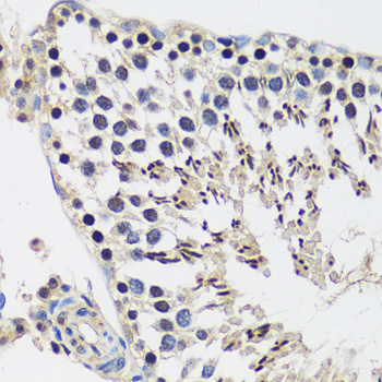
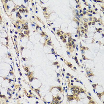
Two anti-KNL1 antibodies list IHC and IF/ICC applications (catalog applications); A32138 has pictured paraffin-section IHC in rat testis and human colon (A32138 image captions).

Real IHC data Immunohistochemistry of paraffin-embedded rat testis using CASC5 antibody at dilution of 1:100 .Perform microwave antigen retrieval with 10 mM PBS buffer pH 7.2 before commencing with IHC staining protocol.
Anti-CASC5 KNL1 Antibody
Cat # A32138

A32138 shows paraffin-section IHC in rat testis and human colon (A32138 image captions) and lists IF/ICC with human and rat reactivity (catalog applications/reactivity). A32138-1 lists rat paraffin-section IHC and human IF/ICC (A32138-1 dilution field), with human, mouse and rat reactivity (catalog reactivity); no images are supplied (A32138-1 image fields).

Which to pick: For tissue IHC, choose A32138 when its pictured rat testis or human colon paraffin sections match your sample (A32138 image captions); the captions specify 1:100 antibody and microwave retrieval in 10 mM PBS, pH 7.2, but do not report the fixative (A32138 image captions). For IF/ICC, A32138-1 lists human use at 5 μg/ml and is polyclonal (A32138-1 dilution field); A32138 lists IF/ICC at 1:50–1:100, with no IF image supplied (A32138 application/dilution/image fields). For broader listed species reactivity, choose A32138-1 for human, mouse and rat (catalog reactivity); its paraffin-section IHC conditions are specifically listed for rat (A32138-1 dilution field).

Each figure is that product's own IHC / IF validation image from its datasheet.

References

  1. UniProt Consortium. UniProt entry Q8NG31 (KNL1_HUMAN, Outer kinetochore KNL1 complex subunit KNL1).
  2. Human Protein Atlas. KNL1 tissue IHC expression (reliability: Enhanced).
  3. Human Protein Atlas. KNL1 subcellular location (ICC-IF): Mainly localized to the nucleoplasm. In addition localized to the nuclear bodies..
  4. Human Protein Atlas. KNL1 antibody validation summary (2 antibodies).
  5. KNL1 is a prognostic and diagnostic biomarker related to immune infiltration in patients with uterine corpus endometrial carcinoma. Frontiers in oncology 2023 — PMC9913269.
  6. KNL1 Regulates Ferroptosis Resistance and Migration in Lung Adenocarcinoma Cells via AMPK-mTOR Signaling. Oncology research 2026 — PMC13040282.
  7. Role of Kinetochore Scaffold 1 (KNL1) in Tumorigenesis and Tumor Immune Microenvironment in Pan-Cancer: Bioinformatics Analyses and Validation of Expression. International journal of general medicine 2023 — PMC10625436.
  8. Microcephaly Modeling of Kinetochore Mutation Reveals a Brain-Specific Phenotype. Cell reports 2018 — PMC6392048.
  9. PubMed PMID:10980622 — UniProt-cited evidence.
  10. PubMed PMID:12087463 — UniProt-cited evidence.
  11. PubMed PMID:12618768 — UniProt-cited evidence.