This website uses cookies to ensure you get the best experience on our website.


- Table of Contents
Plan KPNA2 chromogenic IHC on paraffin sections around nuclear staining in cell subsets (HPA tissue IHC). Use testis pachytene spermatocytes or lymph node germinal center cells as positive controls (HPA tissue IHC), and score nuclei by cell type.

Expected localisation, validated protocols, controls and antibodies — the at-a-glance facts below, then the full design guide.
| Expected localisation | Nuclear staining in tissue (HPA tissue IHC) | |
| Staining pattern | Nuclear staining in subsets of cells in most tissues (HPA tissue IHC) | |
| Antigen retrieval | EDTA pH 8.0 HIER, heat-mediated (datasheet A01776-3) | |
| Positive control | Appendix+4 more · see all | |
| Negative control | Adipose tissue+4 more · see all |
| Fixation | Keep fixation conditions consistent across sections. (standard IHC practice; not target-specific) | |
| Caveat | Staining varies by cell type; score nuclei separately (HPA tissue IHC) | |
| Regulation | High in testis pachytene spermatocytes (HPA tissue IHC) | |
| Isoform / epitope | No isoforms listed; mature chain spans residues 2–529 (UniProt) |
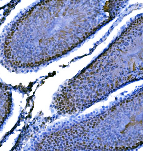
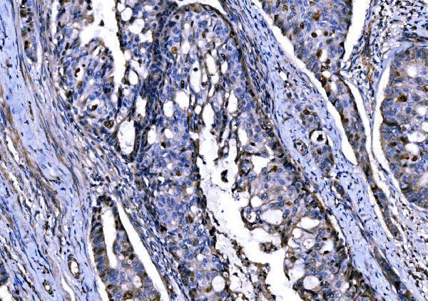
The catalog antibody’s IHC protocol (datasheet: A01776-3) is accompanied by four published KPNA2 tissue-staining protocols (PMC7661224; PMC5403343; PMC9808660; PMC9385962).
| Sample | Paraffin-embedded rat testis tissue; fixative not specified (datasheet A01776-3) |
| Fixation | Image fixative and duration unreported (datasheet A01776-3); verify before use. |
| Sectioning | 4–5 µm sections on charged slides (standard) |
| Deparaffinisation | Xylene, graded ethanol series to water (standard) |
| Antigen retrieval | Heat retrieval: EDTA pH 8.0 (datasheet A01776-3); 20 min, 95–100 °C (standard) |
| Peroxidase block | 3% H2O2, 10 min, room temperature (standard) |
| Blocking | 10% goat serum (datasheet A01776-3) |
| Primary antibody | Rabbit anti-KPNA2, 2-5 μg/ml (datasheet A01776-3) |
| Primary incubation | Overnight at 4 °C (datasheet A01776-3) |
| Detection | HRP-conjugated secondary, DAB chromogen (datasheet A01776-3) |
| Counterstain | Hematoxylin, blue, dehydrate and mount (standard) |
| Expected result | KPNA2-positive staining in lymphoid tissue of appendix (HPA tissue IHC: High). HPA tissue profile: Nuclear expression in subsets of cells in most tissues. No signal in the no-primary control. |
KPNA2 is mainly nuclear in subsets of cells across most tissues (HPA tissue IHC: Enhanced). Its supported nucleoplasmic location has an additional cytosolic component (HPA ICC-IF: supported); UniProt also lists nucleus and cytoplasm, and reports no transmembrane segment (UniProt P52292 topology). Expect the clearest IHC signal in the specific cells HPA rates High, while assessing both compartment and cell identity.
| Strong nuclear staining in germinal center cells, pachytene spermatocytes, or cardiomyocytes. | This fits the reported High staining in those cells (HPA tissue IHC). Score nuclei within the named cell population, since the broader tissue can contain cells with different staining; KPNA2 is reported in subsets of cells in most tissues (HPA tissue IHC). |
| Signal is mainly at cell borders or appears as an exclusive membranous pattern, with little nuclear staining. | Reassess specificity and slide interpretation: the expected tissue pattern is nuclear (HPA tissue IHC), and KPNA2 has no transmembrane segment (UniProt P52292 topology). Some cytosolic signal alone is compatible with the supported ICC-IF location (HPA ICC-IF); do not classify every nonnuclear pixel as artefact. |
| Strong staining appears in adipocytes or prostate glandular cells while expected positive nuclei also stain. | Those cell types were Not detected in the sampled HPA tissue IHC images (HPA tissue IHC). Consider cross-reactivity or endogenous chromogenic activity (standard IHC practice), then compare cell morphology and controls. A Not detected observation is a context-specific reference, not proof that every such cell must be negative. |
| Color spreads across tissue and empty areas, obscuring nuclear boundaries. | Treat the diffuse deposit as background until controls resolve it (standard IHC practice). Check blocking, washes, chromogen development, and a no-primary control (standard IHC practice); the reported KPNA2 tissue pattern is nuclear in subsets of cells (HPA tissue IHC). |
| No nuclear signal appears in an otherwise evaluable known-positive section. | First confirm that the relevant cells are present: HPA rates germinal center cells in lymph node and tonsil, and pachytene spermatocytes in testis, High (HPA tissue IHC). If they are present, review the IHC antibody, retrieval, detection, and run controls (standard IHC practice). |
| Cell population and reference tissue | Choose a reference with identifiable High cells, such as lymph-node germinal center cells or testis pachytene spermatocytes (HPA tissue IHC). HPA reports nuclear expression in subsets of cells in most tissues, so assess the named cells rather than averaging color across a section (HPA tissue IHC). |
| Strength of the IHC reference | The HPA tissue profile has Enhanced reliability, described as high consistency between antibody staining and RNA expression data (HPA tissue IHC). HPA041270 and CAB015460 each have Enhanced IHC validation (HPA antibodies). This supports the reference pattern, but does not establish performance for an unlisted antibody. |
| Antigen retrieval and assay conditions | Retrieval and detection conditions can be checked when IHC staining fails (standard IHC practice). The supplied UniProt and HPA records provide no KPNA2-specific retrieval condition or fixation-sensitivity result; avoid attributing a weak section to a particular fixation effect. |
| Endogenous chromogenic activity | Endogenous enzyme activity or nonspecific reagent binding can create color unrelated to the primary antibody (standard IHC practice). Use a no-primary control and an appropriate blocking step to interpret unexpected deposits (standard IHC practice); HPA cell-level staining alone cannot identify their cause. |
| IF/ICC Q&A: What location should fluorescence show? | Mainly nucleoplasm, with additional cytosol (HPA ICC-IF: supported). This cross-check helps interpret compartment, while the present decision criteria concern chromogenic IHC in paraffin sections (HPA tissue IHC). |
| Situation | Likely cause | Next action |
|---|---|---|
| Known-positive nuclei are blank. | The selected field may lack the named positive cells, or an IHC staining step may have failed (HPA tissue IHC; standard IHC practice). | Locate HPA High cells in the section, then verify the primary-antibody, retrieval, and detection steps against the assay instructions and controls (HPA tissue IHC; standard IHC practice). |
| Only cytoplasmic color is apparent in a presumed positive population. | KPNA2 can have a cytosolic component, but HPA describes the main supported ICC-IF location as nucleoplasm (HPA ICC-IF). | Inspect counterstained nuclei and a positive control before accepting the result; investigate absent nuclear staining with assay controls (standard IHC practice; HPA tissue IHC). |
| Membrane-edge staining dominates. | The pattern conflicts with nuclear tissue staining and the absence of a transmembrane segment (HPA tissue IHC; UniProt P52292 topology). | Compare with a no-primary control and review cell boundaries, chromogen deposit, and antibody specificity (standard IHC practice). |
| Adipocytes or prostate glandular cells stain strongly. | HPA lists those sampled cell populations as Not detected; cross-reactivity or endogenous detection activity is possible (HPA tissue IHC; standard IHC practice). | Check a no-primary control and compare staining with nearby expected positive cells; treat a mismatch as a reason to validate, not an automatic diagnosis of artefact (standard IHC practice; HPA tissue IHC). |
| Diffuse color makes nuclei hard to score. | Background from nonspecific binding, incomplete washing, or excessive chromogen development can obscure compartment boundaries (standard IHC practice). | Review blocking, washes, development, and the no-primary control; score only clearly resolved cellular staining (standard IHC practice). |
| Two sections show different proportions of positive cells. | KPNA2 is reported in subsets of cells in most tissues, and HPA staining levels refer to specific cell types (HPA tissue IHC). | Compare matching cell populations and record their nuclear staining separately from whole-section intensity (HPA tissue IHC; standard IHC practice). |


Comprehensive Human Protein Atlas IHC scoring per tissue (reliability: Enhanced — High consistency between antibody staining and RNA expression data.). Rows are taken directly from the HPA tissue chart — click any row's HPA link to view the source.
| Tissue | Cell type | Level | Evidence | Source |
|---|---|---|---|---|
| Appendix | Lymphoid tissue | High | Protein (IHC) | HPA → |
| Heart muscle | Cardiomyocytes | High | Protein (IHC) | HPA → |
| Lymph node | Germinal center cells | High | Protein (IHC) | HPA → |
| Testis | Pachytene spermatocytes | High | Protein (IHC) | HPA → |
| Tonsil | Germinal center cells | High | Protein (IHC) | HPA → |
| Tissue | Cell type | Level | Evidence | Source |
|---|---|---|---|---|
| Adipose tissue | Adipocytes | Not detected | Protein (IHC) | HPA → |
| Breast | Adipocytes | Not detected | Protein (IHC) | HPA → |
| Ovary | Follicle cells | Not detected | Protein (IHC) | HPA → |
| Prostate | Glandular cells | Not detected | Protein (IHC) | HPA → |
| Smooth muscle | Smooth muscle cells | Not detected | Protein (IHC) | HPA → |
Troubleshoot KPNA2 staining in paraffin sections by checking retrieval, compartment, cell type, controls and scoring before interpreting chromogenic signal.
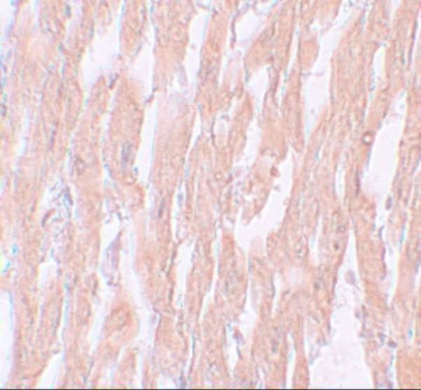
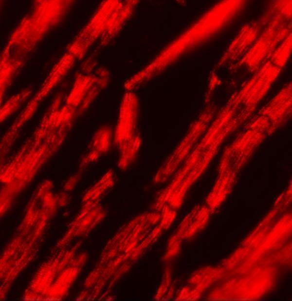
Two anti-KPNA2 antibodies have IHC images from human heart, human tumors, and rat testis; one also has an IF image from human heart (catalog image captions).
A01776-3 has paraffin-section IHC images from rat testis and human colon adenocarcinoma, lymphoma, and lung cancer (A01776-3 IHC captions). A01776 has IHC and IF images from human heart (A01776 image captions).
Which to pick: For paraffin-section IHC, choose A01776-3: its captions document EDTA retrieval at pH 8.0 and primary antibody at 2 μg/ml; the fixative is unreported (A01776-3 IHC captions). For IF, choose A01776, which lists IF and shows staining in human heart at 20 μg/mL; ICC is not listed (A01776 applications and IF caption). Both list human, mouse, and rat reactivity, while the supplied IHC images show human and rat for A01776-3 and human for A01776 (catalog reactivity; IHC captions).