This website uses cookies to ensure you get the best experience on our website.


- Table of Contents
Plan KPNA3 paraffin IHC around nuclear and cytoplasmic staining in most tissues (HPA tissue IHC). Heart cardiomyocytes offer a high-staining reference (HPA tissue IHC); the IHC-validated antibody has a suggested dilution of 1:50–1:100 (datasheet A06131-1).

Expected localisation, validated protocols, controls and antibodies — the at-a-glance facts below, then the full design guide.
| Expected localisation | Nuclear and cytoplasmic in most tissues (HPA tissue IHC) | |
| Staining pattern | Most tissues show nuclear and cytoplasmic staining (HPA tissue IHC) | |
| Antigen retrieval | Citrate pH 6.0 HIER, heat-mediated (datasheet A06131-1) | |
| Positive control | Adrenal gland+4 more · see all | |
| Negative control | Adipose tissue+2 more · see all |
| Fixation | Matched tissue-IHC evidence does not establish the fixation claim. Validate the specimen-specific method before use. (selected-SKU IHC image A06131-1) | |
| Caveat | Staining has medium consistency with RNA expression (HPA tissue IHC) | |
| Regulation | Tissue enhanced in skeletal muscle and tongue (HPA tissue RNA) | |
| Isoform / epitope | No isoforms listed; mature chain spans residues 2–521 (UniProt) |
The catalog antibody has a datasheet IHC-P protocol (datasheet A06131-1). Three published KPNA3 IHC methods provide additional examples (PMC10431729; PMC6692625; PMC7876302).
| Sample | Paraffin-embedded rat stomach tissue; fixative not specified (datasheet A06131-1) |
| Fixation | Image fixative and duration unreported (datasheet A06131-1); verify before use. |
| Sectioning | 4–5 µm sections on charged slides (standard) |
| Deparaffinisation | Xylene, graded ethanol series to water (standard) |
| Antigen retrieval | Heat retrieval: Citrate pH 6.0 (datasheet A06131-1); 20 min, 95–100 °C (standard) |
| Peroxidase block | 3% H2O2, 10 min, room temperature (standard) |
| Blocking | 10% normal serum of the secondary host, 30 min, room temperature (standard) |
| Primary antibody | Rabbit anti-KPNA3, 1:50-1:100 (datasheet A06131-1) |
| Primary incubation | Overnight at 4 °C (standard) |
| Detection | HRP-polymer secondary, DAB chromogen 5–10 min (standard) |
| Counterstain | Hematoxylin, blue, dehydrate and mount (standard) |
| Expected result | KPNA3-positive staining in glandular cells of adrenal gland (HPA tissue IHC: High). HPA tissue profile: Nuclear and cytoplasmic expression in most tissues. No signal in the no-primary control. |
KPNA3 staining in paraffin sections is expected in nuclei and cytoplasm across most tissues, with cell-dependent intensity (HPA tissue IHC: Approved; medium consistency with RNA). Nuclear and cytoplasmic localization is consistent with its protein-import role (UniProt O00505: function and subcellular location). KPNA3 has no transmembrane segment, so a membrane-only pattern is unexpected (UniProt O00505: topology).
| Nuclear staining with some cytoplasmic staining in the expected cells. | This fits the reported IHC pattern in most tissues (HPA tissue IHC: nuclear and cytoplasmic expression). Judge intensity within the identified cell population: HPA reports High staining in heart cardiomyocytes and pancreatic exocrine glandular cells, among others (HPA tissue IHC). |
| Staining is confined to cell membranes, extracellular material, or an unrelated structure. | That distribution conflicts with the reported nuclear and cytoplasmic pattern (HPA tissue IHC) and lack of a transmembrane segment (UniProt O00505: topology). Check morphology, counterstain, and detection controls before interpreting it as KPNA3 (standard IHC practice). |
| Strong staining appears in cells reported as Not detected. | HPA reports adipocytes, ovarian stroma cells, and splenic red-pulp cells as Not detected (HPA tissue IHC). Recheck cell identity and compare a no-primary control; cross-reactivity or endogenous detection activity can mimic a positive result (standard IHC practice). |
| Broad, hazy stain obscures nuclei and cell boundaries. | A diffuse haze cannot establish the reported cellular distribution (HPA tissue IHC: nuclear and cytoplasmic expression). Uneven reagent coverage, inadequate washing, or detection background are possible general IHC causes; inspect the no-primary control and the tissue edges (standard IHC practice). |
| No staining appears in an expected positive cell population. | A blank result in heart cardiomyocytes or adrenal glandular cells needs a technical check before biological interpretation (HPA tissue IHC: High in both). Check tissue preservation, retrieval conditions, primary-antibody application, and detection-system performance (standard IHC practice); no KPNA3-specific fixation sensitivity is supplied. |
| Cell population and tissue | Use the named cells when judging intensity: HPA reports High staining in heart cardiomyocytes, colon endothelial cells, and duodenal glandular cells, but Not detected in adipocytes (HPA tissue IHC). A tissue-wide score can obscure these differences (standard IHC interpretation). |
| Strength of the tissue evidence | The tissue IHC assessment is Approved, with medium consistency between antibody staining and RNA expression (HPA tissue IHC). Treat its cell-level observations as reference patterns, while resolving an unexpected result with controls and morphology (standard IHC practice). |
| Intracellular location and topology | UniProt lists cytoplasm and nucleus and reports no transmembrane segment (UniProt O00505: subcellular location and topology). These observations support intracellular staining and give no basis for calling an isolated membrane outline a KPNA3 pattern. |
| Antibody validation | HPA lists HPA046852 and HPA077643 as IHC Approved; HPA046852 is also ICC Supported (HPA antibodies). These statuses identify the reported application evidence; they do not establish that an untested antibody or every specimen will reproduce the pattern. |
| IF/ICC: where should KPNA3 appear? | Mainly in the nucleoplasm (HPA subcellular ICC-IF: supported), with possible cytosol (HPA subcellular ICC-IF: uncertain). This is a localization answer for IF/ICC, not an IHC-P staining-intensity standard. |
| Epitope and processing limits | UniProt reports no signal peptide or propeptide and lists a 2–521 chain (UniProt O00505: processing). The supplied sources give no antibody epitope or KPNA3-specific retrieval response, so neither a retrieval setting nor a fixation effect can be predicted from this record. |
| Situation | Likely cause | Next action |
|---|---|---|
| The positive-control section is blank. | The assay may have failed at retrieval, primary-antibody application, or detection (standard IHC practice); HPA reports High staining in heart cardiomyocytes (HPA tissue IHC). | Confirm the section contains intact cardiomyocytes, then check reagent application and detection controls; optimize retrieval using the chosen antibody's IHC-P instructions (standard IHC practice). |
| Only a membrane rim is visible. | An isolated membrane pattern disagrees with the reported intracellular localization (HPA tissue IHC; UniProt O00505: topology). Deposited chromogen or nonspecific staining is possible (standard IHC practice). | Compare the no-primary control and inspect the rim at higher magnification. Score KPNA3 only where cellular staining matches the expected compartments (standard IHC practice; HPA tissue IHC). |
| Adipocytes stain strongly. | Adipocytes are reported as Not detected (HPA tissue IHC). Cell misidentification, nonspecific binding, or endogenous detection activity could explain the result (standard IHC practice). | Verify the cells on the counterstained section and compare no-primary and detection controls before assigning a KPNA3-positive score (standard IHC practice). |
| The whole section has diffuse brown haze. | Background may reflect excess detection signal, incomplete washing, or endogenous enzyme activity (standard IHC practice). Haze prevents assessment of the reported nuclear and cytoplasmic pattern (HPA tissue IHC). | Check the no-primary control, washing, and enzyme-blocking step; adjust the general detection workflow if the control also stains (standard IHC practice). |
| The nucleus and cytoplasm differ from the IF/ICC image. | HPA tissue IHC reports nuclear and cytoplasmic staining, while ICC-IF supports nucleoplasm and rates additional cytosol as uncertain (HPA tissue IHC; HPA subcellular ICC-IF). | Interpret the paraffin section against its IHC cell pattern and controls; use the ICC-IF result only as supporting localization evidence (HPA tissue IHC; HPA subcellular ICC-IF). |
| Staining varies across the section. | Uneven section coverage or processing can create a gradient (standard IHC practice). Cell populations also differ in reported KPNA3 staining (HPA tissue IHC). | Check whether the variation follows tissue anatomy or a reagent edge; compare the same identified cell type across well-preserved areas (standard IHC practice; HPA tissue IHC). |


Comprehensive Human Protein Atlas IHC scoring per tissue (reliability: Approved — Medium consistency between antibody staining and RNA expression data.). Rows are taken directly from the HPA tissue chart — click any row's HPA link to view the source.
| Tissue | Cell type | Level | Evidence | Source |
|---|---|---|---|---|
| Adrenal gland | Glandular cells | High | Protein (IHC) | HPA → |
| Colon | Endothelial cells | High | Protein (IHC) | HPA → |
| Duodenum | Glandular cells | High | Protein (IHC) | HPA → |
| Epididymis | Glandular cells | High | Protein (IHC) | HPA → |
| Heart muscle | Cardiomyocytes | High | Protein (IHC) | HPA → |
Troubleshoot KPNA3 staining in paraffin sections by checking retrieval, nuclear and cytoplasmic signal, cell identity, and controls before comparing scores.
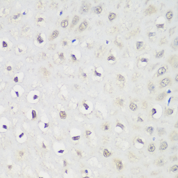
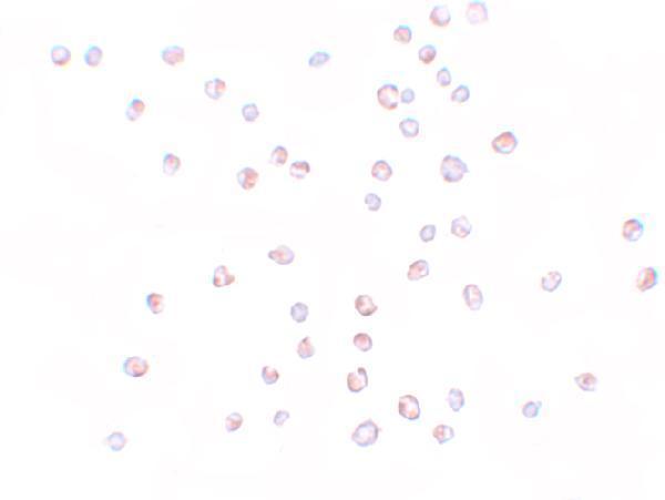
The catalog provides KPNA3 images from paraffin-embedded rat stomach by IHC (A06131-1 image caption) and HeLa cells by ICC (A06131 image caption); both antibodies list human, mouse and rat reactivity (catalog reactivity).
A06131-1 lists IHC and IF applications (catalog applications), with IHC shown in paraffin-embedded rat stomach at 1:100 after citrate retrieval (A06131-1 image caption). A06131 lists IF and ICC applications (catalog applications), with ICC shown in HeLa cells at 10 μg/mL (A06131 image caption).
Which to pick: Choose A06131-1 for paraffin-section IHC because its own image shows that preparation; the fixative is unreported (A06131-1 image caption). Choose A06131 for IF/ICC because those applications are listed and its own image shows ICC staining, while IHC is not listed (A06131 catalog applications; A06131 image caption). Both are rabbit antibodies with no clone specified (catalog host/clone), and both list human, mouse and rat reactivity (catalog reactivity); the pictured IHC evidence is specifically from rat tissue (A06131-1 image caption).