This website uses cookies to ensure you get the best experience on our website.


- Table of Contents
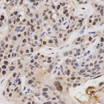
Plan chromogenic KPNA4 IHC around the mainly nuclear tissue pattern (HPA tissue IHC). The guide uses paraffin-section examples from human esophageal cancer and rat heart with catalog antibody A05188-1 at 1:200 (datasheet: IHC captions).

Expected localisation, validated protocols, controls and antibodies — the at-a-glance facts below, then the full design guide.
| Expected localisation | Mainly nuclear in tissues (HPA tissue IHC) | |
| Staining pattern | Mainly nuclear staining across most tissues (HPA tissue IHC) | |
| Antigen retrieval | Tris-EDTA pH 9.0 HIER, 95–98 °C, 20 min (rule: nuclear antigen) | |
| Positive control | Adrenal gland+4 more · see all | |
| Negative control | None in HPA (detected in all 45 tissues); use no-primary + isotype controls |
| Fixation | Matched tissue-IHC evidence does not establish the fixation claim. Validate the specimen-specific method before use. (selected-SKU IHC image A05188-1) | |
| Caveat | Colon glands stain high despite low reported colon expression (HPA tissue IHC; UniProt) | |
| Regulation | High in muscle; low in kidney (UniProt) | |
| Isoform / epitope | No listed isoforms; intracellular chain spans aa 2–521 (UniProt) |
Compare the catalog antibody’s IHC-P protocol with published KPNA4 IHC methods for spinal cord, pancreas, hepatocellular carcinoma, and frontal cortex (PMC12325296; PMC9002131; PMC7950262; PMC6158706).
| Sample | Paraffin-embedded rat heart tissue; fixative not specified (datasheet A05188-1) |
| Fixation | Image fixative and duration unreported (datasheet A05188-1); verify before use. |
| Sectioning | 4–5 µm sections on charged slides (standard) |
| Deparaffinisation | Xylene, graded ethanol series to water (standard) |
| Antigen retrieval | Heat-induced epitope retrieval in Tris-EDTA buffer, pH 9.0, 20 min at 95–98 °C (standard rule: nuclear antigen) |
| Peroxidase block | 3% H2O2, 10 min, room temperature (standard) |
| Blocking | 10% normal serum of the secondary host, 30 min, room temperature (standard) |
| Primary antibody | Rabbit anti-KPNA4, 1:50-1:200 (datasheet A05188-1) |
| Primary incubation | Overnight at 4 °C (standard) |
| Detection | HRP-polymer secondary, DAB chromogen 5–10 min (standard) |
| Counterstain | Hematoxylin, blue, dehydrate and mount (standard) |
| Expected result | KPNA4-positive staining in glandular cells of adrenal gland (HPA tissue IHC: High). HPA tissue profile: Mainly nuclear expression in most tissues. No signal in the no-primary control. |
KPNA4 staining in paraffin sections should be mainly nuclear across most tissues, including glandular and respiratory epithelial cells with reported high staining (HPA tissue IHC: Enhanced reliability; medium consistency with RNA). UniProt also places KPNA4 in the cytoplasm and nucleus and reports no transmembrane segment (UniProt O00629: subcellular location and topology). Interpret staining by compartment and cell type together.
| Distinct nuclear chromogen in bronchial respiratory epithelium or adrenal glandular cells, with visible unstained tissue detail. | This fits the reported high staining in those cells and the mainly nuclear tissue profile (HPA tissue IHC: High; mainly nuclear). Compare intensity within the same run rather than treating every stained cell as equally informative (standard IHC practice). |
| Strong, widespread cytoplasmic staining with little or no nuclear signal in a tissue expected to stain strongly. | Question the IHC result because it departs from HPA’s mainly nuclear tissue pattern (HPA tissue IHC). Some cytoplasmic KPNA4 remains biologically plausible (UniProt O00629: cytoplasm; nucleus), so compartment alone cannot prove an artefact. Check controls and staining conditions (standard IHC practice). |
| Prominent staining in cells outside the reported positive population, while the expected cells are weak. | For example, a slide that misses bronchial respiratory epithelium does not match its reported high staining (HPA tissue IHC: bronchus). Unexpected staining may reflect cross-reactivity or endogenous detection activity; use controls before assigning either cause (standard IHC practice). |
| Uniform chromogen haze across nuclei, cytoplasm and surrounding tissue, obscuring cell boundaries. | This is difficult to score as compartment-specific KPNA4 because the expected tissue pattern is mainly nuclear (HPA tissue IHC). Diffuse background can arise from detection or blocking conditions; inspect the no-primary control and adjust the general IHC workflow (standard IHC practice). |
| No convincing nuclear signal in a control section containing bronchial respiratory epithelium or adrenal glandular cells. | Both populations have reported high KPNA4 staining (HPA tissue IHC: bronchus; adrenal gland). First check that the expected cells are present, then review antibody, retrieval and detection controls (standard IHC practice). A failed run cannot establish absence of KPNA4. |
| Tissue and cell choice | HPA reports high staining in several populations, including adrenal and appendix glandular cells, bronchial respiratory epithelium, and neurons in caudate and cortex (HPA tissue IHC). Nasopharyngeal respiratory epithelium and salivary glandular cells are listed as low, so matched cell identity matters more than the organ name alone (HPA tissue IHC). |
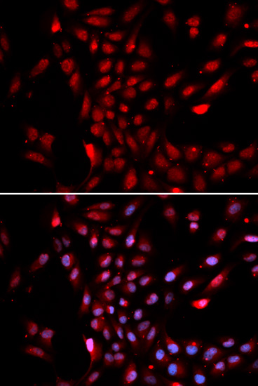
| Compartment evidence across applications | Tissue IHC is mainly nuclear (HPA tissue IHC). ICC-IF places KPNA4 mainly in the nucleoplasm, with additional nuclear membrane localization (HPA subcellular: enhanced nucleoplasm; approved nuclear membrane). Those IF observations support interpretation of localization; they do not specify an IHC-P protocol. |
| Antibody validation | HPA lists HPA045500 and CAB034336 as IHC Enhanced, meaning their pattern has support from independent antibodies or orthogonal data; HPA043154 is IHC Supported (HPA antibodies). The overall tissue reliability is Enhanced but has only medium consistency with RNA, so a single discrepant slide warrants controls (HPA tissue IHC). |
| Expression evidence differs by source | UniProt describes KPNA4 as barely detectable in colon, whereas HPA reports high staining in colon glandular cells (UniProt O00629: tissue specificity; HPA tissue IHC: colon glandular cells, High). Treat colon as a source-dependent example, and prefer an unambiguous HPA high-staining control when assessing the HPA IHC pattern. |
| Molecular context | KPNA4 has no transmembrane segment or signal peptide and functions in nuclear protein import (UniProt O00629: topology; processing; function). These facts support interpreting intracellular staining, but do not establish that any particular retrieval or fixation condition changes the IHC signal. |
| Situation | Likely cause | Next action |
|---|---|---|
| Positive control has no nuclear chromogen. | The section may lack the expected cell population, or the staining run may have failed (standard IHC practice). | Confirm bronchial respiratory epithelium or adrenal glandular cells are present (HPA tissue IHC: High); review the antibody, retrieval and detection steps with run controls (standard IHC practice). |
| Signal appears only after very strong chromogen development. | Weak specific signal and accumulated background can become hard to distinguish (standard IHC practice). | Compare the positive tissue and no-primary control at the same development time, then adjust detection conditions if needed (standard IHC practice). Do not score haze as KPNA4 localization. |
| Cytoplasm dominates while nuclei remain pale. | The pattern differs from mainly nuclear tissue IHC, although cytoplasmic localization is recorded by UniProt (HPA tissue IHC; UniProt O00629: location). | Verify cell identity and compare with a reported high-staining control; review retrieval and antibody conditions without assuming a KPNA4-specific fixation effect (standard IHC practice). |
| Chromogen appears in the no-primary control. | Background may come from the detection system, including endogenous activity when the detection chemistry permits it (standard IHC practice). | Address the relevant endogenous activity and review blocking and detection reagents before interpreting the test section (standard IHC practice). |
| Colon glandular staining differs from the expected control result. | The sources disagree: HPA reports high glandular staining, while UniProt calls colon expression barely detectable (HPA tissue IHC: colon; UniProt O00629: tissue specificity). | Record the discrepancy and assess the same run with another HPA high-staining tissue, such as bronchus or adrenal gland (HPA tissue IHC: High); avoid using colon alone to judge assay success. |
| IF/ICC shows nuclear rim signal as well as nucleoplasmic signal. | HPA records additional nuclear membrane localization in ICC-IF (HPA subcellular: nuclear membrane, approved). | Interpret this alongside the enhanced nucleoplasmic location (HPA subcellular). Use the separate IF/ICC guide for its staining workflow; this IHC section supplies no IF protocol. |


Comprehensive Human Protein Atlas IHC scoring per tissue (reliability: Enhanced — Medium consistency between antibody staining and RNA expression data.). Rows are taken directly from the HPA tissue chart — click any row's HPA link to view the source.
| Tissue | Cell type | Level | Evidence | Source |
|---|---|---|---|---|
| Adrenal gland | Glandular cells | High | Protein (IHC) | HPA → |
| Appendix | Glandular cells | High | Protein (IHC) | HPA → |
| Bronchus | Respiratory epithelial cells | High | Protein (IHC) | HPA → |
| Caudate | Neuronal cells | High | Protein (IHC) | HPA → |
| Cerebellum | Bergmann glia - nucleus | High | Protein (IHC) | HPA → |
| Tissue | Cell type | Level | Evidence | Source |
|---|---|---|---|---|
| None in HPA: KPNA4 is detected in all 45 scored tissues. Use a no-primary (secondary-only) and an isotype control instead. | ||||
Troubleshoot KPNA4 chromogenic IHC by checking nuclear staining, section processing, and controls before comparing signal across tissues (HPA tissue IHC: mainly nuclear).
Validated anti-KPNA4 options show paraffin-section IHC in rat heart and human esophageal cancer (A05188-1 IHC captions), and IF/ICC in HeLa cells (A05188 image captions).
A05188-1 has IHC images from paraffin-embedded rat heart and human esophageal cancer, plus an IF image from U2OS cells (A05188-1 image captions). A05188 has ICC and IF images from HeLa cells and no listed IHC application (A05188 image captions; catalog applications).
Which to pick: Choose A05188-1 for tissue IHC: its paraffin-section captions show 1:200 antibody dilution and microwave retrieval in 10 mM PBS at pH 7.2; the fixative is unreported (A05188-1 IHC captions). Choose A05188 for IF/ICC, supported by HeLa images at 20 μg/mL for IF and 2.5 μg/mL for ICC (A05188 image captions). Both are rabbit antibodies with no clone specified and list Human, Mouse, and Rat reactivity; only A05188-1 lists IHC among its applications (catalog host, clone, reactivity, and applications).