This website uses cookies to ensure you get the best experience on our website.


- Table of Contents
Real validated KRAS Western blot protocols, expected-band and isoform facts, troubleshooting for weak or shifted signal, and recommended anti-KRAS WB antibodies. Everything you need to plan the experiment before you commit precious samples.

Expected bands, validated protocols, controls and antibodies — the at-a-glance facts below, then the full design guide.
| Expected band | ~21.7 kDa | |
| Observed band | ~20 kDa | |
| Gel | 12% (catalog A00046-2) | |
| Positive control | Appendix (IHC candidate; verify WB) +4 more | |
| Negative control | Adipose tissue (IHC candidate; verify WB) |
| PTM | Acetylated; toxin-dependent O-glucosylation | |
| Caveat | Alternative isoforms | |
| Gene-set association | MSigDB C7 membership | |
| Isoform | 2 isoform(s) |
The A00046-2 protocol combines labelled catalog values with standard starting conditions. Published comparisons retain their own sample, reagent and detection scope.
| Sample / lysate | human Hela, human Caco-2, rat brain (catalog A00046-2) |
| Gel % | 12% (catalog A00046-2) |
| Load | 30 ug; reducing conditions (catalog A00046-2) |
| Transfer | a nitrocellulose membrane at 150 mA for 50-90 minutes (catalog A00046-2) |
| Membrane | nitrocellulose membrane (catalog A00046-2) |
| Blocking | 5% non-fat milk/TBS for 1.5 hour at RT (catalog A00046-2) |
| Primary antibody | A00046-2 · 0.5 μg/mL (catalog A00046-2) |
| Primary incubation | overnight at 4°C (catalog A00046-2) |
| Secondary antibody | goat anti-rabbit IgG-HRP, 1:5000 (catalog A00046-2) |
| Secondary incubation | 1.5 hour at RT (catalog A00046-2) |
| Wash | TBS-0.1%Tween 3 times with 5 minutes each (catalog A00046-2) |
| Detection | ECL (catalog A00046-2) |
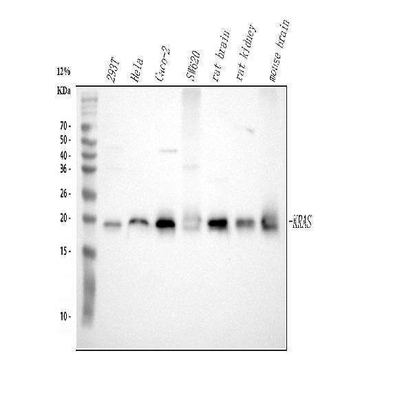
The selected blot caption reports a KRAS band at approximately 20 kDa; the predicted sequence mass is 21.7 kDa. The supplied annotations do not establish why these values differ or demonstrate a migration effect from processing, isoforms, or modifications.
| Band near 20 kDa | Matches the position reported in the selected KRAS blot caption. Confirm identity with antibody specificity and appropriate positive and negative controls. |
| Slightly smaller band | A separate band cannot be assigned to removal of residues 187–189 from position alone; that short processing event does not explain a much larger apparent mass difference. |
| Nearby bands at different positions | KRAS has isoforms 2A and 2B, but the supplied evidence does not show that they resolve as distinct bands. Check antibody recognition and isoform-specific controls. |
| Higher or diffuse band after TcsL exposure | Thr35 O-linked glucose is annotated for P. sordellii toxin TcsL during microbial infection. A single glucose annotation does not establish a visible shift or broad smear; compare exposed and unexposed samples before assigning the signal. |
| Predicted KRAS mass | The sequence-based mass is 21.7 kDa. The selected blot caption separately reports a band at approximately 20 kDa; the reason for the difference is undetermined. |
| Propeptide at residues 187–189 | C-terminal processing is annotated, but loss of three residues is too small to explain the approximately 1.7 kDa difference between the predicted mass and the captioned band. No distinct processed band is demonstrated. |
| Isoforms 2A and 2B | Alternative splicing changes the C-terminal sequence. The supplied evidence does not establish distinct, resolvable positions on this blot. |
| TcsL-dependent O-glucosylation at Thr35 | This annotation applies during microbial infection with P. sordellii toxin TcsL. The supplied evidence does not demonstrate a migration effect from the single attached glucose. |
| Situation | Likely cause | Next action |
|---|---|---|
| No band in lysate | KRAS signal may be weak in the sampled lysate, or detection may be inadequate. | Check protein loading, antibody performance, and a suitable positive control; consider a membrane-enriched fraction. |
| Band higher than expected | Band position alone does not identify a KRAS modification; the supplied Thr35 annotation does not demonstrate a visible shift. | Verify the size marker and antibody specificity. If TcsL exposure occurred, compare exposed and unexposed samples. |
| Band lower than expected | The supplied evidence does not explain the difference between the predicted 21.7 kDa mass and the captioned approximately 20 kDa band; removal of residues 187–189 is too small to account for it. | Compare with the captioned KRAS position and confirm identity using an appropriate control. |
| Broad smear instead of sharp band | A smear has several possible technical or sample-related causes; a single Thr35 glucose annotation does not establish one. | Check sample integrity, loading, and electrophoresis conditions. If relevant, compare TcsL-exposed and unexposed samples. |
| Multiple bands | The presence of KRAS isoforms 2A and 2B does not establish that they form separate resolvable bands. | Check which isoforms the antibody recognizes and use isoform-specific or KRAS-depletion controls to assign bands. |
| Fragments below expected size | Smaller signals may reflect degradation or nonspecific antibody binding; their identity is unconfirmed. | Check sample handling, compare a fresh lysate, and verify the signals with a KRAS-specific control. |
Comprehensive Human Protein Atlas IHC scoring per tissue. Rows are taken directly from the HPA tissue chart — click any row's HPA link to view the source.
| Tissue | Cell type | Level | Evidence | Source |
|---|---|---|---|---|
| Appendix | glandular cells | High | Protein (IHC) | HPA → |
| Colon | glandular cells | High | Protein (IHC) | HPA → |
| Duodenum | glandular cells | High | Protein (IHC) | HPA → |
| Gallbladder | glandular cells | High | Protein (IHC) | HPA → |
| Rectum | glandular cells | High | Protein (IHC) | HPA → |
| Tissue | Cell type | Level | Evidence | Source |
|---|---|---|---|---|
| Adipose tissue | adipocytes | Not detected | Protein (IHC) | HPA → |
| Adrenal gland | glandular cells | Not detected | Protein (IHC) | HPA → |
| Breast | adipocytes | Not detected | Protein (IHC) | HPA → |
| Caudate | glial cells | Not detected | Protein (IHC) | HPA → |
| Cervix | glandular cells | Not detected | Protein (IHC) | HPA → |
Deeper troubleshooting and optimisation questions for KRAS, answered from its protein features.
Catalog antibodies with Western blot application and product-specific WB images. Evaluate suitability with the reported sample, controls and experimental conditions.
The catalog reports one anti-KRAS antibody, A00046-2, with a Western blot analysis image. Its caption identifies human HeLa and Caco-2 whole cell lysates and rat and mouse brain tissue lysates, with a KRAS band at approximately 20 kDa.
Which to pick: A00046-2 is the only listed option for KRAS; its Western blot documents those specific sample contexts. Check the full product details for other intended samples.