KRT15 / Keratin, type I cytoskeletal 15 · IHC design guide

Design Immunohistochemistry for KRT15

Plan chromogenic KRT15 IHC around basal and glandular cell staining (HPA tissue IHC). Start M06791-1 at 1:50 (datasheet: M06791-1) and score cytoplasmic and membranous staining by cell type (HPA tissue IHC).

Evidence assembled Oct 2026 · For research use; verify linked source records and product datasheet before use
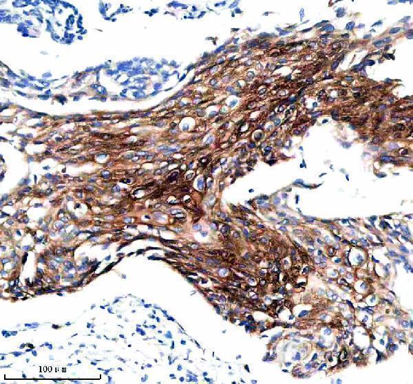
Immunohistochemistry protocol sheet for KRT15 (IHC for KRT15): expected localisation Cytoplasmic, with membranous staining in some epithelia (HPA tissue IHC), antibody M06791-1, validated IHC image, and IHC protocol steps
Printable KRT15 IHC protocol sheet — expected localisation Cytoplasmic, with membranous staining in some epithelia (HPA tissue IHC), antibody M06791-1, controls and protocol steps. Open the full KRT15 IHC guide →

KRT15 Immunohistochemistry Experimental Design Guide

Expected localisation, validated protocols, controls and antibodies — the at-a-glance facts below, then the full design guide.

Must know before staining
Expected localisation Cytoplasmic, with membranous staining in some epithelia (HPA tissue IHC)
Staining pattern Basal and glandular cells: cytoplasmic ± membranous staining (HPA tissue IHC)
Antigen retrieval EDTA pH 8.0 HIER, heat-mediated (datasheet M06791-1)
Positive control ⓘ Breast+4 more · see all
Negative control ⓘ Adipose tissue+4 more · see all
Important caveats
Reasons your staining may differ from the expected pattern.
Fixation Keep fixation consistent across paraffin sections (standard IHC practice; not target-specific)
Caveat Presumed off-target staining was observed and disregarded (HPA tissue IHC)
Regulation Patchy adult skin basal expression (UniProt)
Isoform / epitope 2 isoforms; epitope differences are unspecified (UniProt)
Section 1

Recommended KRT15 IHC & IF Protocols

The catalog antibody’s IHC-P protocol (datasheet M06791-1) is accompanied by three published KRT15 IHC protocols (PMC9302346; PMC11473747; PMC7054907).

Recommended immunohistochemistry (IHC-P) protocol parameters
SampleParaffin-embedded human cervical cancer tissue; fixative not specified (datasheet M06791-1)
FixationImage fixative and duration unreported (datasheet M06791-1); verify before use.
Sectioning4–5 µm sections on charged slides (standard)
DeparaffinisationXylene, graded ethanol series to water (standard)
Antigen retrievalHeat retrieval: EDTA pH 8.0 (datasheet M06791-1); 20 min, 95–100 °C (standard)
Peroxidase block3% H2O2, 10 min, room temperature (standard)
Blocking10% goat serum (datasheet M06791-1)
Primary antibodyRabbit monoclonal (clone DOB-11) anti-KRT15, 1:50 (datasheet M06791-1)
Primary incubationOvernight at 4 °C (datasheet M06791-1)
DetectionHRP-conjugated secondary, DAB chromogen (datasheet M06791-1)
CounterstainHematoxylin, blue, dehydrate and mount (standard)
Expected resultKRT15-positive staining in glandular cells of breast (HPA tissue IHC: High). HPA tissue profile: Cytoplasmic and membranous expression in basal cells of the respiratory epithelium, prostate, squamous epithelium and glandular cells of breast. Cytoplasmic expression also in hair follicle and eccrine glands. No signal in the no-primary control.
💡Decision noteStart with heat-mediated EDTA retrieval at pH 8.0 (datasheet M06791-1); the cited excerpts do not specify retrieval conditions (PMC9302346; PMC11473747; PMC7054907).
Section 2

What Is the Expected KRT15 Staining Pattern?

KRT15 is a cytoplasmic intermediate filament protein with no transmembrane segment (UniProt P19012: subcellular location and topology). In paraffin section IHC, expect staining in selected epithelial cells, including respiratory basal cells, squamous epithelium and hair follicle outer root sheath cells (HPA: tissue IHC; UniProt P19012: tissue specificity). HPA rates its tissue IHC evidence Enhanced, while noting presumed off-target staining that was disregarded (HPA: tissue IHC reliability).

What am I looking at on my slide?
Basal respiratory cells, squamous epithelium or hair outer root sheath cells stain.This fits the reported distribution (HPA: tissue IHC; UniProt P19012: tissue specificity). Compare signal within the relevant cell layer: esophageal squamous cells and bronchial respiratory cells have High HPA staining, whereas oral mucosal squamous cells have Medium staining (HPA: tissue IHC).
Staining appears exclusively nuclear or outlines only the cell surface.Neither is the expected IHC pattern for a cytoplasmic keratin (UniProt P19012: subcellular location). HPA describes some membranous IHC expression, so assess border staining together with cytoplasmic signal rather than rejecting it alone (HPA: tissue IHC profile).
Strong signal appears in adipocytes or marrow hematopoietic cells.These cell types are Not detected in HPA tissue IHC (HPA: adipose tissue and bone marrow). Treat the result as suspect; cross-reactivity or endogenous detection activity are possible explanations under general IHC practice, subject to control slides.
Color spreads across cells and tissue without a recognizable epithelial pattern.A diffuse deposit does not match the reported cell-specific distribution (HPA: tissue IHC). In general IHC practice, nonspecific antibody binding, endogenous detection activity or excessive chromogen development can produce background; the image alone cannot identify which occurred.
A known-positive bronchus or hair section has no signal.HPA reports High staining in bronchial respiratory epithelial cells and hair outer root sheath cells (HPA: tissue IHC). Check that the expected cells are present before interpreting absence; a failed positive control can also indicate a general IHC detection or antibody performance problem.
💡Expected KRT15 appearanceCall positive IHC when staining follows the cytoplasm of the appropriate epithelial cells, with conspicuous signal in HPA High examples such as bronchus or hair; isolated nuclear staining or strong staining in HPA Not detected adipocytes is suspect (UniProt P19012: cytoplasm; HPA: tissue IHC).
How each factor affects the staining
Cell layer and tissueDistribution varies by site: adult epidermal basal staining is discontinuous, whereas fetal epidermal basal staining is continuous (UniProt P19012: tissue specificity). HPA tissue IHC levels also differ among sampled cell types; score the cells present, not an entire section as uniformly positive.
Intracellular filament locationKRT15 forms an epithelial intermediate filament network with KRT5 and lacks a transmembrane segment (UniProt P19012: function and topology). Interpret cytoplasmic, cell-associated staining in that context; topology alone does not establish an antigen retrieval requirement.
Antibody evidence and isoformsTwo listed antibodies have Enhanced IHC validation, and HPA's tissue assessment notes presumed off-target binding (HPA: antibodies; tissue IHC reliability). UniProt lists two isoforms, but these sources provide no epitope map or isoform-specific staining result (UniProt P19012: isoforms).
IF/ICC Q: What location should be expected?A: HPA supports intermediate filaments as the main ICC-IF location and lists nucleoplasm as an uncertain additional location (HPA: subcellular). Use that distinction when reviewing IF images; this IHC section provides no IF/ICC protocol.
Why is my staining missing, weak or wrong?
SituationLikely causeNext action
No staining in a bronchus positive control.The expected respiratory epithelial cells may be absent, or the IHC detection run may have failed (HPA: bronchus High; general IHC practice).Verify the sampled epithelial cells on the section, then review run controls, antibody application and detection steps. Repeat with a known-positive section if the run controls fail (general IHC practice).
Only nuclei stain in an otherwise suitable section.Predominantly nuclear IHC conflicts with UniProt's cytoplasmic location (UniProt P19012). HPA's uncertain nucleoplasmic observation comes from ICC-IF, not tissue IHC (HPA: subcellular).Review morphology and counterstain, then compare an independent IHC-validated antibody where available (HPA: two Enhanced IHC antibodies; general IHC practice).
Adipocytes or marrow cells stain strongly.Both are Not detected in the supplied HPA tissue IHC entries; off-target binding or endogenous detection activity is possible (HPA: tissue IHC; general IHC practice).Run appropriate reagent omission and detection controls, and compare the epithelial positive control. Interpret persistent unexpected staining cautiously (general IHC practice).
Broad background obscures epithelial cell boundaries.The pattern cannot be assigned to KRT15 from appearance alone; nonspecific binding or detection background is possible (HPA: cell-specific tissue profile; general IHC practice).Check negative controls, blocking and wash performance, and chromogen development. Reassess whether staining still follows the expected epithelial cells (general IHC practice; HPA: tissue IHC).
Adult skin shows patchy basal staining.Discontinuous basal expression in adult epidermis is reported for KRT15 (UniProt P19012: tissue specificity).Score positive basal cells and their location before changing the assay; compare with a suitable positive tissue if the entire run looks weak (UniProt P19012: tissue specificity; general IHC practice).
IF shows some nucleoplasmic signal.HPA lists nucleoplasm as an uncertain additional ICC-IF location, while intermediate filaments are supported as the main location (HPA: subcellular).Report filament signal separately from uncertain nucleoplasmic signal. Consult the separate IF/ICC guide for its assay workflow (HPA: subcellular).

Sample controls for KRT15 IHC & IF

🧪Run esophagus first: squamous epithelial cells should stain (HPA: High in esophageal squamous epithelial cells). Use adipose tissue as a negative, with adipocytes at background (HPA: Not detected in adipocytes); non-epithelial stromal cells on the esophagus slide should also remain at background.
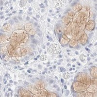
Positive control tissue: Breast (Glandular cells, HPA High)
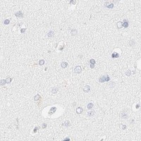
Negative control tissue: Adipose tissue (HPA Not detected)
ICC-IF cell lines (HPA subcellular resource): HPA ICC-IF images show KRT15 in HaCaT, with annotated localisation: Intermediate filaments (supported) (HPA subcellular).
Technical controls: Include a no-primary (secondary-only) control, a concentration-matched rabbit isotype control, and KRT15-knockout material as a biological negative (M06791-1 caption: rabbit primary antibody). For HRP/DAB detection, quench endogenous peroxidase and check the esophagus slide for background staining (M06791-1 caption: HRP/DAB detection).
⚠️Feasibility: No target-specific fixation window or fixation effect is reported in the supplied evidence, and the selected M06791-1 tissue-IHC caption does not state a fixative. Heat retrieval in EDTA at pH 8.0 is documented for that paraffin-section example, but its necessity across specimens is unreported (M06791-1 caption: heat retrieval in EDTA, pH 8.0). Whether frozen sections or IF are easier is unreported; when assessing IF, expect an intermediate-filament pattern and treat nucleoplasmic signal cautiously (HPA: intermediate filaments supported; nucleoplasm uncertain).

HPA tissue IHC evidence for KRT15

Comprehensive Human Protein Atlas IHC scoring per tissue (reliability: Enhanced — High consistency between antibody staining and RNA expression data. Presumed off target binding observed and disregarded.). Rows are taken directly from the HPA tissue chart — click any row's HPA link to view the source.

Positive expression · recommended positive controls

TissueCell typeLevelEvidenceSource
Breast Glandular cells High Protein (IHC) HPA →
Bronchus Respiratory epithelial cells High Protein (IHC) HPA →
Esophagus Squamous epithelial cells High Protein (IHC) HPA →
Hair Cells in external root sheath High Protein (IHC) HPA →
Skin Secretory cells High Protein (IHC) HPA →

Undetected expression · recommended negative controls

TissueCell typeLevelEvidenceSource
Adipose tissue Adipocytes Not detected Protein (IHC) HPA →
Adrenal gland Glandular cells Not detected Protein (IHC) HPA →
Bone marrow Hematopoietic cells Not detected Protein (IHC) HPA →
Caudate Glial cells Not detected Protein (IHC) HPA →
Cerebellum Cells in granular layer Not detected Protein (IHC) HPA →
Section 3

Advanced KRT15 IHC Tips

Use the catalog antibody’s paraffin-section example as the starting point, then judge KRT15 staining against epithelial cell identity and its expected filament pattern.

What retrieval should I try first if KRT15 staining is weak?
Start with heat-mediated antigen retrieval in EDTA at pH 8.0 for paraffin sections (datasheet M06791-1). The catalog example detected KRT15 in a paraffin-embedded human cervical cancer section after this retrieval, but its fixative was unreported (datasheet M06791-1). If staining remains weak, compare a more or less intense retrieval treatment on adjacent sections while keeping the antibody at the documented 1:50 dilution (datasheet M06791-1; general IHC practice). Judge any gain by sharper cytoplasmic epithelial staining and preserved morphology; retrieval that increases diffuse background or damages tissue has not improved the assay (UniProt P19012 subcellular; general IHC practice).
Could fixation explain weak or uneven KRT15 staining?
The selected paraffin-section caption does not report a fixative, so target-specific KRT15 sensitivity to fixation is unknown (datasheet M06791-1). Record the fixative, fixation duration and processing history for each specimen before attributing a weak signal to fixation (general IHC practice). Compare similarly processed sections with the documented EDTA pH 8.0 retrieval and 1:50 antibody dilution, changing one condition at a time (datasheet M06791-1; general IHC practice). Include an expected positive epithelial region on each run, and assess both staining and morphology; an uneven pattern alone cannot establish a KRT15-specific fixation effect (UniProt P19012 tissue specificity; general IHC practice).
How should I assess a membranous or nuclear KRT15 signal?
Expect predominantly cytoplasmic filament-associated staining: KRT15 is a cytoplasmic intermediate-filament protein without a transmembrane segment (UniProt P19012 subcellular and topology; HPA subcellular: intermediate filaments supported). HPA tissue IHC also reports cytoplasmic and membranous expression in several epithelial populations, so assess apparent membrane staining alongside the filament pattern and cell boundaries (HPA tissue IHC profile). Treat an isolated nuclear pattern cautiously because HPA lists nucleoplasmic localisation as uncertain (HPA subcellular: nucleoplasm uncertain). Check the same compartment in an expected positive epithelial region, then inspect the no-primary control before scoring unusual localisation as KRT15 (UniProt P19012 tissue specificity; general IHC practice).
Could isoforms or epitope accessibility explain discordant KRT15 staining?
KRT15 has 2 annotated isoforms, but the supplied antibody caption does not identify its epitope or establish recognition of both (UniProt P19012 isoforms; datasheet M06791-1). Its intermediate-filament rod spans residues 105–417, and several annotated phosphoserines lie near the amino terminus; neither fact establishes this antibody’s binding site (UniProt P19012 domains and modified residues). If two assays disagree, verify each reagent’s stated immunogen and isoform coverage before assigning the difference to splicing or phosphorylation (general IHC practice). Compare adjacent, similarly processed sections and expected epithelial compartments; keep retrieval at EDTA pH 8.0 for the catalog-antibody comparison (datasheet M06791-1; general IHC practice).
How can I check KRT15 by multiplex IF alongside chromogenic IHC?
For a separate IF/ICC assay, pair KRT15 with an independently validated epithelial or basal-cell marker to confirm the identity of stained cells (UniProt P19012 tissue specificity; general IF practice). Select spectrally separated fluorophores after checking tissue autofluorescence and single-color controls, especially where weak filament signal could be obscured (general IF practice). Because KRT15 has no transmembrane segment and resides mainly in cytoplasmic intermediate filaments, permeabilise cells sufficiently to expose intracellular epitopes while preserving filament morphology (UniProt P19012 topology; HPA subcellular: intermediate filaments supported; general IF practice). The paraffin-section 1:50 dilution and EDTA pH 8.0 retrieval document IHC conditions only; optimise IF staining independently (datasheet M06791-1; general IF practice).
What should I change when DAB background obscures KRT15?
Separate diffuse DAB deposit from cellular staining by inspecting a no-primary section and the expected epithelial pattern (general IHC practice; UniProt P19012 tissue specificity). The catalog example used 10% goat serum block, 1:50 primary overnight at 4°C, and an HRP-based DAB readout (datasheet M06791-1). For excessive signal, check peroxidase quenching, washing, secondary-antibody background and DAB development on matched sections, changing one condition at a time (general chromogenic IHC practice). Retain an expected positive region during optimisation: HPA reports high staining in bronchial respiratory epithelial cells and hair external-root-sheath cells, although its tissue reliability note acknowledges presumed off-target binding (HPA tissue IHC).
How should I score KRT15 staining across paraffin sections? ⚠ ANSWER MARKED FOR VERIFICATION
Define the epithelial compartment before scoring, because KRT15 distribution varies by site and epithelial layer (UniProt P19012 tissue specificity). For each region, report the percentage of positive eligible cells and intensity on a 0–3 scale; an H-score ranges from 0–300 when intensity is weighted by the percentage at each level (general IHC practice). Alternatively, report positive-cell density per mm² of viable epithelial area, keeping the denominator consistent across specimens (general IHC practice). Exclude folds, necrosis and section edges, and compare matched processing batches or an internal positive compartment before interpreting differences in score (general IHC practice).
When is KRT15 staining credible rather than an artefact?
A credible result has cytoplasmic, preferably filament-associated signal in an anatomically appropriate epithelial population, such as the hair external root sheath or bronchial basal cells (UniProt P19012 tissue specificity; HPA subcellular: intermediate filaments supported). Staining confined to nuclei, necrotic debris or section edges warrants review against morphology and a no-primary control (HPA subcellular: nucleoplasm uncertain; general IHC practice). Check for endogenous peroxidase or nonspecific DAB deposit when staining appears outside the expected cells (general chromogenic IHC practice). HPA reports enhanced tissue-IHC reliability but also notes presumed off-target binding, so reconcile any surprising pattern with cell identity and an independent control (HPA tissue IHC reliability description; general IHC practice).
Boster reagents

Best KRT15 / Keratin, type I cytoskeletal 15 IHC Antibodies

The catalog covers KRT15 IHC in human, mouse, and rat paraffin sections, with real IHC images for M06791-1 (M06791-1 IHC captions). IF/ICC is listed without an IF image (catalog applications/image alts).

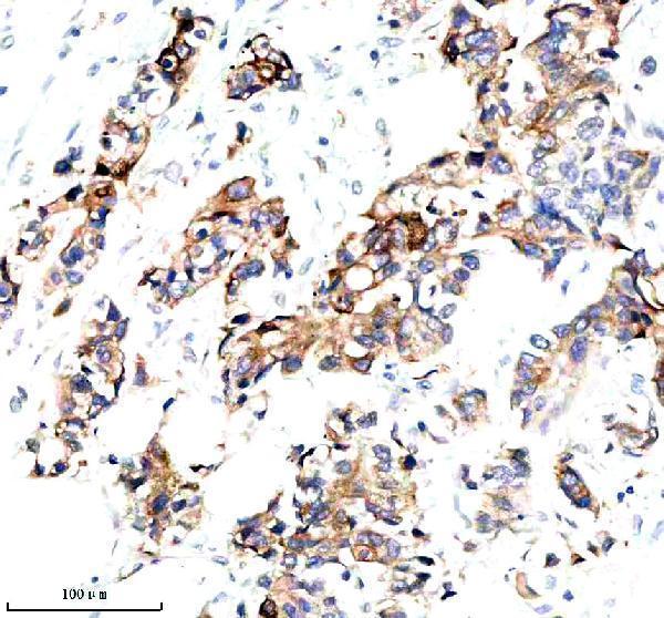
Real IHC data IHC analysis of KRT15 using anti-KRT15 antibody (M06791-1). KRT15 was detected in a paraffin-embedded section of human cervical cancer tissue. Heat mediated antigen retrieval was performed in EDTA buffer (pH 8.0, epitope retrieval solution). The tissue section was blocked with 10% goat serum. The tissue section was then incubated with 1:50 rabbit anti-KRT15 Antibody (M06791-1) overnight at 4°C. Peroxidase Conjugated Goat Anti-rabbit IgG was used as secondary antibody and incubated for 30 minutes at 37°C. The tissue section was developed using HRP Conjugated Rabbit IgG Super Vision Assay Kit (Catalog # SV0002) with DAB as the chromogen.
Anti-Cytokeratin 15 Rabbit Monoclonal Antibody
Cat # M06791-1

M06791-1 is the SKU with a rendered IHC card; its caption shows KRT15 staining in a human cervical cancer paraffin section (rendered card caption). Additional IHC captions show human breast cancer and mouse and rat skin paraffin sections; IF/ICC is listed, but no IF image is supplied (M06791-1 IHC captions; catalog applications/IF image alts).

Which to pick: Choose M06791-1 for tissue IHC when a paraffin-section example is useful; it is a rabbit monoclonal with a listed 1:50 IHC dilution (catalog clone/host/dilution; M06791-1 IHC captions). For IF/ICC and work across human, mouse, and rat, M06791-1 is the listed option, with a 1:50 IF dilution but no supplied IF figure (catalog applications/reactivity/dilution/IF image alts). A06791-4 is a polyclonal IHC option listed for human and rat without an IHC image caption; the M06791-1 captions establish paraffin sections, but do not report the fixative (catalog A06791-4 applications/reactivity/image alts; M06791-1 IHC captions).

Each figure is that product's own IHC / IF validation image from its datasheet.

References

  1. UniProt Consortium. UniProt entry P19012 (K1C15_HUMAN, Keratin, type I cytoskeletal 15).
  2. Human Protein Atlas. KRT15 tissue IHC expression (reliability: Enhanced).
  3. Human Protein Atlas. KRT15 subcellular location (ICC-IF): Mainly localized to the intermediate filaments. In addition localized to the nucleoplasm..
  4. Human Protein Atlas. KRT15 antibody validation summary (2 antibodies).
  5. Upregulated keratin 15 links to the occurrence of lymphovascular invasion, stromal cervical invasion as well as unfavorable survival profile in endometrial cancer patients. Medicine 2022 — PMC9302346.
  6. Keratin-15 high expression links with lymph node metastasis and poor survival prognosis in epithelial ovarian cancer patients. Discover oncology 2024 — PMC11473747.
  7. The value of desmosomal plaque-related markers to distinguish squamous cell carcinoma and adenocarcinoma of the lung. Upsala journal of medical sciences 2020 — PMC7054907.
  8. Single-Cell Transcriptome Sequence Profiling on the Morphogenesis of Secondary Hair Follicles in Ordos Fine-Wool Sheep. International journal of molecular sciences 2024 — PMC10779399.
  9. PubMed PMID:2468493 — UniProt-cited evidence.
  10. PubMed PMID:2452170 — UniProt-cited evidence.
  11. PubMed PMID:10623642 — UniProt-cited evidence.