This website uses cookies to ensure you get the best experience on our website.


- Table of Contents
Plan KRT16 paraffin-section IHC around cytoplasmic staining in squamous epithelial cells, using tonsil as a medium-staining reference (HPA tissue IHC). Keep fixation consistent (standard IHC practice) and interpret results with HPA’s low antibody–RNA concordance in mind (HPA tissue IHC).

Expected localisation, validated protocols, controls and antibodies — the at-a-glance facts below, then the full design guide.
| Expected localisation | Cytoplasmic in squamous epithelial cells (HPA tissue IHC) | |
| Staining pattern | Cytoplasmic staining in squamous epithelial cells (HPA tissue IHC) | |
| Antigen retrieval | No retrieval / mild HIER, citrate pH 6.0 if weak (rule: intermediate filament) | |
| Positive control | Tonsil | |
| Negative control | Adipose tissue+4 more · see all |
| Fixation | Keep paraffin-section fixation consistent (standard IHC practice; not target-specific); Matched tissue-IHC evidence does not establish the fixation claim. Validate the specimen-specific method before use. (selected-SKU IHC image M03393) | |
| Caveat | Low consistency between antibody staining and RNA expression (HPA tissue IHC) | |
| Regulation | Tissue-enhanced RNA expression (HPA tissue RNA) | |
| Isoform / epitope | No isoforms or processing annotated; epitope site unknown (UniProt) |
The catalog antibody’s IHC-P protocol is accompanied by published KRT16 protocols for tumor and skin sections (PMC8974228; PMC11900751; PMC13283874).
| Sample | Paraffin-embedded human skin tissue; fixative not specified (datasheet M03393) |
| Fixation | Image fixative and duration unreported (datasheet M03393); verify before use. |
| Sectioning | 4–5 µm sections on charged slides (standard) |
| Deparaffinisation | Xylene, graded ethanol series to water (standard) |
| Antigen retrieval | No retrieval needed for most intermediate-filament epitopes; if staining is weak, mild HIER in citrate buffer, pH 6.0, 10 min at 95 °C (standard rule: intermediate filament) |
| Peroxidase block | 3% H2O2, 10 min, room temperature (standard) |
| Blocking | 10% normal serum of the secondary host, 30 min, room temperature (standard) |
| Primary antibody | Rabbit monoclonal (clone EDC-11) anti-KRT16, 1:50 (datasheet M03393) |
| Primary incubation | Overnight at 4 °C (standard) |
| Detection | HRP-polymer secondary, DAB chromogen 5–10 min (standard) |
| Counterstain | Hematoxylin, blue, dehydrate and mount (standard) |
| Expected result | KRT16-positive staining in squamous epithelial cells of tonsil (HPA tissue IHC: Medium). HPA tissue profile: Cytoplasmic expression in squamous epithelial cells. No signal in the no-primary control. |
In paraffin IHC, expect KRT16 in the cytoplasm of squamous epithelial cells (HPA: tissue IHC profile), including medium staining in tonsillar squamous epithelium (HPA: Tonsil, Medium). KRT16 is a keratin intermediate filament protein with no transmembrane segment (UniProt P08779: function and topology). Interpret the pattern cautiously: HPA rates its tissue IHC profile Approved but reports low consistency between antibody staining and RNA expression (HPA: reliability description).
| Squamous epithelial cytoplasm stains in tonsil, with a moderate chromogenic signal. | This matches the reported positive cell population and level (HPA: Tonsil squamous epithelial cells, Medium). Score the epithelial compartment and its staining separately from neighboring cells; a tissue-wide score would obscure the cell-specific reference pattern (HPA: tissue IHC profile). |
| Strong nuclear or sharply membrane-restricted staining predominates in squamous cells. | That distribution conflicts with the reported cytoplasmic IHC profile (HPA: cytoplasmic expression in squamous epithelial cells) and the intermediate filament location seen by ICC-IF (HPA: subcellular summary). Check counterstain, detection deposits, and antibody controls before calling it KRT16 (standard IHC practice). |
| Adipocytes stain strongly, especially when the expected squamous pattern is absent. | Adipocytes were reported as Not detected in adipose tissue (HPA: Adipose tissue adipocytes, Not detected). Consider nonspecific binding or endogenous detection activity (standard IHC practice). Cell identity matters: HPA also reports Low staining in lung macrophages and skin fibroblasts (HPA: tissue IHC low entries). |
| Weak color spreads across epithelium, stroma, and empty areas without cell boundaries. | A widespread haze cannot establish the reported squamous cytoplasmic pattern (HPA: tissue IHC profile). Review the no-primary control, blocking, washes, and chromogen development for background sources (standard IHC practice); assess any remaining signal by cell type and compartment. |
| Tonsillar squamous epithelium has no detectable signal. | This misses a reported Medium positive reference (HPA: Tonsil squamous epithelial cells, Medium). Check section quality, retrieval, primary-antibody conditions, and detection with appropriate controls (standard IHC practice). One negative section does not resolve the profile's reported antibody–RNA inconsistency (HPA: reliability description). |
| IHC reference population | Tonsillar squamous epithelial cells provide a Medium positive reference; oral mucosal squamous cells are reported Low (HPA: tissue IHC entries). Compare the same cell type, since a Low reference can be difficult to judge at a glance (standard IHC practice). |
| Interpretation of negative tissue | HPA reports Not detected in adipose-tissue adipocytes and bronchial respiratory epithelial cells (HPA: tissue IHC negative entries). Those calls concern the named cells; they do not certify every cell in either section as KRT16-negative. |
| Evidence strength | The tissue IHC profile is Approved, with low consistency between antibody staining and RNA expression (HPA: reliability and reliability description). Treat a matching image as support for the observed pattern, then use controls to investigate discordant samples (standard IHC practice). |
| Antibody validation by application | CAB000136 is listed as IHC Approved, while HPA000539 is ICC Approved with no IHC status supplied (HPA: antibody validation entries). Use the IHC validation when judging paraffin-section staining; the ICC entry does not establish IHC performance. |
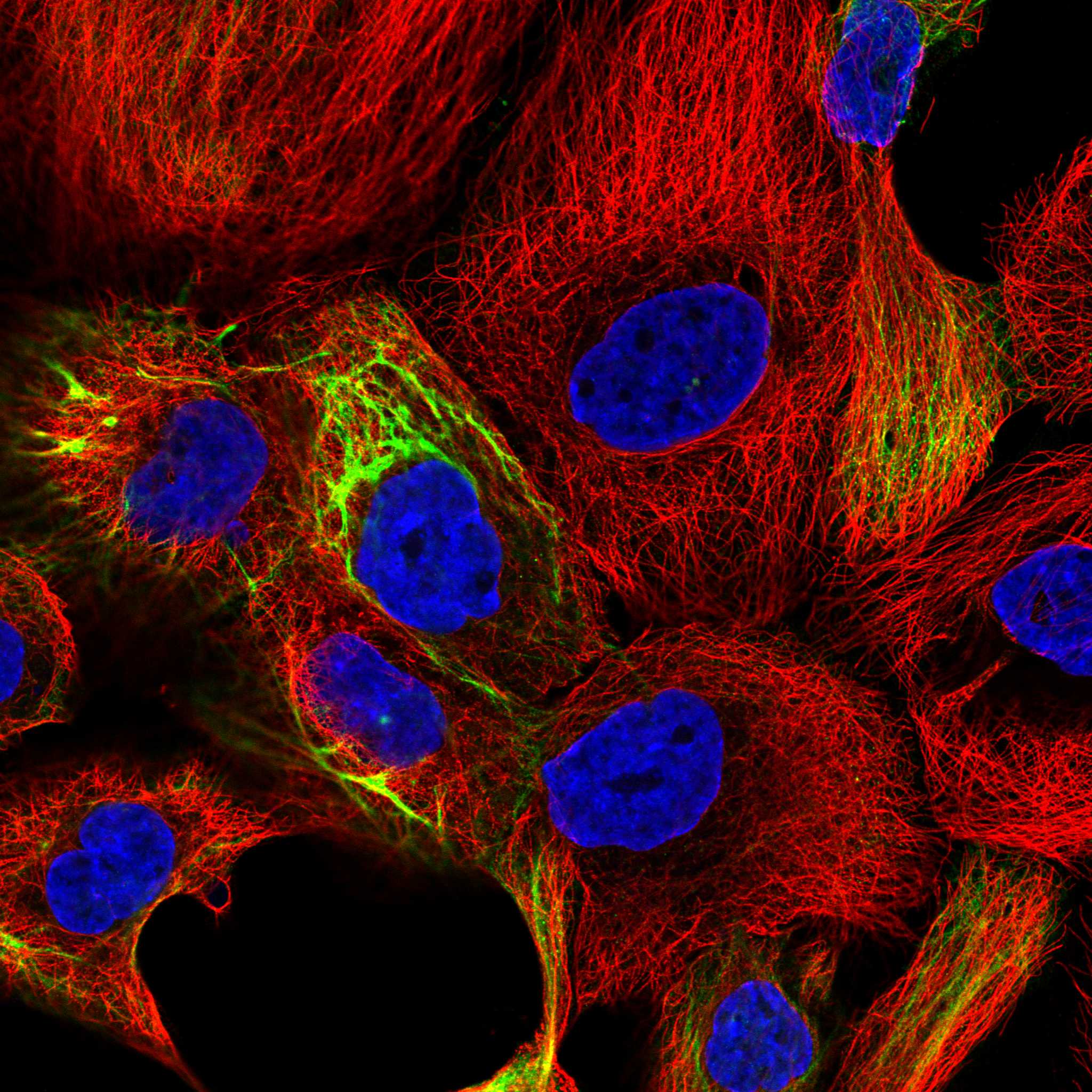
| IF/ICC Q: Where should fluorescence appear? | A: HPA reports intermediate filament localization in ICC-IF, based on antibodies targeting proteins from multiple genes (HPA: subcellular summary and caution). This supports a filamentous cytoplasmic expectation, with that specificity caveat; it is not a paraffin IHC protocol. |
| Molecular context | KRT16 forms epithelial keratin intermediate filaments with type II keratins, including KRT6A or KRT6B (UniProt P08779: function and subunit). UniProt lists no transmembrane segment or signal peptide (UniProt P08779: topology and processing), consistent with an intracellular readout. |
| Situation | Likely cause | Next action |
|---|---|---|
| No stain in tonsillar squamous epithelium | A Medium signal is reported there (HPA: Tonsil); inadequate retrieval, primary incubation, or detection can produce a technical miss (standard IHC practice). | Run a known-positive section and detection controls alongside the sample; review retrieval and antibody conditions against the validated IHC procedure (standard IHC practice). |
| Nuclear signal dominates | Nuclear dominance conflicts with the cytoplasmic tissue profile (HPA: tissue IHC profile). Counterstain overlap or nonspecific detection can confuse interpretation (standard IHC practice). | Inspect the no-primary control and counterstain; score KRT16 only where signal follows the squamous cytoplasm (standard IHC practice; HPA: tissue IHC profile). |
| Strong staining in adipocytes | Adipocytes are Not detected in the HPA adipose-tissue reference (HPA: tissue IHC negative entry); nonspecific binding or endogenous detection activity is possible (standard IHC practice). | Confirm cell identity and compare a no-primary control; check blocking and the detection system before assigning target staining (standard IHC practice). |
| Diffuse brown haze obscures the epithelium | Background from insufficient washing or detection development can obscure a cell-specific pattern (standard IHC practice; HPA: squamous cytoplasmic profile). | Compare control and stained sections, review washes and chromogen timing, then reassess whether epithelial cytoplasm is distinguishable (standard IHC practice; HPA: tissue IHC profile). |
| Signal appears weaker than expected in oral mucosa | Oral mucosal squamous cells are reported Low, compared with Medium in tonsillar squamous cells (HPA: tissue IHC entries). | Use tonsillar squamous epithelium as the stronger reported reference; compare cell-specific staining under the same run conditions (HPA: tissue IHC entries; standard IHC practice). |
| ICC images and paraffin IHC seem different | ICC-IF resolves an intermediate filament location, while the tissue IHC report describes cytoplasmic staining (HPA: subcellular and tissue IHC summaries). | Judge each image against its own application and validation entry: CAB000136 is IHC Approved; HPA000539 is ICC Approved (HPA: antibody validation entries). |

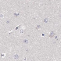
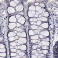
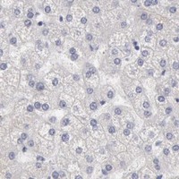
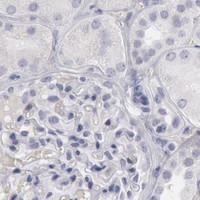

Comprehensive Human Protein Atlas IHC scoring per tissue (reliability: Approved — Low consistency between antibody staining and RNA expression data.). Rows are taken directly from the HPA tissue chart — click any row's HPA link to view the source.
| Tissue | Cell type | Level | Evidence | Source |
|---|---|---|---|---|
| Tonsil | Squamous epithelial cells | Medium | Protein (IHC) | HPA → |
| Tissue | Cell type | Level | Evidence | Source |
|---|---|---|---|---|
| Adipose tissue | Adipocytes | Not detected | Protein (IHC) | HPA → |
| Adrenal gland | Glandular cells | Not detected | Protein (IHC) | HPA → |
| Appendix | Glandular cells | Not detected | Protein (IHC) | HPA → |
| Bone marrow | Hematopoietic cells | Not detected | Protein (IHC) | HPA → |
| Breast | Adipocytes | Not detected | Protein (IHC) | HPA → |
Troubleshooting KRT16 staining in paraffin sections focuses on retrieval, cytoplasmic filament localisation, cell identity, and controls for chromogenic background.
One human-reactive anti-KRT16 antibody has an IHC image from paraffin-embedded human skin (catalog reactivity: Human; catalog IHC image caption). IF/ICC is listed without an IF image (catalog applications; catalog IF image alts: none).
M03393 is listed for human IHC, IF and ICC (catalog applications; catalog reactivity: Human). Its IHC image shows paraffin-embedded human skin; no IF image is supplied (catalog IHC image caption; catalog IF image alts: none).
Which to pick: For human paraffin-section IHC, choose M03393: it is a rabbit monoclonal with an IHC image from paraffin-embedded human skin (catalog host: Rabbit; catalog clone: EDC-11; catalog IHC image caption). The caption does not report the fixative (catalog IHC image caption). M03393 is also listed for human IF/ICC, but no IF image is supplied; its Human-only reactivity provides no basis for a cross-species choice (catalog applications; catalog IF image alts: none; catalog reactivity: Human).