This website uses cookies to ensure you get the best experience on our website.


- Table of Contents
Plan chromogenic KRT17 IHC in paraffin sections around the selective cytoplasmic and membranous tissue pattern (HPA tissue IHC). Use breast myoepithelial cells as a positive reference and adipose tissue adipocytes as a negative reference (HPA tissue IHC).

Expected localisation, validated protocols, controls and antibodies — the at-a-glance facts below, then the full design guide.
| Expected localisation | Cytoplasmic and membranous (HPA tissue IHC) | |
| Staining pattern | Selective cytoplasmic and membranous staining in squamous epithelia (HPA tissue IHC) | |
| Antigen retrieval | EDTA pH 8.0 HIER, heat-mediated (datasheet M02289-1) | |
| Positive control | Breast+4 more · see all | |
| Negative control | Adipose tissue+4 more · see all |
| Fixation | Keep fixation consistent across paraffin sections. (standard IHC practice; not target-specific) | |
| Caveat | Staining and RNA have medium consistency (HPA tissue IHC) | |
| Regulation | Expression varies by epithelial site (UniProt) | |
| Isoform / epitope | 0 isoforms; full-length cytoplasmic chain (UniProt) |
The catalog antibody protocol is paired with published KRT17 IHC methods for oral lichen planus, cervical lesions, and colon adenocarcinoma (PMC11259633; PMC4026928; PMC8809968).
| Sample | Paraffin-embedded human bladder cancer tissue; fixative not specified (datasheet M02289-1) |
| Fixation | Image fixative and duration unreported (datasheet M02289-1); verify before use. |
| Sectioning | 4–5 µm sections on charged slides (standard) |
| Deparaffinisation | Xylene, graded ethanol series to water (standard) |
| Antigen retrieval | Heat retrieval: EDTA pH 8.0 (datasheet M02289-1); 20 min, 95–100 °C (standard) |
| Peroxidase block | 3% H2O2, 10 min, room temperature (standard) |
| Blocking | 10% goat serum (datasheet M02289-1) |
| Primary antibody | Rabbit monoclonal (clone DIB-11) anti-KRT17, 1:50 recommended; image 1 μg/ml (datasheet M02289-1) |
| Primary incubation | Overnight at 4 °C (datasheet M02289-1) |
| Detection | HRP-conjugated secondary, DAB chromogen (datasheet M02289-1) |
| Counterstain | Hematoxylin, blue, dehydrate and mount (standard) |
| Expected result | KRT17-positive staining in myoepithelial cells of breast (HPA tissue IHC: High). HPA tissue profile: Selective cytoplasmic and membranous expression mainly in squamous epithelia, urinary bladder, salivary gland, breast, prostate and respiratory tract. No signal in the no-primary control. |
KRT17 is a cytoplasmic keratin that assembles into epithelial intermediate filaments and has no transmembrane segment (UniProt Q04695). In paraffin sections, expect selective staining of epithelial populations, including breast myoepithelial cells, skin sebaceous glands and urinary bladder urothelial cells (HPA: tissue IHC). HPA rates the tissue staining profile Enhanced, while noting medium consistency between antibody staining and RNA expression (HPA: tissue IHC).
| Selective cytoplasmic staining in breast myoepithelial cells, skin sebaceous glands or bladder urothelial cells. | This fits KRT17's intermediate filament location (UniProt Q04695; HPA: subcellular IF). HPA reports High staining in these named populations (HPA: tissue IHC). Score the relevant cells and their cytoplasmic signal, rather than treating every cell in a positive tissue as positive (general IHC practice). |
| Predominantly nuclear staining, with little staining in the expected epithelial cells. | A nuclear dominant pattern conflicts with KRT17's cytoplasmic location and HPA's intermediate filament assignment (UniProt Q04695; HPA: subcellular IF). Treat it as suspect; compare a known positive section, a negative reagent control and the antibody's validation images before interpreting it as KRT17 (general IHC practice). |
| Strong signal in adipocytes or bone marrow hematopoietic cells, especially when expected epithelial staining is absent. | HPA reports KRT17 as Not detected in those cell populations (HPA: tissue IHC). Consider antibody cross-reactivity or endogenous detection activity; check a negative reagent control and whether staining follows tissue structures independently of the primary antibody (general IHC practice). |
| Diffuse color across epithelium, stroma and empty areas of the section. | A broad, poorly localized deposit does not resemble HPA's selective tissue profile (HPA: tissue IHC). It can arise from nonspecific binding, insufficient washing or detection background (general IHC practice). Judge the section against a control processed with the same detection reagents (general IHC practice). |
| No signal in a breast myoepithelial or bladder urothelial positive control. | HPA reports High staining in these cells (HPA: tissue IHC). If tissue identity and cell preservation are sound, review primary antibody use, retrieval conditions and detection performance before calling the specimen negative (general IHC practice). HPA's Enhanced profile still has medium RNA–staining consistency (HPA: tissue IHC). |
| Cell population and tissue context (HPA: tissue IHC). | High staining is reported in breast myoepithelial cells, prostate and salivary gland glandular cells, skin sebaceous glands and bladder urothelial cells; respiratory epithelial cells in bronchus and nasopharynx are Medium (HPA: tissue IHC). Select and score the named population, since a tissue label alone does not specify which cells should stain (general IHC practice). |
| Compartment and filament architecture (UniProt Q04695; HPA: subcellular IF). | KRT17 is cytoplasmic, lacks a transmembrane segment and contains an intermediate filament rod; HPA localizes it to intermediate filaments in ICC-IF (UniProt Q04695; HPA: subcellular IF). HPA's tissue summary also describes cytoplasmic and membranous expression, so an apparent cell edge component alone is not a reason to reject otherwise selective epithelial staining (HPA: tissue IHC). |
| Evidence and antibody validation (HPA: tissue IHC; HPA: antibodies). | The HPA tissue profile is Enhanced but has medium consistency with RNA expression (HPA: tissue IHC). HPA lists IHC validation as Enhanced for HPA000453, HPA045062 and CAB000029, and Supported for HPA000452 (HPA: antibodies). Check the actual primary antibody's IHC validation before extending these observations to it (general IHC practice). |
| Apparent tissue disagreement (UniProt Q04695; HPA: tissue IHC). | UniProt reports expression in exocervix, while HPA reports Not detected in the sampled cervix squamous epithelial cells (UniProt Q04695; HPA: tissue IHC). Preserve the sampled cell and tissue context when interpreting this difference; neither entry establishes that every cervical section must stain (general IHC practice). |
| IF/ICC Q&A: What pattern should microscopy show? | An intermediate filament pattern is expected in cells that express KRT17 (HPA: subcellular IF; UniProt Q04695). HPA reports Enhanced ICC validation for HPA000453 and HPA045062 (HPA: antibodies). IF/ICC imaging supports localization, but its result should be interpreted separately from chromogenic paraffin section staining (general IHC practice). |
| Situation | Likely cause | Next action |
|---|---|---|
| A known positive epithelial control has no color. | Primary antibody, retrieval or detection may have failed (general IHC practice). HPA reports High staining in breast myoepithelial and bladder urothelial cells (HPA: tissue IHC). | Confirm the expected cells are present, then check antibody application, the catalog antibody's IHC-P instructions, retrieval conditions and detection controls (general IHC practice). Avoid assigning a target specific fixation effect without supporting evidence. |
| The section shows only nuclear color. | This conflicts with cytoplasmic and intermediate filament localization (UniProt Q04695; HPA: subcellular IF). Background or an unrelated target is possible (general IHC practice). | Compare the positive tissue pattern and a negative reagent control; reassess specificity before scoring nuclear staining as KRT17 (general IHC practice). |
| Color appears in HPA negative cell populations. | Adipocytes and bone marrow hematopoietic cells are Not detected in HPA tissue IHC; cross-reactivity or endogenous detection activity may explain color there (HPA: tissue IHC; general IHC practice). | Run a control without primary antibody and inspect the detection chemistry for endogenous activity; verify that the intended epithelial cells stain selectively (general IHC practice). |
| The entire section has diffuse chromogen. | Nonspecific binding, excess reagent or inadequate washing can obscure a selective pattern (general IHC practice). | Review blocking, primary antibody concentration, washing and chromogen development against matched controls; retain conditions that preserve the selective epithelial pattern (general IHC practice; HPA: tissue IHC). |
| A bronchial section stains less strongly than a breast control. | HPA reports Medium staining in bronchial respiratory epithelial cells and High staining in breast myoepithelial cells (HPA: tissue IHC). | Score each named cell population against its own expected level and a matched control; do not use the breast intensity as a universal threshold (HPA: tissue IHC; general IHC practice). |
| A cervical section is negative despite a positive control elsewhere. | HPA reports cervix squamous epithelial cells as Not detected, while UniProt reports expression in exocervix (HPA: tissue IHC; UniProt Q04695). | Record the sampled region and cell type, confirm the control worked, and report the observed result with both source specific expectations rather than treating either entry as universal (general IHC practice; HPA: tissue IHC; UniProt Q04695). |
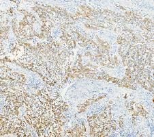
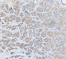
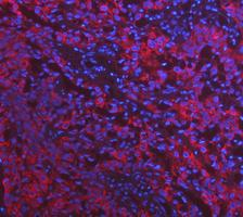


Comprehensive Human Protein Atlas IHC scoring per tissue (reliability: Enhanced — Medium consistency between antibody staining and RNA expression data. Antibody staining in cells/structures not annotated, view images.). Rows are taken directly from the HPA tissue chart — click any row's HPA link to view the source.
| Tissue | Cell type | Level | Evidence | Source |
|---|---|---|---|---|
| Breast | Myoepithelial cells | High | Protein (IHC) | HPA → |
| Prostate | Glandular cells | High | Protein (IHC) | HPA → |
| Salivary gland | Glandular cells | High | Protein (IHC) | HPA → |
| Skin | Sebaceous glands | High | Protein (IHC) | HPA → |
| Urinary bladder | Urothelial cells | High | Protein (IHC) | HPA → |
| Tissue | Cell type | Level | Evidence | Source |
|---|---|---|---|---|
| Adipose tissue | Adipocytes | Not detected | Protein (IHC) | HPA → |
| Adrenal gland | Glandular cells | Not detected | Protein (IHC) | HPA → |
| Appendix | Glandular cells | Not detected | Protein (IHC) | HPA → |
| Bone marrow | Hematopoietic cells | Not detected | Protein (IHC) | HPA → |
| Caudate | Glial cells | Not detected | Protein (IHC) | HPA → |
Troubleshoot KRT17 staining in paraffin sections by checking retrieval, tissue context, cytoplasmic pattern, and controls before interpreting chromogenic signal.
The IHC-validated antibody has paraffin-section images from human bladder and breast cancer tissue; IF images cover human bladder and lung cancer tissue and HeLa cells (catalog image captions).
The rendered card is M02289-1, with IHC images from paraffin-embedded human bladder and breast cancer sections (M02289-1 IHC captions). M02289-1 also has IF images from paraffin-embedded human bladder and lung cancer sections and an IF image of HeLa cells (M02289-1 IF captions).
Which to pick: For tissue IHC, pick M02289-1: it is a rabbit monoclonal with paraffin-section IHC images; the captions do not report the fixative (catalog; M02289-1 IHC captions). For IF/ICC, M02289-1 lists both applications and has tissue IF images plus a HeLa-cell IF image (catalog; M02289-1 IF captions). For human, mouse or rat samples, both M02289-1 and the rabbit polyclonal A02289-1 list reactivity, but only M02289-1 has supplied IHC and IF images (catalog).