This website uses cookies to ensure you get the best experience on our website.


- Table of Contents
This KRT6A IHC-P guide covers the cytoplasmic staining expected in squamous and respiratory epithelia (HPA tissue IHC). Use cervical squamous epithelium as a positive control (HPA tissue IHC), and account for staining that may reflect proteins from more than one gene (HPA tissue IHC).

Expected localisation, validated protocols, controls and antibodies — the at-a-glance facts below, then the full design guide.
| Expected localisation | Cytoplasm in squamous and respiratory epithelia (HPA tissue IHC) | |
| Staining pattern | Squamous and respiratory epithelial cells show cytoplasmic staining (HPA tissue IHC) | |
| Antigen retrieval | EDTA pH 8.0 HIER, heat-mediated (datasheet M30943-1) | |
| Positive control | Cervix+4 more · see all | |
| Negative control | Adipose tissue+4 more · see all |
| Fixation | Keep fixation consistent across sections (standard IHC practice; not target-specific) | |
| Caveat | Staining may reflect proteins from more than one gene (HPA tissue IHC) | |
| Regulation | Wound-edge induction (by similarity) (UniProt) | |
| Isoform / epitope | No annotated isoforms; chain starts at residue 2 (UniProt) |
Compare the catalog antibody’s IHC-P protocol (datasheet M30943-1) with the four published KRT6A IHC protocols listed below (cited PMCs).
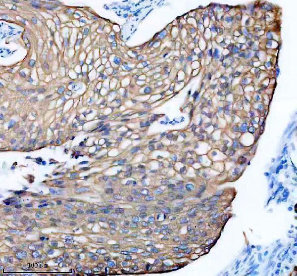
| Sample | Paraffin-embedded human cervical cancer tissue; fixative not specified (datasheet M30943-1) |
| Fixation | Image fixative and duration unreported (datasheet M30943-1); verify before use. |
| Sectioning | 4–5 µm sections on charged slides (standard) |
| Deparaffinisation | Xylene, graded ethanol series to water (standard) |
| Antigen retrieval | Heat retrieval: EDTA pH 8.0 (datasheet M30943-1); 20 min, 95–100 °C (standard) |
| Peroxidase block | 3% H2O2, 10 min, room temperature (standard) |
| Blocking | 10% goat serum (datasheet M30943-1) |
| Primary antibody | Rabbit monoclonal (clone GAO-11) anti-KRT6A, 1:50-1:200 (datasheet M30943-1) |
| Primary incubation | Overnight at 4 °C (datasheet M30943-1) |
| Detection | HRP-conjugated secondary, DAB chromogen (datasheet M30943-1) |
| Counterstain | Hematoxylin, blue, dehydrate and mount (standard) |
| Expected result | KRT6A-positive staining in squamous epithelial cells of cervix (HPA tissue IHC: High). HPA tissue profile: Distinct cytoplasmic expression in squamous and respiratory epithelia. No signal in the no-primary control. |
KRT6A is a cytoplasmic keratin with no transmembrane segment (UniProt P02538). In IHC, expect staining in squamous epithelial cells of cervix, esophagus, oral mucosa, tonsil and vagina, and respiratory epithelial cells of nasopharynx (HPA tissue IHC: High). HPA rates its tissue pattern Supported, while cautioning that the antibodies may detect proteins from more than one gene (HPA tissue IHC: reliability).
| Distinct cytoplasmic staining in squamous epithelium, with adjacent cell types comparatively unstained. | This matches the reported distribution in cervix, esophagus, oral mucosa, tonsil and vagina (HPA tissue IHC: High in squamous epithelial cells). Assess staining by cell type and compartment: KRT6A is cytoplasmic (UniProt P02538), and HPA describes a distinct epithelial pattern (HPA tissue IHC: profile). |
| Predominantly nuclear staining, with little epithelial cytoplasmic staining. | Nuclear dominance conflicts with the recorded cytoplasmic location (UniProt P02538) and intermediate filament localization seen by ICC-IF (HPA subcellular: intermediate filaments). Treat it as a suspect pattern; review controls, detection background and scoring before calling the section KRT6A positive (general IHC practice). |
| Strong staining in cells outside the expected epithelial population. | Staining in, for example, adipocytes or hematopoietic cells conflicts with HPA observations for those cells (HPA tissue IHC: Not detected in adipocytes and hematopoietic cells). Cross-reactivity or endogenous detection activity is possible (general IHC practice); the HPA multi-gene antibody caution makes target identity especially important (HPA tissue IHC: reliability). |
| Diffuse color across epithelium, stroma and blank areas, without clear cell boundaries. | A widespread haze does not reproduce HPA's distinct epithelial cytoplasmic pattern (HPA tissue IHC: profile). Inspect no-primary and detection controls, washes and blocking for nonspecific or endogenous signal (general IHC practice). Do not score intensity until the epithelial signal can be distinguished from background (general IHC practice). |
| No epithelial signal in a section chosen as a positive control. | First confirm that the sampled region contains the relevant cells: HPA reports High staining in oral mucosa squamous cells and nasopharyngeal respiratory cells (HPA tissue IHC). Then check the run controls and antibody detection; absence of signal alone cannot establish biological absence (general IHC practice). |
| Compartment and structure | KRT6A is cytoplasmic and forms keratin intermediate filament networks with type I keratins (UniProt P02538). HPA ICC-IF places the signal on intermediate filaments (HPA subcellular). In chromogenic IHC, evaluate cytoplasmic distribution; a filament-like pattern may be easier to resolve by IF microscopy (general microscopy practice). |
| Tissue and cell selection | HPA reports High staining in several squamous epithelia and nasopharyngeal respiratory cells, but Medium staining in bronchial respiratory cells (HPA tissue IHC). Choose a documented High site when assessing assay performance; compare the correct epithelial cells rather than assigning one intensity to the entire section (general IHC practice). |
| Antibody specificity | Two listed antibodies, HPA045697 and HPA061168, have IHC Supported status (HPA antibodies). HPA also cautions that its staining may reflect proteins from more than one gene (HPA tissue IHC: reliability). Agreement with the expected pattern supports interpretation, but these data alone do not prove that every stained cell contains KRT6A specifically. |
| Biological state | KRT6A is rapidly induced in wound-edge keratinocytes, according to a similarity-based UniProt annotation (UniProt P02538: function). A sampled repair region may therefore differ from resting epithelium; record whether the section contains an epithelial wound edge before comparing regions (UniProt P02538: function; general IHC practice). |
| IF/ICC: what should I expect? | Expect cytoplasmic intermediate filament localization (HPA subcellular: intermediate filaments; UniProt P02538: cytoplasm). HPA lists ICC-IF images and marks both listed antibodies ICC Approved (HPA subcellular; HPA antibodies). Use the separate IF/ICC guide for its workflow; these records supply no IF dilution or permeabilisation setting. |
| Fixation and retrieval evidence | Target-specific fixation effects are not established by the supplied assay evidence. Verify with a matched IHC source before attributing a result to fixation. |
| Situation | Likely cause | Next action |
|---|---|---|
| A known-positive epithelial region is blank. | The sampled area may lack the documented positive cells, or staining or detection may have failed (HPA tissue IHC: positive cell types; general IHC practice). | Confirm epithelial identity on the section, then inspect the positive run control and the chosen antibody's IHC-P instructions for retrieval, dilution and detection (general IHC practice). |
| Only nuclei appear positive. | Nuclear dominance conflicts with cytoplasmic KRT6A and HPA intermediate filament localization (UniProt P02538; HPA subcellular). | Check the no-primary control, counterstain and detection background; require interpretable epithelial cytoplasmic signal before scoring a positive result (general IHC practice). |
| Unrelated cells stain as strongly as the epithelium. | Cross-reactivity or endogenous detection activity may contribute (general IHC practice); HPA cautions that antibodies may target more than one gene (HPA tissue IHC: reliability). | Compare the suspect cells with HPA cell-level observations and review no-primary and detection controls. Report unresolved staining as uncertain rather than assigning it specifically to KRT6A (HPA tissue IHC; general IHC practice). |
| The whole section has diffuse chromogenic background. | Nonspecific binding, insufficient washing or endogenous detection activity can obscure a distinct epithelial pattern (general IHC practice; HPA tissue IHC: profile). | Review blocking, washes and detection controls under the assay's standard IHC workflow; reassess only where cytoplasmic epithelial signal separates clearly from background (general IHC practice). |
| Bronchial staining looks weaker than a squamous positive control. | HPA reports Medium staining in bronchial respiratory cells and High staining in several squamous epithelial populations (HPA tissue IHC). | Score bronchial respiratory cells against their documented level and local controls. Do not use equal intensity across the two tissues as the criterion for a successful run (HPA tissue IHC; general IHC practice). |
| A section differs from the HPA reference pattern. | Differences in sampled epithelial cells or a repair region can affect interpretation (HPA tissue IHC: cell-level pattern; UniProt P02538: wound-edge induction by similarity). | Document the tissue region and cell type, check run controls, and compare matching epithelial populations. If staining remains discordant, report the limitation and the HPA multi-gene antibody caution (general IHC practice; HPA tissue IHC: reliability). |


Comprehensive Human Protein Atlas IHC scoring per tissue (reliability: Supported — High consistency between antibody staining and RNA expression data. Caution, targets protein from more than one gene.). Rows are taken directly from the HPA tissue chart — click any row's HPA link to view the source.
| Tissue | Cell type | Level | Evidence | Source |
|---|---|---|---|---|
| Cervix | Squamous epithelial cells | High | Protein (IHC) | HPA → |
| Esophagus | Squamous epithelial cells | High | Protein (IHC) | HPA → |
| Nasopharynx | Respiratory epithelial cells | High | Protein (IHC) | HPA → |
| Oral mucosa | Squamous epithelial cells | High | Protein (IHC) | HPA → |
| Skin | Cells in corneal layer | High | Protein (IHC) | HPA → |
| Tissue | Cell type | Level | Evidence | Source |
|---|---|---|---|---|
| Adipose tissue | Adipocytes | Not detected | Protein (IHC) | HPA → |
| Adrenal gland | Glandular cells | Not detected | Protein (IHC) | HPA → |
| Appendix | Glandular cells | Not detected | Protein (IHC) | HPA → |
| Bone marrow | Hematopoietic cells | Not detected | Protein (IHC) | HPA → |
| Breast | Adipocytes | Not detected | Protein (IHC) | HPA → |
Use the catalog antibody’s paraffin-section IHC conditions as the starting point, then judge staining against KRT6A’s expected epithelial and cytoplasmic pattern.
Human and rat IHC/IF reactivity is cataloged (catalog: applications/reactivity); real IHC data show M30943-1 on a paraffin-embedded human cervical cancer section (M30943-1 image caption).
M30943-1 is listed for human IHC and IF/ICC (catalog: applications/reactivity), with an IHC image from paraffin-embedded human cervical cancer tissue (M30943-1 image caption). A04012-2 is listed for human and rat IHC and IF/ICC (catalog: applications/reactivity); no IHC or IF image is supplied for it (catalog: image alts).
Which to pick: For human tissue IHC, choose M30943-1 for its paraffin-section IHC image (M30943-1 image caption); its fixative is unreported (M30943-1 image caption). For human IF/ICC, M30943-1 is a monoclonal option listed for both applications (catalog: clone/applications), although no IF image is supplied (catalog: image alts). For rat IHC or IF/ICC, choose A04012-2 based on its listed rat reactivity and applications (catalog: reactivity/applications); it is polyclonal (catalog: dilution_raw).