This website uses cookies to ensure you get the best experience on our website.


- Table of Contents
Plan chromogenic IHC for LAMA4 using its basement membrane and vascular cell staining profile (HPA tissue IHC). This guide covers fixation, controls and interpretation for a secreted extracellular matrix protein (UniProt).

Expected localisation, validated protocols, controls and antibodies — the at-a-glance facts below, then the full design guide.
| Expected localisation | Basement membranes; endothelial and smooth muscle cells (HPA tissue IHC) | |
| Staining pattern | Basement membrane staining with endothelial and smooth muscle staining (HPA tissue IHC) | |
| Antigen retrieval | Citrate pH 6.0 HIER, 95–98 °C, 20 min (rule: cytoplasmic / membrane antigen) | |
| Positive control | Smooth muscle+1 more · see all | |
| Negative control | Adrenal gland+4 more · see all |
| Fixation | Keep formalin fixation consistent across sections. (standard IHC practice; not target-specific) | |
| Caveat | Secreted protein may stain away from RNA-positive cells (HPA tissue IHC) | |
| Regulation | Expression varies between adult and fetal tissues (UniProt) | |
| Isoform / epitope | Three isoforms; signal peptide 1–24 is removed, so map the epitope (UniProt) |
The catalog antibody’s IHC-P protocol is paired with published chromogenic IHC methods for canine hemangiosarcoma and human tissue arrays (PMC7310061; PMC4875769).
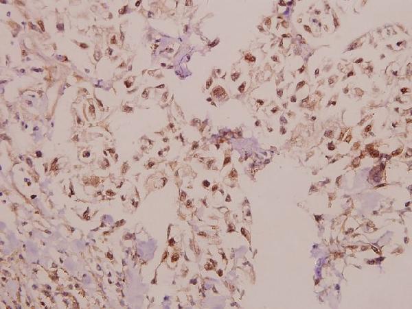
| Sample | Paraffin-embedded human colorectal carcinoma tissue; fixative not specified (datasheet A04999-1) |
| Fixation | Image fixative and duration unreported (datasheet A04999-1); verify before use. |
| Sectioning | 4–5 µm sections on charged slides (standard) |
| Deparaffinisation | Xylene, graded ethanol series to water (standard) |
| Antigen retrieval | Heat-induced epitope retrieval in citrate buffer, pH 6.0, 20 min at 95–98 °C (standard rule: cytoplasmic / membrane antigen) |
| Peroxidase block | 3% H2O2, 10 min, room temperature (standard) |
| Blocking | 10% normal serum of the secondary host, 30 min, room temperature (standard) |
| Primary antibody | Rabbit anti-LAMA4, 1:50-1:200 (datasheet A04999-1) |
| Primary incubation | Overnight at 4 °C (standard) |
| Detection | HRP-polymer secondary, DAB chromogen 5–10 min (standard) |
| Counterstain | Hematoxylin, blue, dehydrate and mount (standard) |
| Expected result | LAMA4-positive staining in smooth muscle cells of smooth muscle (HPA tissue IHC: High). HPA tissue profile: Distinct expression in endothelial cells, smooth muscle cells, basement membranes and decidual cells. No signal in the no-primary control. |
LAMA4 is a secreted basement membrane and extracellular matrix protein with no transmembrane segment (UniProt Q16363 topology). In paraffin section IHC, expect staining along basement membranes and in regions associated with endothelial and smooth muscle cells; decidual cells may also stain (HPA tissue IHC). HPA rates tissue IHC reliability as Enhanced while noting medium consistency with RNA data, partly because a secreted protein can appear away from its site of production (HPA tissue IHC).
| Basement membrane outlines stain near vessels or smooth muscle, with little nuclear signal. | This fits LAMA4's extracellular basement membrane location (UniProt Q16363 subcellular location) and HPA's distinct staining in basement membranes, endothelial cells and smooth muscle cells (HPA tissue IHC). Judge the pattern by its location as well as its intensity. |
| Smooth muscle cells stain strongly; decidual cells show a weaker positive pattern. | These are useful reference patterns: HPA reports High staining in smooth muscle cells and Medium staining in placental decidual cells (HPA tissue IHC). A weaker decidual result can still be consistent with the reference; assess it alongside the expected basement membrane distribution. |
| Signal is predominantly nuclear or confined to an unexpected compartment. | A predominantly nuclear IHC pattern conflicts with the secreted extracellular location and lack of a transmembrane segment (UniProt Q16363 topology). Treat it as suspect and review controls and detection background (general IHC practice). HPA reports plasma membrane and cytosol in ICC-IF, which is a separate application (HPA subcellular). |
| Strong staining appears in an unexpected cell population, such as adrenal glandular cells. | HPA reports adrenal glandular cells as Not detected (HPA tissue IHC). Check whether the signal follows a basement membrane rather than the cells themselves (UniProt Q16363 subcellular location). If it is truly cellular, assess antibody cross-reactivity and endogenous detection activity with appropriate controls (general IHC practice). |
| Diffuse haze obscures tissue boundaries, or a reference positive section has no specific signal. | Haze prevents a reliable location call; absent staining in smooth muscle cells also conflicts with an HPA High reference (HPA tissue IHC). Compare positive and negative controls, then inspect background, primary antibody dilution, retrieval and detection performance (general IHC practice). |
| Extracellular location | LAMA4 is secreted and is a major basement membrane component, without a transmembrane segment (UniProt Q16363 subcellular location and topology). Interpret staining in relation to tissue boundaries; an outline around cells need not mean that the outlined cells produced the protein (HPA tissue IHC reliability note). |
| Tissue reference and reliability | HPA reports High staining in smooth muscle cells, Medium staining in placental decidual cells and Not detected in several specified cell populations (HPA tissue IHC). Its Enhanced reliability carries a medium RNA–staining consistency caveat because secreted protein and RNA locations can differ (HPA tissue IHC). |
| Antibody validation | HPA lists HPA015693 and CAB078156 as IHC Enhanced (HPA antibodies). This supports comparison against HPA's tissue pattern, but it does not establish the performance of a different catalog antibody, a particular retrieval setting or fixation sensitivity (HPA antibodies; general IHC practice). |
| Processing and variants | UniProt lists a signal peptide at residues 1–24, a mature chain at 25–1823 and three isoforms (UniProt Q16363 processing and isoforms). If the catalog antibody's epitope is known, check its position against these annotations; epitope coverage is unknown from the supplied record. |
| IF/ICC question: Should its cell signal define the IHC result? | No. HPA reports approved plasma membrane and cytosol locations in ICC-IF images from A-431, U-251MG and U2OS (HPA subcellular). For paraffin section IHC, use the basement membrane and cell distribution reported in HPA tissue IHC; the applications need separate interpretation. |
| Situation | Likely cause | Next action |
|---|---|---|
| No signal in a smooth muscle reference section. | HPA reports High smooth muscle cell staining, so a blank result needs a technical check before interpretation (HPA tissue IHC). | Verify the positive control, primary antibody use, retrieval and chromogenic detection steps; adjust one general IHC variable at a time (general IHC practice). No LAMA4-specific retrieval condition is supplied. |
| Placental decidual staining is faint. | HPA reports Medium staining in decidual cells, below its High rating for smooth muscle cells (HPA tissue IHC). | Compare with a smooth muscle positive reference and check whether faint signal has the expected location before changing conditions (HPA tissue IHC; general IHC practice). |
| Whole-section brown haze hides basement membrane outlines. | Non-specific background or endogenous detection activity can obscure a localized chromogenic signal (general IHC practice). | Inspect a no-primary control, blocking and detection steps, then optimize primary antibody dilution against a positive reference (general IHC practice). |
| Strong glandular cell signal appears in adrenal gland or appendix. | HPA reports those glandular cell populations as Not detected; cross-reactivity or endogenous detection activity is possible (HPA tissue IHC; general IHC practice). | Check a no-primary control and the exact cellular location. Compare the pattern with an independently validated IHC antibody where available (general IHC practice; HPA antibodies). |
| Most of the apparent signal is nuclear. | That compartment conflicts with UniProt's extracellular basement membrane annotation (UniProt Q16363 subcellular location). | Check the no-primary control, counterstain and chromogenic detection background before assigning LAMA4 positivity (general IHC practice). |
| IHC and ICC-IF appear to show different locations. | HPA reports basement membrane and tissue-associated IHC staining, but approved plasma membrane and cytosol locations in ICC-IF (HPA tissue IHC; HPA subcellular). | Interpret each result within its application and controls. Do not use the ICC-IF location alone to score paraffin section IHC (HPA tissue IHC; HPA subcellular). |


Comprehensive Human Protein Atlas IHC scoring per tissue (reliability: Enhanced — Medium consistency between antibody staining and RNA expression data. At least one protein variant secreted, tissue location of RNA and protein might differ and correlation is complex. Antibody staining in cells/structures not annotated, view images.). Rows are taken directly from the HPA tissue chart — click any row's HPA link to view the source.
| Tissue | Cell type | Level | Evidence | Source |
|---|---|---|---|---|
| Smooth muscle | Smooth muscle cells | High | Protein (IHC) | HPA → |
| Placenta | Decidual cells | Medium | Protein (IHC) | HPA → |
| Tissue | Cell type | Level | Evidence | Source |
|---|---|---|---|---|
| Adrenal gland | Glandular cells | Not detected | Protein (IHC) | HPA → |
| Appendix | Glandular cells | Not detected | Protein (IHC) | HPA → |
| Bone marrow | Hematopoietic cells | Not detected | Protein (IHC) | HPA → |
| Breast | Adipocytes | Not detected | Protein (IHC) | HPA → |
| Bronchus | Respiratory epithelial cells | Not detected | Protein (IHC) | HPA → |
Troubleshoot LAMA4 chromogenic IHC by assessing basement membrane staining alongside cell type, tissue architecture, and controls (UniProt Q16363 localisation; HPA tissue IHC).
A04999-1 has IHC data from a paraffin-embedded human colorectal carcinoma section (IHC image caption); IF is a listed application, with Human, Mouse and Rat reactivity (catalog applications and reactivity).
A04999-1 was shown in IHC on paraffin-embedded human colorectal carcinoma tissue at 1:50 (IHC image caption). IF is listed without an IF figure, and Human, Mouse and Rat are listed as reactive species (catalog applications and reactivity; catalog IF images).
Which to pick: For paraffin-section IHC, choose A04999-1: its polyclonal antibody has an IHC image from paraffin-embedded human colorectal carcinoma tissue (IHC image caption). For IF/ICC, A04999-1 lists IF, but provides no IF figure or separate ICC validation (catalog applications and IF images). For cross-species work, A04999-1 lists Human, Mouse and Rat reactivity; the pictured IHC sample is human, and its fixative is unreported (catalog reactivity; IHC image caption).