This website uses cookies to ensure you get the best experience on our website.


- Table of Contents
Plan chromogenic LAP3 IHC in paraffin sections using the catalog antibody’s documented tissue workflow (datasheet A01921-1). Compare cytoplasmic staining with high expression in hepatocytes and kidney proximal tubules (HPA tissue IHC).

Expected localisation, validated protocols, controls and antibodies — the at-a-glance facts below, then the full design guide.
| Expected localisation | Cytoplasm in most tissues; brain and muscle are exceptions (HPA tissue IHC) | |
| Staining pattern | Cytoplasmic in many cells; high in hepatocytes and renal tubules (HPA tissue IHC) | |
| Antigen retrieval | EDTA pH 8.0 HIER, heat-mediated (datasheet A01921-1) | |
| Positive control | Kidney+4 more · see all | |
| Negative control | Caudate+4 more · see all |
| Fixation | Keep fixation consistent across sections (standard IHC practice; not target-specific) | |
| Caveat | Staining has medium consistency with RNA expression (HPA tissue IHC) | |
| Regulation | Staining regulation is unreported (UniProt) | |
| Isoform / epitope | 2 isoforms; epitope coverage is unspecified (UniProt) |
The catalog antibody’s IHC-P protocol (datasheet: A01921-1) is accompanied by three published LAP3 IHC protocols (PMC5790517; PMC10882082; PMC9977676).
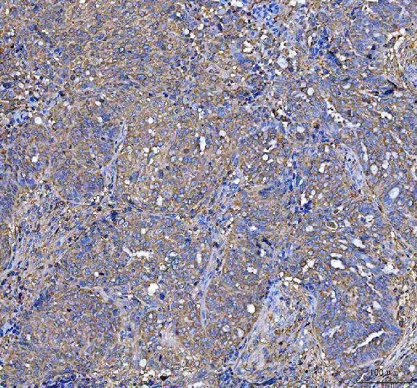
| Sample | Paraffin-embedded human ovarian serous adenocarcinoma tissue; fixative not specified (datasheet A01921-1) |
| Fixation | Image fixative and duration unreported (datasheet A01921-1); verify before use. |
| Sectioning | 4–5 µm sections on charged slides (standard) |
| Deparaffinisation | Xylene, graded ethanol series to water (standard) |
| Antigen retrieval | Heat retrieval: EDTA pH 8.0 (datasheet A01921-1); 20 min, 95–100 °C (standard) |
| Peroxidase block | 3% H2O2, 10 min, room temperature (standard) |
| Blocking | 10% goat serum (datasheet A01921-1) |
| Primary antibody | Rabbit anti-LAP3, 2-5 μg/ml (datasheet A01921-1) |
| Primary incubation | Overnight at 4 °C (datasheet A01921-1) |
| Detection | HRP-conjugated secondary, DAB chromogen (datasheet A01921-1) |
| Counterstain | Hematoxylin, blue, dehydrate and mount (standard) |
| Expected result | LAP3-positive staining in proximal tubules (microvilli) of kidney (HPA tissue IHC: High). HPA tissue profile: Cytoplasmic expression in essentially all tissues, except for brain and muscle tissue. No signal in the no-primary control. |
LAP3 is a cytoplasmic protein with no transmembrane segment (UniProt P28838: subcellular location and topology). In paraffin-section IHC, expect cytoplasmic staining across many tissues, with high staining reported in kidney proximal-tubule microvilli, liver hepatocytes, lung endothelial cells, stomach glandular cells and late spermatids (HPA: tissue IHC). HPA rates the tissue profile Enhanced and describes medium consistency between antibody staining and RNA expression (HPA: reliability description).
| Cytoplasmic staining in hepatocytes or stomach glandular cells; signal may vary between cells (HPA: High in both cell types). | This fits the reported IHC distribution and UniProt location (HPA: tissue IHC; UniProt P28838: Cytoplasm). Judge localisation alongside the cell identity; staining intensity alone does not establish specificity (general IHC practice). |
| Strong nuclear or sharply membrane-confined signal dominates the section. | That compartment conflicts with the reported cytoplasmic pattern and lack of a transmembrane segment (HPA: tissue IHC; UniProt P28838: topology). Treat it as suspect and check controls and detection conditions (general IHC practice). |
| Strong staining is confined to cardiomyocytes, skeletal myocytes or glial cells in sampled brain regions. | Those cells are reported as Not detected in the corresponding HPA sections (HPA: heart muscle, skeletal muscle, caudate and hippocampus). Consider antibody cross-reactivity or endogenous detection activity; confirm cell identity and controls (general IHC practice). |
| Weak, diffuse color covers tissue, stroma and blank areas without a clear cellular pattern. | This is difficult to score as LAP3 because the expected pattern is cellular and mainly cytoplasmic (HPA: tissue IHC; UniProt P28838: Cytoplasm). Nonspecific binding or detection background are possible (general IHC practice). |
| No staining appears in an otherwise interpretable liver or kidney section. | HPA reports High staining in hepatocytes and proximal-tubule microvilli (HPA: liver and kidney). Review tissue preservation, retrieval, primary-antibody application and detection controls before calling the sample negative (general IHC practice). |
| Cell type and tissue | LAP3 staining is broad but uneven: HPA reports High in hepatocytes, lung endothelial cells and stomach glandular cells, Medium in adrenal, appendix and breast glandular cells, and Not detected in several brain and muscle cell populations (HPA: tissue IHC). Compare like cell types when scoring. |
| Subcellular location | UniProt places LAP3 in the cytoplasm and reports no transmembrane segment or signal peptide (UniProt P28838: location and topology). HPA tissue IHC is cytoplasmic overall, while kidney proximal-tubule microvilli are specifically reported High (HPA: tissue IHC). |
| Antibody validation | Both listed antibodies have Enhanced IHC status (HPA: HPA029606 and HPA029607). HPA describes Enhanced IHC as a pattern supported by independent antibodies or orthogonal data (HPA: antibody-validation summary); it does not make every stain in every specimen specific. |
| Isoforms and processing | UniProt lists two isoforms and a chain spanning residues 1–519, with no annotated signal peptide or propeptide (UniProt P28838: isoforms and processing). The supplied HPA IHC profile does not assign its tissue pattern to either isoform (HPA: tissue IHC). |
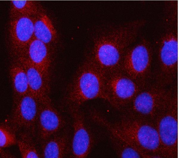
| IF/ICC cross-check | Q: What localisation should IF/ICC show? A: Mainly cytosol; HPA also approves calyx, mid piece and principal piece localisation in its subcellular record (HPA: ICC-IF). This supports a compartment check, while the IHC tissue profile governs paraffin-section interpretation (HPA: tissue IHC). |
| Retrieval and detection conditions | Antigen retrieval and detection conditions can alter IHC signal and background in general (general IHC practice). Target-specific fixation sensitivity or an optimal retrieval condition is unreported in the supplied UniProt and HPA records; avoid inferring either from staining levels. |
| Situation | Likely cause | Next action |
|---|---|---|
| No signal in liver hepatocytes or kidney proximal tubules. | A technical failure is possible because both are reported High (HPA: liver and kidney); the supplied records do not identify a LAP3-specific fixation effect. | Check section quality, retrieval, primary-antibody application and detection using appropriate controls (general IHC practice). Reassess the expected cells before scoring a negative result (HPA: tissue IHC). |
| Dominant nuclear staining accompanies little cytoplasmic signal. | The pattern conflicts with the reported cytoplasmic localisation (UniProt P28838: Cytoplasm; HPA: tissue IHC). Nonspecific antibody or detection signal is possible (general IHC practice). | Compare a positive tissue and a primary-omission control, then review antibody dilution and detection conditions (general IHC practice). Interpret only a reproducible, cell-appropriate pattern. |
| Strong signal appears in cardiomyocytes or skeletal myocytes. | HPA reports Not detected in those cells (HPA: heart muscle and skeletal muscle). Cross-reactivity or endogenous detection activity could explain discordant color (general IHC practice). | Verify cell identity and examine a primary-omission control; address endogenous detection activity if that control is colored (general IHC practice). Compare with an HPA-supported positive tissue. |
| Color is diffuse across tissue and blank slide areas. | A noncellular pattern does not match cytoplasmic LAP3 staining (HPA: tissue IHC; UniProt P28838: Cytoplasm). Inadequate blocking, overly strong primary staining or detection background may contribute (general IHC practice). | Inspect primary-omission and reagent controls; optimize blocking, washing, antibody dilution and detection exposure as appropriate (general IHC practice). Score cells only after background is controlled. |
| Kidney staining looks apical rather than evenly cytoplasmic. | High proximal-tubule microvillar staining is specifically reported (HPA: kidney); apical signal in those cells need not indicate a membrane-spanning LAP3 protein (UniProt P28838: no transmembrane segment). | Confirm proximal-tubule morphology and compare adjacent cell types before rejecting the pattern (HPA: kidney; general IHC practice). Assess whether staining remains associated with cells. |
| Weak staining in squamous epithelium seems inconsistent with a strong liver control. | HPA lists squamous epithelial cells in oral mucosa, esophagus and vagina as Low, versus High hepatocyte staining (HPA: tissue IHC). This difference can be expected. | Score each tissue against its reported cell-specific level and verify the liver control and assay background (HPA: tissue IHC; general IHC practice). Avoid imposing one intensity threshold across tissues. |
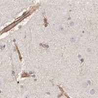


Comprehensive Human Protein Atlas IHC scoring per tissue (reliability: Enhanced — Medium consistency between antibody staining and RNA expression data.). Rows are taken directly from the HPA tissue chart — click any row's HPA link to view the source.
| Tissue | Cell type | Level | Evidence | Source |
|---|---|---|---|---|
| Kidney | Proximal tubules (microvilli) | High | Protein (IHC) | HPA → |
| Liver | Hepatocytes | High | Protein (IHC) | HPA → |
| Lung | Endothelial cells | High | Protein (IHC) | HPA → |
| Stomach | Glandular cells | High | Protein (IHC) | HPA → |
| Testis | Elongated or late spermatids | High | Protein (IHC) | HPA → |
| Tissue | Cell type | Level | Evidence | Source |
|---|---|---|---|---|
| Caudate | Glial cells | Not detected | Protein (IHC) | HPA → |
| Cerebellum | Cells in granular layer | Not detected | Protein (IHC) | HPA → |
| Heart muscle | Cardiomyocytes | Not detected | Protein (IHC) | HPA → |
| Hippocampus | Glial cells | Not detected | Protein (IHC) | HPA → |
| Skeletal muscle | Myocytes | Not detected | Protein (IHC) | HPA → |
Troubleshoot LAP3 chromogenic IHC by checking retrieval, cytoplasmic localisation, controls and cell-level scoring; IF considerations are covered separately.
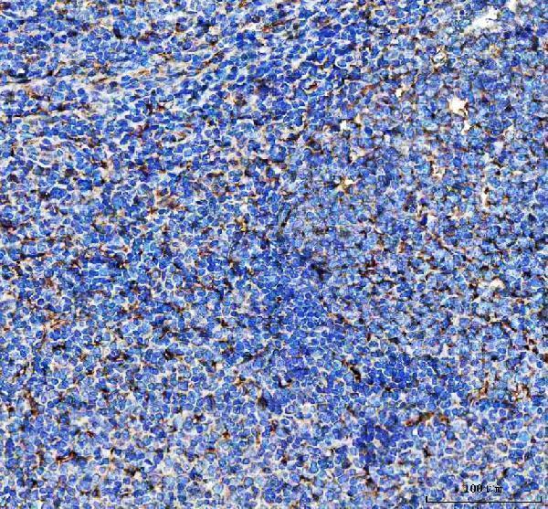
A01921-1 has pictured IHC in human ovarian serous adenocarcinoma and tonsil paraffin sections, plus IF/ICC in U2OS cells (A01921-1 image captions).
A01921-1 will render with pictured human paraffin-section IHC; its separate IF/ICC image shows U2OS cells (A01921-1 image captions). M01921 will render as a human-reactive IHC antibody without a supplied IHC image (catalog: M01921 applications, reactivity, image list).
Which to pick: For tissue IHC, choose A01921-1 when pictured human paraffin-section evidence matters: its captions report EDTA retrieval at pH 8.0 and 2 μg/ml primary antibody, but do not report the fixative (A01921-1 IHC captions). M01921 is a human-reactive rabbit monoclonal listed for IHC, with no supplied IHC image (catalog: M01921 host, clone, applications, reactivity, image list). For IF/ICC or a mouse or rat reactive antibody, choose A01921-1; its IF/ICC image is in U2OS cells, while its pictured tissue IHC is human only (catalog: A01921-1 applications and reactivity; A01921-1 image captions).