This website uses cookies to ensure you get the best experience on our website.


- Table of Contents
Plan LARP1 staining in paraffin sections using the reported cytoplasmic tissue pattern (HPA tissue IHC). This guide pairs tissue controls with the catalog antibody’s chromogenic IHC workflow (HPA tissue IHC; datasheet A04488-2).

Expected localisation, validated protocols, controls and antibodies — the at-a-glance facts below, then the full design guide.
| Expected localisation | General cytoplasmic staining (HPA tissue IHC) | |
| Staining pattern | Cytoplasmic signal in glandular and neuronal cells (HPA tissue IHC) | |
| Antigen retrieval | EDTA pH 8.0 HIER, heat-mediated (datasheet A04488-2) | |
| Positive control | Appendix+4 more · see all | |
| Negative control | Adipose tissue+2 more · see all |
| Fixation | Keep fixation consistent across sections (standard IHC practice; not target-specific); Matched tissue-IHC evidence does not establish the fixation claim. Validate the specimen-specific method before use. (selected-SKU IHC image A04488-2) | |
| Caveat | Staining and RNA expression have medium consistency (HPA tissue IHC) | |
| Regulation | Nutrients regulate LARP1 activity (UniProt) | |
| Isoform / epitope | Two isoforms; antibody epitope coverage is unknown (UniProt; datasheet A04488-2) |
The catalog antibody’s IHC-P protocol (datasheet A04488-2) is paired with published LARP1 IHC protocols for colorectal cancer (PMC5126195) and clear cell renal cell carcinoma (PMC7933513).
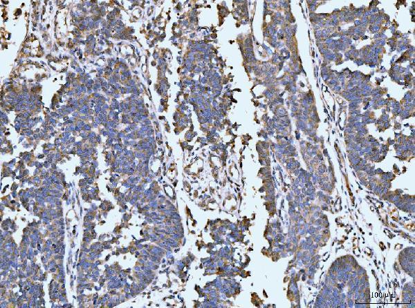
| Sample | Paraffin-embedded human rectal cancer tissue; fixative not specified (datasheet A04488-2) |
| Fixation | Image fixative and duration unreported (datasheet A04488-2); verify before use. |
| Sectioning | 4–5 µm sections on charged slides (standard) |
| Deparaffinisation | Xylene, graded ethanol series to water (standard) |
| Antigen retrieval | Heat retrieval: EDTA pH 8.0 (datasheet A04488-2); 20 min, 95–100 °C (standard) |
| Peroxidase block | 3% H2O2, 10 min, room temperature (standard) |
| Blocking | 10% goat serum (datasheet A04488-2) |
| Primary antibody | Rabbit anti-LARP1, 2-5 μg/ml (datasheet A04488-2) |
| Primary incubation | Overnight at 4 °C (datasheet A04488-2) |
| Detection | HRP-conjugated secondary, DAB chromogen (datasheet A04488-2) |
| Counterstain | Hematoxylin, blue, dehydrate and mount (standard) |
| Expected result | LARP1-positive staining in glandular cells of appendix (HPA tissue IHC: High). HPA tissue profile: General cytoplasmic expression. No signal in the no-primary control. |
LARP1 is a cytoplasmic protein with no transmembrane segment; it can occur in cytoplasmic granules (UniProt Q6PKG0). In paraffin-section IHC, expect predominantly cytoplasmic staining in cells such as colonic glandular cells and cerebellar Purkinje cells (HPA: High in both). HPA describes general cytoplasmic expression, but rates its tissue IHC evidence Approved with medium staining–RNA consistency and pending external verification (HPA tissue IHC).
| Cytoplasmic chromogen in colonic or duodenal glandular cells, with little signal in adjacent unstained structures. | This fits the reported IHC compartment and two High cell populations (HPA: general cytoplasmic expression; High in colonic and duodenal glandular cells). Assess the named cells, rather than assigning one intensity to the entire tissue. |
| Predominantly nuclear staining in a paraffin section, without a convincing cytoplasmic component. | Recheck interpretation and staining controls: tissue IHC is described as generally cytoplasmic (HPA tissue IHC). Nuclear signal alone does not establish the expected IHC pattern; HPA separately reports nucleoplasmic localization in ICC-IF (HPA subcellular). |
| Strong chromogen in adipocytes, skeletal myocytes, or cervical glandular cells. | These cells are reported Not detected in HPA tissue IHC (HPA: adipocytes, skeletal myocytes, cervical glandular cells). Consider antibody cross-reactivity or endogenous detection activity (general IHC practice); the HPA result alone cannot identify which caused the signal. |
| Diffuse chromogen across cells and surrounding tissue, obscuring cell boundaries. | An even haze is difficult to reconcile with a cell-resolved cytoplasmic pattern (HPA: general cytoplasmic expression). Inspect the negative and detection controls for nonspecific binding, incomplete blocking, or detection background (general IHC practice). |
| No staining in colonic glandular cells or cerebellar Purkinje cells. | Both are reported High, so a blank result warrants a technical review (HPA: High in colonic glandular and Purkinje cells). Check section integrity, the antibody and detection controls, and retrieval conditions (general IHC practice); HPA does not establish LARP1-specific retrieval sensitivity. |
| Which compartment should guide IHC scoring? | Use the cytoplasm as the primary tissue-IHC reference (HPA: general cytoplasmic expression; UniProt Q6PKG0: cytoplasm). UniProt also describes granules resembling stress granules, but neither source requires puncta in every paraffin section (UniProt Q6PKG0). |
| How strong is the tissue-pattern evidence? | HPA calls tissue IHC Approved while reporting medium staining–RNA consistency and pending external verification (HPA tissue IHC). Two listed antibodies have Approved IHC status; this is not an Enhanced validation claim (HPA: HPA051397, HPA054819). |
| Do isoforms or modifications define a different stain? | UniProt lists 2 isoforms and multiple modified residues (UniProt Q6PKG0). Without an antibody epitope or isoform-specific validation in the supplied evidence, these facts cannot assign a stain to one isoform or explain loss of signal. |
| Does antigen retrieval have a LARP1-specific predicted effect? | Retrieval is a standard variable to optimize in paraffin-section IHC (general IHC practice). Neither HPA nor UniProt supplies a LARP1-specific retrieval or fixation effect, so changes in signal cannot be predicted from the tissue levels or protein topology. |
| Should IF/ICC show the same compartment as tissue IHC? | IF/ICC has its own guide. HPA reports nucleoplasm and endoplasmic reticulum as Approved ICC-IF locations and cytosol as Supported (HPA subcellular); those observations should not be substituted for the general cytoplasmic tissue-IHC profile (HPA tissue IHC). |
| Situation | Likely cause | Next action |
|---|---|---|
| Known High cells are blank while the counterstain is visible. | Possible failure of antibody binding or chromogenic detection (general IHC practice); the HPA High designation does not establish a fixation cause (HPA tissue IHC). | Check a documented positive section and detection control, then review antibody use and retrieval conditions (general IHC practice). Judge the result in the named glandular or Purkinje cells (HPA: High). |
| Only nuclei appear positive in tissue IHC. | Compartment assignment or nonspecific staining may be involved (general IHC practice). Tissue IHC is generally cytoplasmic, although ICC-IF also reports nucleoplasm (HPA tissue IHC; HPA subcellular). | Compare nuclear and cytoplasmic boundaries under the counterstain and inspect controls (general IHC practice). Record the discrepancy instead of treating the ICC-IF location as tissue-IHC confirmation. |
| Adipocytes or skeletal myocytes stain strongly. | Unexpected signal in cells reported Not detected may reflect cross-reactivity or endogenous chromogenic activity (HPA tissue IHC; general IHC practice). | Review the negative and detection-only controls and cell identity; adjust blocking or detection conditions if controls show background (general IHC practice). |
| A uniform brown haze prevents cytoplasmic scoring. | Nonspecific background or excess chromogen can obscure the cell-resolved pattern (general IHC practice; HPA: general cytoplasmic expression). | Inspect the negative control and detection timing, then optimize blocking, antibody concentration, and washes (general IHC practice). Score only after cell boundaries can be resolved. |
| Staining differs between High tissues or within a section. | HPA assigns intensity to named cell populations, and its tissue IHC has medium staining–RNA consistency (HPA tissue IHC). Tissue-wide averages may conceal which cells contribute signal. | Score the specified cell population and cytoplasmic compartment separately in each section (HPA tissue IHC; general IHC practice). Document deviations without assigning an unsupported LARP1-specific fixation effect. |


Comprehensive Human Protein Atlas IHC scoring per tissue (reliability: Approved — Medium consistency between antibody staining and RNA expression data. Pending external verification.). Rows are taken directly from the HPA tissue chart — click any row's HPA link to view the source.
| Tissue | Cell type | Level | Evidence | Source |
|---|---|---|---|---|
| Appendix | Glandular cells | High | Protein (IHC) | HPA → |
| Cerebellum | Purkinje cells | High | Protein (IHC) | HPA → |
| Cerebral cortex | Neuronal cells | High | Protein (IHC) | HPA → |
| Colon | Glandular cells | High | Protein (IHC) | HPA → |
| Duodenum | Glandular cells | High | Protein (IHC) | HPA → |
Troubleshoot LARP1 staining in paraffin sections by checking retrieval, controls, cell compartment, and scoring before interpreting changes in signal.
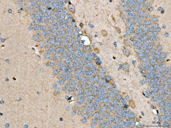
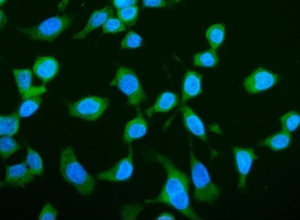
A04488-2 has IHC images from paraffin sections of human rectal cancer and mouse brain, plus an ICC/IF image from CACO-2 cells (A04488-2 image captions).
A04488-2 was shown in IHC on paraffin sections of human rectal cancer and mouse brain (A04488-2 IHC captions). A04488-2 was also shown in ICC/IF on CACO-2 cells (A04488-2 IF caption).
Which to pick: For tissue IHC, choose A04488-2: its own images show paraffin sections with EDTA retrieval at pH 8.0; the fixative is unreported (A04488-2 IHC captions). For IF/ICC, choose A04488-2 based on its CACO-2 image and listed IF/ICC applications (A04488-2 IF caption; catalog applications). It is also the listed human/mouse option (catalog reactivity: Human, Mouse); clonality is unreported.