LATS1 / Serine/threonine-protein kinase LATS1 · IHC design guide

Design Immunohistochemistry for LATS1

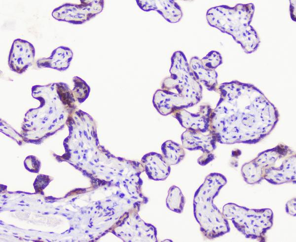
Plan LATS1 paraffin IHC around mainly cytoplasmic staining and a placenta positive control (HPA tissue IHC). The catalog antibody A01051-2 is specified at 0.5–1 μg/mL for IHC-P, with 1 μg/mL used in its placenta example (datasheet A01051-2).

Evidence assembled Oct 2026 · For research use; verify linked source records and product datasheet before use
Immunohistochemistry protocol sheet for LATS1 (IHC for LATS1): expected localisation Cytoplasmic staining (HPA tissue IHC); mitotic localization is molecular (UniProt), antibody A01051-2, validated IHC image, and IHC protocol steps
Printable LATS1 IHC protocol sheet — expected localisation Cytoplasmic staining (HPA tissue IHC); mitotic localization is molecular (UniProt), antibody A01051-2, controls and protocol steps. Open the full LATS1 IHC guide →

LATS1 Immunohistochemistry Experimental Design Guide

Expected localisation, validated protocols, controls and antibodies — the at-a-glance facts below, then the full design guide.

Must know before staining
Expected localisation Cytoplasmic staining (HPA tissue IHC); mitotic localization is molecular (UniProt)
Staining pattern Mainly cytoplasmic in most tissues; high in trophoblasts (HPA tissue IHC)
Antigen retrieval Citrate pH 6 HIER, heat-mediated (datasheet A01051-2)
Positive control ⓘ Adrenal gland+4 more · see all
Negative control ⓘ Spleen
Important caveats
Reasons your staining may differ from the expected pattern.
Fixation Keep section fixation consistent (standard IHC practice; not target-specific)
Caveat Antibody staining has low consistency with tissue RNA (HPA tissue IHC)
Regulation Expression regulation is unreported (UniProt)
Isoform / epitope 2 isoforms; no transmembrane segment; epitope coverage unreported (UniProt)
Section 1

Recommended LATS1 IHC & IF Protocols

The catalog antibody’s IHC-P protocol (datasheet A01051-2) is accompanied by three published LATS1 chromogenic IHC protocols (PMC5903336; PMC5746657; PMC10068324).

Recommended immunohistochemistry (IHC-P) protocol parameters
SampleParaffin-embedded human placenta tissue; fixative not specified (datasheet A01051-2)
FixationImage fixative and duration unreported (datasheet A01051-2); verify before use.
Sectioning4–5 µm sections on charged slides (standard)
DeparaffinisationXylene, graded ethanol series to water (standard)
Antigen retrievalHeat retrieval: Citrate pH 6, 20 min (datasheet A01051-2)
Peroxidase block3% H2O2, 10 min, room temperature (standard)
Blocking10% goat serum (datasheet A01051-2)
Primary antibodyRabbit anti-LATS1, 0.5-1μg/ml (datasheet A01051-2)
Primary incubationOvernight at 4 °C (datasheet A01051-2)
DetectionStreptavidin-biotin complex (SABC), DAB chromogen (datasheet A01051-2)
CounterstainHematoxylin, blue, dehydrate and mount (standard)
Expected resultLATS1-positive staining in glandular cells of adrenal gland (HPA tissue IHC: High). HPA tissue profile: Mainly cytoplasmic expression in most tissues. No signal in the no-primary control.
💡Decision noteStart with heat-mediated citrate retrieval at pH 6 for the catalog antibody (datasheet A01051-2); two published protocols also used microwave retrieval at pH 6 (PMC5903336; PMC5746657).
Section 2

What Is the Expected LATS1 Staining Pattern?

LATS1 is mainly cytoplasmic in tissue IHC, with high staining reported in hepatocytes, cardiomyocytes, kidney tubule cells and several glandular cell populations (HPA: tissue IHC). It has no transmembrane segment and also localizes to centrosomes and mitotic structures (UniProt O95835: topology and subcellular location). Treat the tissue pattern as a guide: HPA rates its IHC evidence Approved but reports low consistency between antibody staining and RNA expression (HPA: reliability).

What am I looking at on my slide?
Cytoplasmic staining in hepatocytes or cardiomyocytes.This matches high cell-specific staining in liver and heart muscle and the mainly cytoplasmic tissue pattern (HPA: tissue IHC). Evaluate the intended cells against neighboring cells and the negative control; intensity alone does not establish antibody specificity (general IHC practice).
Predominantly nuclear staining, with little cytoplasmic signal.That differs from the reported main tissue pattern (HPA: tissue IHC). Review the control, chromogen deposits and compartment scoring before calling it LATS1; UniProt describes cytoskeletal and mitotic locations, which should not be used to explain a broadly nuclear IHC pattern (UniProt O95835: subcellular location).
Strong staining in an unexpected cell population.Check the cell identity first: HPA reports low staining in glia, adipocytes, chondrocytes and lymph node non-germinal center cells, and no detection in splenic red-pulp cells (HPA: tissue IHC). Signal in those cells may reflect antibody cross-reactivity or endogenous detection activity; it requires controls before interpretation (general IHC practice).
Similar color across cells, stroma and empty areas.A diffuse deposit lacks the cell-specific cytoplasmic distribution described for LATS1 (HPA: tissue IHC). Check the no-primary control for endogenous detection activity and review blocking, washing and chromogen development (general IHC practice).
No staining in hepatocytes in an otherwise interpretable section.HPA reports high hepatocyte staining, so absence there calls for a technical check before scoring another specimen negative (HPA: tissue IHC). Confirm tissue identity and assess controls, antibody dilution, retrieval and detection using the chosen IHC workflow (general IHC practice).
💡Expected LATS1 appearanceA positive IHC result is predominantly cytoplasmic staining in an HPA high-staining cell population, such as hepatocytes, with intensity judged against controls; uniform stromal color or predominantly nuclear color is suspect (HPA: tissue IHC; general IHC practice).
How each factor affects the staining
Tissue and cell selectionUse the named high-staining cells as practical positive references; splenic red-pulp cells are reported as not detected, but this does not make the entire spleen a negative control (HPA: tissue IHC).
Compartment and cell-cycle stateUniProt places LATS1 at centrosomes during interphase and the spindle, spindle poles and midbody during mitosis; those small structures may require IF-level resolution, whereas HPA describes tissue IHC as mainly cytoplasmic (UniProt O95835: subcellular location; HPA: tissue IHC).
Antibody evidenceHPA031804 has Approved IHC status, and HPA flags low staining-to-RNA consistency; treat the pattern as supportive evidence rather than definitive proof of target identity (HPA: antibody validation and tissue IHC).
Target formUniProt lists 2 isoforms, a single annotated protein chain and no transmembrane segment; the supplied evidence gives no antibody epitope, so isoform-specific staining or epitope accessibility cannot be predicted (UniProt O95835: isoforms, processing and topology).
Target-specific fixation sensitivityNo fixation-effect evidence is supplied by UniProt or HPA here. Retrieval choices belong to the general IHC workflow and should be checked with controls, without attributing a staining change to a documented LATS1 fixation effect (general IHC practice).
Why is my staining missing, weak or wrong?
SituationLikely causeNext action
Positive tissue is blank.The cause is unresolved; hepatocytes are reported high by HPA, while retrieval, antibody dilution and detection are workflow variables (HPA: tissue IHC; general IHC practice).Verify the tissue and control performance, then review the catalog antibody's IHC-P instructions and each detection step before interpreting study sections (general IHC practice).
Kidney tubules stain strongly but another reference suggests no kidney expression.HPA reports high staining in tubule cells, whereas UniProt says adult kidney was an exception among tissues examined for expression; these records disagree at the tissue level (HPA: tissue IHC; UniProt O95835: tissue specificity).Report the discordance and assess specificity with independent controls; avoid treating kidney staining alone as proof of LATS1 (HPA: tissue IHC; general IHC practice).
Nuclei dominate the slide.A widespread nuclear pattern does not match HPA's mainly cytoplasmic tissue IHC profile (HPA: tissue IHC).Check the no-primary control, examine whether color is truly nuclear, and repeat scoring by compartment; do not assign a mitotic localization without identifiable mitotic structures (UniProt O95835: subcellular location; general IHC practice).
Splenic red-pulp cells stain strongly.HPA reports LATS1 as not detected in those cells; cross-reactivity or endogenous detection activity are possible explanations, not established causes (HPA: tissue IHC; general IHC practice).Compare with a no-primary control and a high-staining reference cell population, then review blocking and detection chemistry (HPA: tissue IHC; general IHC practice).
Background obscures cell boundaries.Diffuse deposit can follow incomplete blocking, washing or detection control in chromogenic IHC (general IHC practice).Use the no-primary control to assess endogenous activity; optimize blocking, washes and chromogen development before comparing cell-specific staining (general IHC practice).
Can punctate IF signal be called LATS1?UniProt describes centrosomal and mitotic locations, but HPA supplies no ICC-IF images, main location or ICC validation for the listed antibody (UniProt O95835: subcellular location; HPA: subcellular and antibody records).Assess IF localization with appropriate controls in the separate IF/ICC guide; do not infer an IF-validated staining pattern from the tissue IHC result (HPA: subcellular and antibody records; general IF practice).

Sample controls for LATS1 IHC & IF

🧪Run adrenal gland first: glandular cells should stain (HPA: High in adrenal gland glandular cells); use adjacent non-glandular cells showing only counterstain or background as internal negative candidates, rather than assuming all such cells lack LATS1. Run spleen as the negative tissue, assessing red-pulp cells (HPA: Not detected in spleen red-pulp cells).
Positive control tissue: Adrenal gland (Glandular cells, HPA High)
Negative control tissue: Spleen (HPA Not detected)
ICC-IF cell lines (HPA subcellular resource): HPA carries no ICC-IF cell line for LATS1; derive a cell-line control from the positive tissue's cell type (Glandular cells) and confirm it by RNA or western blot first.
Technical controls: Include no-primary (secondary-only), concentration-matched rabbit IgG isotype, and LATS1 knockout tissue or a validated peptide-block control (caption: rabbit primary antibody; standard IHC control practice). Quench endogenous peroxidase and assess endogenous biotin background when using the caption’s biotin–SABC/DAB detection system (caption: biotinylated secondary, SABC and DAB; standard IHC practice).
⚠️Feasibility: A target-specific fixation window or fixation effect is unreported, and the selected A01051-2 paraffin-section caption does not state the fixative (caption: fixative unreported). That caption uses heat retrieval in citrate buffer at pH 6 for 20 minutes, so begin with that condition while checking retrieval dependence in the selected tissue (caption: citrate retrieval). The supplied evidence does not establish whether frozen sections or IF are easier, or document an adrenal-specific artefact (supplied HPA and caption evidence).

HPA tissue IHC evidence for LATS1

Comprehensive Human Protein Atlas IHC scoring per tissue (reliability: Approved — Low consistency between antibody staining and RNA expression data.). Rows are taken directly from the HPA tissue chart — click any row's HPA link to view the source.

Positive expression · recommended positive controls

TissueCell typeLevelEvidenceSource
Adrenal gland Glandular cells High Protein (IHC) HPA →
Endometrium Glandular cells High Protein (IHC) HPA →
Gallbladder Glandular cells High Protein (IHC) HPA →
Heart muscle Cardiomyocytes High Protein (IHC) HPA →
Kidney Cells in tubules High Protein (IHC) HPA →

Undetected expression · recommended negative controls

TissueCell typeLevelEvidenceSource
Spleen Cells in red pulp Not detected Protein (IHC) HPA →
Section 3

Advanced LATS1 IHC Tips

Use compartment, cell type, and matched controls to troubleshoot LATS1 staining in paraffin sections (UniProt O95835; HPA tissue IHC).

How should I adjust retrieval when LATS1 staining is weak?
Start with heat-mediated citrate retrieval at pH 6 for 20 minutes in paraffin sections (datasheet A01051-2). The selected antibody stained human placenta after this retrieval, followed by 1 μg/mL primary antibody overnight at 4°C (datasheet A01051-2). If staining remains weak, compare a second retrieval duration on adjacent sections while holding antibody concentration and detection constant, and inspect tissue morphology for heat damage (standard IHC practice). Score trophoblastic cells against a no-primary control; placenta is a useful comparison because trophoblastic staining is reported as high, although the HPA tissue profile has low consistency with RNA data (HPA tissue IHC).
Could fixation explain weak or patchy LATS1 staining?
The selected tissue caption identifies paraffin-embedded placenta but does not state its fixative, so target-specific LATS1 fixation sensitivity is unknown (datasheet A01051-2). Record the fixative, fixation duration, processing history, and section age for each specimen before comparing staining across blocks (standard IHC practice). Run sections from differently processed blocks in the same staining batch with the same citrate pH 6 retrieval and 1 μg/mL antibody concentration used in the caption (datasheet A01051-2; standard IHC practice). If results differ, assess morphology and a no-primary control alongside the LATS1 stain before attributing the difference to fixation (standard IHC practice).
Which staining compartments are plausible for LATS1 in tissue sections?
Expect predominantly cytoplasmic tissue staining, while LATS1 also localizes to centrosomes during interphase and to spindle structures and the midbody during mitosis (HPA tissue IHC; UniProt O95835 subcellular). Because LATS1 has no transmembrane segment, a continuous cell-surface rim would warrant checking for nonspecific staining (UniProt O95835 topology; standard IHC practice). Compare cytoplasmic signal in trophoblastic cells with neighboring structures and a no-primary section before accepting a focal pattern (HPA: high in placental trophoblastic cells; standard IHC practice). Mitotic puncta may be uncommon in routine sections, so their absence alone should not overturn a reproducible cytoplasmic result (UniProt O95835 subcellular; standard IHC practice).
How can epitope uncertainty affect interpretation of LATS1 staining?
LATS1 has 2 recorded isoforms, but the supplied antibody caption does not identify the recognized epitope or establish isoform coverage (UniProt O95835 isoforms; datasheet A01051-2). Its UBA domain spans residues 100–141, its kinase domain 705–1010, and recorded phosphorylation sites include Thr-246 and Ser-278 (UniProt O95835 domains; modified residues). Check the antibody's disclosed immunogen or epitope before claiming that a stain represents both isoforms or a phosphorylation state (standard IHC practice). If epitope information remains unavailable, report staining as antibody-detected LATS1 signal and compare orthogonal evidence without assigning an isoform or modification (standard IHC practice).
How should I investigate LATS1 by IF alongside tissue IHC?
For a separate IF experiment, multiplex LATS1 with an independently validated trophoblast marker when examining placenta, where trophoblastic cells show high tissue staining (HPA tissue IHC; standard IF practice). Choose a fluorophore channel after checking tissue autofluorescence in an unstained section, and include single-stain controls to assess spectral bleed-through (standard IF practice). LATS1 lacks a transmembrane segment and has intracellular locations, so evaluate permeabilisation for access to intracellular epitopes while checking that cell structure remains intact (UniProt O95835 topology and subcellular; standard IF practice). The supplied evidence does not establish IF performance for antibody A01051-2; assess IF specificity independently of its paraffin-section result (datasheet A01051-2; standard IF practice).
What should I check when DAB background obscures LATS1?
The selected paraffin-section example used 10% goat serum, a biotinylated secondary antibody, a streptavidin-biotin complex, and DAB (datasheet A01051-2). Include a no-primary section and inspect it for diffuse secondary-associated signal, endogenous peroxidase activity, or detection-system background (standard IHC practice). Apply a peroxidase block as a general chromogenic IHC step, then compare background after washing and blocking while keeping the 1 μg/mL primary concentration fixed initially (standard IHC practice; datasheet A01051-2). If background persists, compare lower primary concentrations on adjacent sections and judge signal against morphology and expected cytoplasmic staining (standard IHC practice; HPA tissue IHC).
How should I score LATS1 across differently cellular sections? ⚠ ANSWER MARKED FOR VERIFICATION
Define the cell population and compartment before scoring: the HPA tissue profile describes mainly cytoplasmic staining and reports high signal in placental trophoblastic cells (HPA tissue IHC). For comparable sections, record the percentage of positive cells and cytoplasmic intensity, then calculate an H-score using percentages at each intensity grade (standard IHC practice). Normalise positive-cell counts to the number of evaluable target cells, or report positive-cell density per mm² of viable annotated tissue when cell counting is impractical (standard IHC practice). Keep retrieval, detection, exposure to DAB, and scoring thresholds consistent across batches, and exclude folds and necrotic regions (standard IHC practice).
How can I distinguish LATS1 staining from an artefact?
A plausible result follows the mainly cytoplasmic tissue pattern and occurs in an expected cell population, such as placental trophoblastic cells (HPA tissue IHC). Treat a continuous membrane rim, signal confined to section edges or necrosis, and staining reproduced in a no-primary control as reasons to investigate artefact (UniProt O95835 topology; standard IHC practice). Check endogenous peroxidase and detection-system background before calling a DAB deposit positive, especially when the deposit lacks matching cellular detail (standard IHC practice). Interpret a weak or absent stain cautiously: HPA labels its tissue data approved while reporting low consistency between antibody staining and RNA expression (HPA tissue IHC).
Boster reagents

Best LATS1 / Serine/threonine-protein kinase LATS1 IHC Antibodies

The catalog provides human paraffin-section IHC images for A01051-2 (catalog IHC captions) and IF or ICC application listings for antibodies reactive with human, mouse, and rat (catalog applications/reactivity).

Real IHC data IHC analysis of LATS1 1using anti-LATS1 antibody (A01051-2). LATS1 was detected in paraffin-embedded section of human placenta tissue. Heat mediated antigen retrieval was performed in citrate buffer (pH6, epitope retrieval solution) for 20 mins. The tissue section was blocked with 10% goat serum. The tissue section was then incubated with 1μg/ml rabbit anti-LATS1 Antibody (A01051-2) overnight at 4°C. Biotinylated goat anti-rabbit IgG was used as secondary antibody and incubated for 30 minutes at 37°C. The tissue section was developed using Strepavidin-Biotin-Complex (SABC)(Catalog # SA1022) with DAB as the chromogen.
Anti-LATS1 Antibody ®
Cat # A01051-2

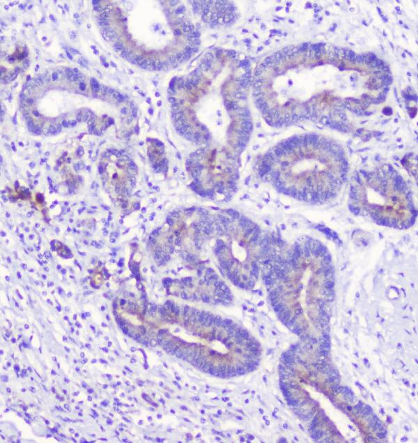
A01051-2 has IHC images from human placenta, cholangiocarcinoma, and rectal cancer paraffin sections and is listed for ICC and frozen-section IHC (A01051-2 IHC captions/applications). M01051-1 is a rabbit monoclonal antibody listed for IF in human, mouse, and rat, with no IF image supplied (M01051-1 catalog description/applications/reactivity/image alts).

Which to pick: Choose A01051-2 for tissue IHC: its own captions document human paraffin sections; the fixative is unreported (A01051-2 IHC captions). Choose A01051-2 for ICC or M01051-1 for IF, which is listed as a rabbit monoclonal antibody (catalog applications; M01051-1 catalog description). Both list human, mouse, and rat reactivity, but the supplied IHC images document human tissue only (catalog reactivity; A01051-2 IHC captions).

Each figure is that product's own IHC / IF validation image from its datasheet.

References

  1. UniProt Consortium. UniProt entry O95835 (LATS1_HUMAN, Serine/threonine-protein kinase LATS1).
  2. Human Protein Atlas. LATS1 tissue IHC expression (reliability: Approved).
  3. Human Protein Atlas. LATS1 subcellular location (ICC-IF): Highest expression in HEL: 27.0 nTPM.
  4. Human Protein Atlas. LATS1 antibody validation summary (1 antibodies).
  5. The Immunoexpression of YAP1 and LATS1 Proteins in Clear Cell Renal Cell Carcinoma: Impact on Patients' Survival. BioMed research international 2018 — PMC5903336.
  6. Exploring the role of YAP1 and TAZ in pancreatic acinar cells and the therapeutic potential of VT-104 in pancreatic inflammation. Journal of pancreatology 2025 — PMC11925344.
  7. Clinicopathological Significance of Large Tumor Suppressor (LATS) Expression in Gastric Cancer. Journal of gastric cancer 2017 — PMC5746657.
  8. Inhibition of EZH2 exerts antitumorigenic effects in renal cell carcinoma via LATS1. FEBS open bio 2023 — PMC10068324.
  9. PubMed PMID:9988268 — UniProt-cited evidence.
  10. PubMed PMID:10518011 — UniProt-cited evidence.
  11. PubMed PMID:15489334 — UniProt-cited evidence.