This website uses cookies to ensure you get the best experience on our website.


- Table of Contents
Plan chromogenic IHC for LAYN using the reported cytoplasmic tissue pattern and high staining in breast myoepithelial cells, smooth muscle cells and stomach glandular cells (HPA tissue IHC). Interpret that staining alongside LAYN’s annotated membrane location and extracellular and cytoplasmic regions (UniProt).

Expected localisation, validated protocols, controls and antibodies — the at-a-glance facts below, then the full design guide.
| Expected localisation | Cytoplasmic staining across tissues (HPA tissue IHC) | |
| Staining pattern | Cytoplasmic staining in cells across tissues (HPA tissue IHC) | |
| Antigen retrieval | Citrate pH 6.0 HIER, 95–98 °C, 20 min (rule: cytoplasmic / membrane antigen) | |
| Positive control | Breast+4 more · see all | |
| Negative control | None in HPA (detected in all 45 tissues); use no-primary + isotype controls |
| Fixation | Matched tissue-IHC evidence does not establish the fixation claim. Validate the specimen-specific method before use. (selected-SKU IHC image A10853) | |
| Caveat | IHC and RNA show medium consistency; verify the pattern (HPA tissue IHC) | |
| Regulation | Low tissue specificity (HPA tissue RNA) | |
| Isoform / epitope | 3 isoforms; check whether the epitope is extracellular or cytoplasmic (UniProt) |
The catalog antibody’s IHC-P protocol is accompanied by two published LAYN IHC methods (PMC10908921; PMC9747853).
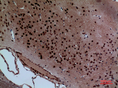
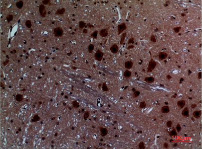
| Sample | Paraffin-embedded Rat Brain tissue; fixative not specified (datasheet A10853) |
| Fixation | Image fixative and duration unreported (datasheet A10853); verify before use. |
| Sectioning | 4–5 µm sections on charged slides (standard) |
| Deparaffinisation | Xylene, graded ethanol series to water (standard) |
| Antigen retrieval | Heat-induced epitope retrieval in citrate buffer, pH 6.0, 20 min at 95–98 °C (standard rule: cytoplasmic / membrane antigen) |
| Peroxidase block | 3% H2O2, 10 min, room temperature (standard) |
| Blocking | 10% normal serum of the secondary host, 30 min, room temperature (standard) |
| Primary antibody | Rabbit anti-LAYN, 1:100 (datasheet A10853) |
| Primary incubation | Overnight at 4 °C (standard) |
| Detection | HRP-polymer secondary, DAB chromogen 5–10 min (standard) |
| Counterstain | Hematoxylin, blue, dehydrate and mount (standard) |
| Expected result | LAYN-positive staining in myoepithelial cells of breast (HPA tissue IHC: High). HPA tissue profile: Cytoplasmic expression in all tissues. No signal in the no-primary control. |
LAYN is a transmembrane protein with an extracellular domain and cytoplasmic tail; UniProt reports colocalization with TLN1 at membrane ruffles (UniProt Q6UX15 topology). In tissue IHC, HPA reports cytoplasmic staining across tissues, with high staining in breast myoepithelial cells, smooth muscle cells, and stomach glandular cells (HPA tissue IHC). HPA rates its tissue staining Approved, with medium consistency against RNA data and external verification pending (HPA tissue IHC).
| Cytoplasmic staining is strongest in breast myoepithelial cells, smooth muscle cells, or stomach glandular cells. | This matches HPA's high staining in those cell types (HPA tissue IHC). Judge the signal in its tissue context; HPA also reports cytoplasmic expression across tissues, so staining elsewhere is not automatically unexpected (HPA tissue IHC). |
| A membrane edge or ruffle is visible alongside cytoplasmic staining. | Membrane staining is biologically plausible because LAYN spans the membrane and colocalizes with TLN1 at membrane ruffles (UniProt Q6UX15 topology). HPA's tissue IHC description is cytoplasmic, however, so a membrane-only result needs cautious interpretation (HPA tissue IHC). |
| Signal is confined to nuclei, with no convincing cytoplasmic or membrane staining. | A nuclear-only pattern conflicts with HPA's cytoplasmic tissue profile and UniProt's membrane topology (HPA tissue IHC; UniProt Q6UX15 topology). Treat it as a possible artefact and check controls before assigning it to LAYN. |
| Unexpected cells stain more strongly than the documented high-staining cell types. | The mismatch raises possible cross-reactivity or endogenous detection activity; neither can be established from appearance alone (HPA tissue IHC; standard IHC practice). HPA's low-staining examples include skeletal muscle myocytes and lymph-node germinal-center cells (HPA tissue IHC). |
| Color spreads across tissue or appears where cellular boundaries cannot be judged. | Diffuse background prevents a reliable call on LAYN localization (standard IHC practice). Compare a no-primary control and review blocking, washes, and detection conditions before interpreting weak staining (standard IHC practice). |
| Cellular compartment | HPA describes cytoplasmic tissue staining, while UniProt places LAYN at the membrane, with an extracellular region at residues 22–235 and a cytoplasmic region at 257–382 (HPA tissue IHC; UniProt Q6UX15 topology). Interpret membrane accent in that context. |
| Tissue and cell choice | Breast myoepithelial cells, smooth muscle cells, and stomach glandular cells are high-staining examples (HPA tissue IHC). Salivary glandular cells and skeletal muscle myocytes are low-staining examples; low means limited signal, not a guaranteed blank control (HPA tissue IHC). |
| Evidence strength | HPA rates the tissue IHC profile Approved, reports medium consistency with RNA expression, and notes that external verification is pending (HPA tissue IHC). The listed antibody, HPA040087, is IHC Approved; the supplied record does not label its IHC validation Enhanced (HPA antibodies). |
| Epitope and isoforms | UniProt records 3 LAYN isoforms, a signal peptide at residues 1–21, and one glycosylation site at residue 117 (UniProt Q6UX15). Without an antibody epitope map, these facts do not predict which isoforms or regions the IHC antibody detects. |
| Situation | Likely cause | Next action |
|---|---|---|
| No signal in a documented high-staining tissue | The result conflicts with HPA's high staining in the relevant cell type (HPA tissue IHC); failed detection or unsuitable assay conditions are possibilities (standard IHC practice). | Confirm that the expected cells are present, then review the no-primary and detection controls, antigen retrieval conditions, and antibody dilution (standard IHC practice). No LAYN-specific retrieval condition is supplied. |
| Only a sharp membrane outline stains | Membrane localization is plausible from LAYN topology, but HPA describes tissue IHC staining as cytoplasmic (UniProt Q6UX15 topology; HPA tissue IHC). | Check whether cytoplasmic signal is also present in documented high-staining cells and compare controls before calling an isolated outline a confirmed LAYN pattern (HPA tissue IHC; standard IHC practice). |
| An unexpected cell population dominates the slide | Possible explanations include antibody cross-reactivity or endogenous detection activity (standard IHC practice). HPA reports low rather than absent staining in several cell types (HPA tissue IHC). | Check a no-primary control and compare the cell type with HPA's tissue profile; do not treat low-staining examples as definitively negative (standard IHC practice; HPA tissue IHC). |
| Diffuse color obscures cell boundaries | Background from blocking, washing, or detection can obscure specific cellular staining (standard IHC practice). | Review blocking and washes, inspect a no-primary control, and adjust detection conditions before scoring localization or intensity (standard IHC practice). |
| Nuclear-only signal persists | A nuclear-only result conflicts with the reported cytoplasmic tissue pattern and membrane topology (HPA tissue IHC; UniProt Q6UX15 topology). | Verify the counterstain and inspect controls; score LAYN only where a convincing cellular pattern supports the call (standard IHC practice). |
| What should IF/ICC show? | HPA reports an approved cytosolic ICC-IF location, with images from A-431, U-251MG, and U2OS cells (HPA subcellular). | Use cytosolic staining as the HPA ICC-IF comparison and interpret any membrane accent alongside UniProt's membrane localization (HPA subcellular; UniProt Q6UX15 topology). |
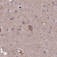
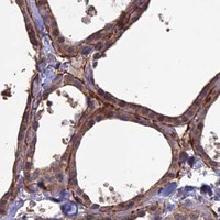
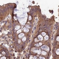


Comprehensive Human Protein Atlas IHC scoring per tissue (reliability: Approved — Medium consistency between antibody staining and RNA expression data. Pending external verification.). Rows are taken directly from the HPA tissue chart — click any row's HPA link to view the source.
| Tissue | Cell type | Level | Evidence | Source |
|---|---|---|---|---|
| Breast | Myoepithelial cells | High | Protein (IHC) | HPA → |
| Smooth muscle | Smooth muscle cells | High | Protein (IHC) | HPA → |
| Stomach | Glandular cells | High | Protein (IHC) | HPA → |
| Adipose tissue | Adipocytes | Medium | Protein (IHC) | HPA → |
| Adrenal gland | Glandular cells | Medium | Protein (IHC) | HPA → |
| Tissue | Cell type | Level | Evidence | Source |
|---|---|---|---|---|
| None in HPA: LAYN is detected in all 45 scored tissues. Use a no-primary (secondary-only) and an isotype control instead. | ||||
Troubleshoot LAYN staining in paraffin section IHC by checking retrieval, compartment, cell identity, controls, and scoring (UniProt Q6UX15; HPA tissue IHC).
A10853 has paraffin-section IHC images from rat and mouse brain (image captions); IF is listed without an image (catalog). Human, mouse and rat reactivity are listed (catalog).
A10853 will render with IHC images captioned as paraffin-embedded rat and mouse brain (image captions). Its listed applications include IHC and IF, with human, mouse and rat reactivity; no IF image is supplied (catalog).
Which to pick: For tissue IHC, choose A10853: its own images show paraffin-embedded rat and mouse brain, while the fixative is unreported (image captions); its listed IHC dilution is 1:100 (catalog). For IF/ICC, A10853 lists IF at 1:50, but the payload supplies no IF image or ICC validation (catalog). For cross-species work, A10853 lists human, mouse and rat reactivity, though its pictured IHC evidence covers only rat and mouse brain; clonality is unspecified (catalog; image captions).