This website uses cookies to ensure you get the best experience on our website.


- Table of Contents
Plan chromogenic IHC for LCP2 in paraffin sections using cytoplasmic staining in lymphoid tissue as the expected pattern (HPA tissue IHC). Use high-staining bone marrow hematopoietic cells or lymph node non-germinal center cells as positive tissue references (HPA tissue IHC).

Expected localisation, validated protocols, controls and antibodies — the at-a-glance facts below, then the full design guide.
| Expected localisation | Cytoplasmic staining in lymphoid tissue (HPA tissue IHC) | |
| Staining pattern | Cytoplasm of lymphoid-tissue cells (HPA tissue IHC) | |
| Antigen retrieval | Citrate pH 6.0 HIER, 95–98 °C, 20 min (rule: cytoplasmic / membrane antigen) | |
| Positive control | Bone marrow+4 more · see all | |
| Negative control | Adipose tissue+4 more · see all |
| Fixation | Keep paraffin-section fixation consistent. (standard IHC practice; not target-specific) | |
| Caveat | Cell composition can change staining across lymphoid regions (HPA tissue IHC) | |
| Regulation | Expression regulation not specified (UniProt) | |
| Isoform / epitope | No isoforms; one cytoplasmic chain, no extracellular segment (UniProt) |
Compare the catalog antibody’s IHC-P protocol with three published LCP2 protocols for lung adenocarcinoma, salivary gland, and placental tissue (PMC8649447; PMC10545362; PMC11979283).
| Sample | Paraffin-embedded human breast carcinoma tissue; fixative not specified (datasheet A04880) |
| Fixation | Image fixative and duration unreported (datasheet A04880); verify before use. |
| Sectioning | 4–5 µm sections on charged slides (standard) |
| Deparaffinisation | Xylene, graded ethanol series to water (standard) |
| Antigen retrieval | Heat-induced epitope retrieval in citrate buffer, pH 6.0, 20 min at 95–98 °C (standard rule: cytoplasmic / membrane antigen) |
| Peroxidase block | 3% H2O2, 10 min, room temperature (standard) |
| Blocking | 10% normal serum of the secondary host, 30 min, room temperature (standard) |
| Primary antibody | Rabbit anti-LCP2, 1:50-1:200 (datasheet A04880) |
| Primary incubation | Overnight at 4 °C (standard) |
| Detection | HRP-polymer secondary, DAB chromogen 5–10 min (standard) |
| Counterstain | Hematoxylin, blue, dehydrate and mount (standard) |
| Expected result | LCP2-positive staining in hematopoietic cells of bone marrow (HPA tissue IHC: High). HPA tissue profile: Cytoplasmic expression in lymphoid tissues. No signal in the no-primary control. |
LCP2 should appear predominantly in the cytoplasm of hematopoietic and lymphoid cells, with especially strong staining in bone marrow hematopoietic cells and selected lymphoid tissue compartments (UniProt Q13094: Cytoplasm; HPA tissue IHC: High in these cells). It has no transmembrane segment, so a crisp surface rim is unexpected (UniProt Q13094: topology). HPA rates its tissue IHC pattern Enhanced, reflecting high consistency between antibody staining and RNA expression (HPA tissue IHC: Enhanced).
| Strong cytoplasmic stain in bone marrow hematopoietic cells (HPA tissue IHC: High). | This fits an expected positive control: the signal occupies the compartment assigned by UniProt, in a cell population scored High by HPA (UniProt Q13094: Cytoplasm; HPA tissue IHC: bone marrow). Compare staining with nearby cells on the same section before judging overall intensity. |
| Cytoplasmic stain in lymph node or tonsil non-germinal center cells, or spleen red-pulp cells (HPA tissue IHC: High). | These are supported positive patterns; staining need not be uniform across a lymphoid section (HPA tissue IHC: cell-specific High scores). Assess the annotated cell population, since a tissue-wide score could conceal a correct, restricted pattern. |
| Dominant nuclear stain or a sharp cell-surface rim. | Treat this as a compartment mismatch: UniProt places LCP2 in the cytoplasm and reports no transmembrane segment (UniProt Q13094: subcellular location and topology). Review controls and morphology before interpreting the signal as LCP2; the source record does not support either pattern as expected. |
| Prominent stain in adipocytes or squamous epithelium (HPA tissue IHC: Not detected in listed examples). | Check for cross-reactivity or detection-system activity when these cells stain: HPA reports no signal in adipocytes from adipose tissue or breast, and squamous epithelium from esophagus or oral mucosa (HPA tissue IHC: Not detected). A different cell population in the same specimen may still stain. |
| Diffuse color across cells and extracellular space, or no signal in bone marrow hematopoietic cells. | Diffuse deposition limits compartment scoring; absence in a known-positive population makes a negative result difficult to trust (standard IHC practice; HPA tissue IHC: bone marrow hematopoietic cells High). Check a no-primary control for background and a known-positive section for assay performance before drawing a biological conclusion. |
| Cell-specific tissue context (HPA tissue IHC). | Use the named cell population when choosing controls: bone marrow hematopoietic cells, lymph node and tonsil non-germinal center cells, and spleen red-pulp cells score High (HPA tissue IHC). A negative score for adipocytes does not make every cell in an adipose-containing specimen negative (HPA tissue IHC: cell-level annotations). |
| Compartment and topology (UniProt Q13094). | LCP2 is cytoplasmic, has no signal peptide or transmembrane segment, and is recorded as one chain spanning residues 1–533 (UniProt Q13094). Those annotations support cytoplasmic interpretation; they do not identify the antibody's epitope or prove a retrieval requirement. |
| Antibody validation (HPA antibodies). | HPA lists three antibodies with Enhanced IHC status: HPA036396, HPA036397 and CAB004574 (HPA antibodies: IHC Enhanced). This supports the reported tissue pattern, but does not establish that an unlisted catalog antibody will give the same staining or specify its working dilution. |
| Antigen retrieval (general IHC practice). | For paraffin IHC, follow the IHC-validated antibody's stated retrieval conditions where available and judge the outcome against positive and no-primary controls (general IHC practice). The supplied UniProt and HPA records do not report LCP2-specific retrieval or fixation sensitivity. |
| IF/ICC Q&A: What pattern should be checked? | Look for cytosolic signal: HPA calls the cytosol its supported main location and lists HEL and serum-starved hTERT-RPE1 among cells with ICC-IF images (HPA subcellular: Cytosol supported). This is an interpretation cue for the separate IF/ICC guide, not an IHC-P protocol option. |
| Situation | Likely cause | Next action |
|---|---|---|
| No stain in bone marrow hematopoietic cells (HPA tissue IHC: High). | The expected positive control failed; a biological negative cannot be separated from an assay failure on that result alone (HPA tissue IHC: High; general IHC practice). | Confirm the IHC-validated antibody, its stated dilution and retrieval conditions, and that the chromogenic detection reagents worked; repeat alongside a known-positive section (general IHC practice). |
| Weak, patchy signal in an otherwise expected positive population (HPA tissue IHC: High). | Cell selection or section-to-section assay variation may account for the appearance; HPA's cell-specific score does not set an intensity threshold for every specimen (HPA tissue IHC: cell annotations; general IHC practice). | Compare the same annotated cell population in a known-positive section, then review the antibody's IHC-P conditions and counterstain so faint cytoplasmic signal can be judged (general IHC practice). |
| Predominantly nuclear stain or a continuous membrane rim. | The compartment conflicts with cytoplasmic LCP2 and its lack of a transmembrane segment (UniProt Q13094: location and topology). | Check staining without primary antibody, inspect cell boundaries and nuclei, and seek a concordant cytoplasmic pattern with an independently validated antibody if available (general IHC practice; HPA antibodies: Enhanced IHC entries). |
| Strong stain in adipocytes or listed squamous epithelial cells (HPA tissue IHC: Not detected). | Possible nonspecific antibody binding or detection activity; the HPA negative calls apply to those specific cell populations (HPA tissue IHC: Not detected; general IHC practice). | Compare a no-primary control and an HPA-supported positive cell population, then reassess the IHC-validated antibody and detection controls before assigning LCP2 expression (general IHC practice; HPA tissue IHC). |
| Diffuse chromogen obscures cell borders. | Background deposition prevents a reliable cytoplasmic call; it does not establish LCP2 expression (general IHC practice; UniProt Q13094: Cytoplasm). | Inspect a no-primary control and review blocking, wash steps and detection conditions; score only cells whose signal can be localized clearly (general IHC practice). |
| Moderate glandular or lung macrophage staining seems inconsistent with a lymphoid-focused pattern. | HPA also reports Medium staining in adrenal and gallbladder glandular cells and lung macrophages (HPA tissue IHC: Medium); a non-lymphoid site alone does not establish cross-reactivity. | Identify the stained cell type and compartment first, compare the relevant HPA cell-level annotation, and use controls if the pattern remains discordant (HPA tissue IHC: Medium; UniProt Q13094: Cytoplasm; general IHC practice). |


Comprehensive Human Protein Atlas IHC scoring per tissue (reliability: Enhanced — High consistency between antibody staining and RNA expression data. Antibody staining in cells/structures not annotated, view images.). Rows are taken directly from the HPA tissue chart — click any row's HPA link to view the source.
| Tissue | Cell type | Level | Evidence | Source |
|---|---|---|---|---|
| Bone marrow | Hematopoietic cells | High | Protein (IHC) | HPA → |
| Lymph node | Non-germinal center cells | High | Protein (IHC) | HPA → |
| Spleen | Cells in red pulp | High | Protein (IHC) | HPA → |
| Tonsil | Non-germinal center cells | High | Protein (IHC) | HPA → |
| Adrenal gland | Glandular cells | Medium | Protein (IHC) | HPA → |
| Tissue | Cell type | Level | Evidence | Source |
|---|---|---|---|---|
| Adipose tissue | Adipocytes | Not detected | Protein (IHC) | HPA → |
| Breast | Adipocytes | Not detected | Protein (IHC) | HPA → |
| Cerebellum | Cells in granular layer | Not detected | Protein (IHC) | HPA → |
| Cervix | Glandular cells | Not detected | Protein (IHC) | HPA → |
| Esophagus | Squamous epithelial cells | Not detected | Protein (IHC) | HPA → |
Troubleshoot LCP2 staining by checking cytoplasmic localisation, immune cell identity and matched controls (UniProt Q13094; HPA tissue IHC).
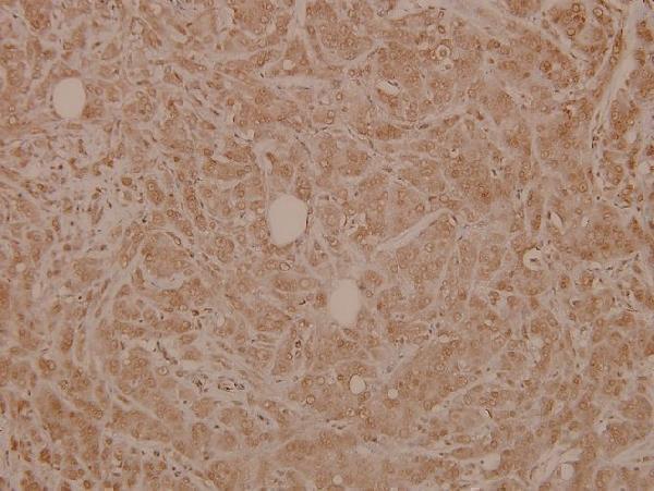
The catalog antibody has real IHC data from paraffin-embedded human breast carcinoma (image caption). Human, mouse, and rat reactivity is listed; no IF application or image is supplied (catalog).
A04880 has an IHC image from paraffin-embedded human breast carcinoma at 1:100 (image caption). Its listed applications are IHC and WB, and its listed reactivity is human, mouse, and rat (catalog).
Which to pick: Choose A04880 for paraffin-section tissue IHC because its own image shows that preparation; the fixative is unreported (image caption). No SKU in the payload lists IF/ICC or shows an IF image (catalog). A04880 is the cross-species candidate because human, mouse, and rat reactivity is listed, though its IHC image shows only human tissue (catalog; image caption).