This website uses cookies to ensure you get the best experience on our website.


- Table of Contents
Plan LDB3 paraffin-section IHC using heart and skeletal muscle as positive tissues, with cytoplasmic staining expected in their muscle cells (HPA tissue IHC). This guide covers consistent fixation, chromogenic detection, the catalog antibody’s 1:50–1:200 IHC dilution (datasheet A04633), and isoform-aware interpretation (UniProt).

Expected localisation, validated protocols, controls and antibodies — the at-a-glance facts below, then the full design guide.
| Expected localisation | Cytoplasmic in heart and skeletal muscle (HPA tissue IHC) | |
| Staining pattern | Cardiomyocyte cytoplasm high; skeletal myocyte cytoplasm medium (HPA tissue IHC) | |
| Antigen retrieval | Citrate pH 6.0 HIER, heat-mediated (datasheet A04633) | |
| Positive control | Heart muscle+2 more · see all | |
| Negative control | Adipose tissue+4 more · see all |
| Fixation | Keep fixation consistent across paraffin sections. (standard IHC practice; not target-specific) | |
| Caveat | Staining may detect proteins from more than one gene (HPA tissue IHC) | |
| Regulation | Heart high; skeletal muscle medium (HPA tissue IHC) | |
| Isoform / epitope | Seven isoforms; check epitope coverage; no extracellular domain (UniProt) |
The catalog antibody’s IHC-P protocol is paired with two published LDB3 IHC protocols (PMC9822979; PMC9636405).
| Sample | Paraffin-embedded mouse stomach tissue; fixative not specified (datasheet A04633) |
| Fixation | Image fixative and duration unreported (datasheet A04633); verify before use. |
| Sectioning | 4–5 µm sections on charged slides (standard) |
| Deparaffinisation | Xylene, graded ethanol series to water (standard) |
| Antigen retrieval | Heat retrieval: Citrate pH 6.0 (datasheet A04633); 20 min, 95–100 °C (standard) |
| Peroxidase block | 3% H2O2, 10 min, room temperature (standard) |
| Blocking | 10% normal serum of the secondary host, 30 min, room temperature (standard) |
| Primary antibody | Rabbit anti-LDB3, 1:50-1:200 (datasheet A04633) |
| Primary incubation | Overnight at 4 °C (standard) |
| Detection | HRP-polymer secondary, DAB chromogen 5–10 min (standard) |
| Counterstain | Hematoxylin, blue, dehydrate and mount (standard) |
| Expected result | LDB3-positive staining in cardiomyocytes of heart muscle (HPA tissue IHC: High). HPA tissue profile: Selective cytoplasmic expression in heart and skeletal muscle. No signal in the no-primary control. |
LDB3 is a cytoplasmic, Z-line-associated protein with no transmembrane segment (UniProt O75112). In paraffin-section IHC, expect staining chiefly in cardiomyocytes and skeletal myocytes, with possible staining in late spermatids (HPA tissue IHC: High, Medium, and Medium, respectively). HPA rates the tissue pattern Supported, while cautioning that the antibody targets proteins from more than one gene (HPA tissue IHC).
| Strong cytoplasmic staining in cardiomyocytes, with less intense staining in skeletal myocytes. | This fits the reported High cardiomyocyte and Medium skeletal-myocyte signals (HPA tissue IHC). A striated pattern would be compatible with LDB3 at sarcomeric Z-lines (UniProt O75112), but HPA's tissue summary establishes cytoplasmic expression, not a required striation pattern (HPA tissue IHC). |
| Medium staining in elongated or late spermatids, or weak staining in placental trophoblastic cells. | These are reported outside the principal muscle pattern: Medium in late spermatids and Low in trophoblastic cells (HPA tissue IHC). Judge them by their stated cell types and levels; neither finding alone overrides the stronger heart and skeletal-muscle pattern (HPA tissue IHC). |
| Predominantly nuclear staining in cardiomyocytes or skeletal myocytes. | A nuclear-dominant IHC result conflicts with the expected cytoplasmic muscle pattern (HPA tissue IHC; UniProt O75112). Investigate background and antibody specificity before scoring it as LDB3. An uncertain nucleoplasmic ICC-IF location does not establish nuclear muscle IHC staining (HPA subcellular). |
| Strong staining in adipocytes or other cells listed as Not detected. | This disagrees with the listed HPA cell-level observations, including adipocytes in adipose tissue (HPA tissue IHC). Check whether staining follows tissue structures or detection background; cross-reactivity or endogenous chromogen activity are possible general IHC explanations, not demonstrated causes in this specimen. |
| Broad, even colour across cells and surrounding tissue, without a clear cytoplasmic muscle pattern. | Treat this as background until controls show otherwise. The selective cytoplasmic heart and skeletal-muscle profile provides the comparison (HPA tissue IHC). General IHC causes include incomplete blocking, concentrated antibody, or residual detection activity; appearance alone cannot identify which occurred. |
| Tissue and cell selection | Heart muscle cardiomyocytes provide the clearest reported positive reference (High); skeletal-muscle myocytes are Medium (HPA tissue IHC). Adipose-tissue adipocytes are listed as Not detected (HPA tissue IHC). Compare named cell populations, because the tissue-level label alone does not identify the stained cells. |
| Intracellular location | LDB3 has no transmembrane segment and is assigned to cytoplasm, cytoskeleton, myofibrils and sarcomeric Z-lines (UniProt O75112). Interpret muscle IHC against the cytoplasmic HPA profile; do not require a resolved Z-line pattern in every paraffin section (HPA tissue IHC). |
| Antibody evidence and specificity | The tissue-IHC reliability is Supported, with an explicit caution that staining may target proteins from more than one gene (HPA tissue IHC). HPA048955 is IHC Supported but ICC Uncertain (HPA antibodies). These ratings support pattern comparison; they do not prove that every stained structure is LDB3. |
| Isoform coverage | UniProt lists 7 LDB3 isoforms (UniProt O75112). The supplied record gives no antibody epitope or isoform coverage, so a difference between specimens cannot be assigned to an isoform from this evidence. Confirm the catalog antibody's epitope information before making an isoform-specific claim. |
| Detection chemistry | For chromogenic IHC, endogenous enzyme activity or nonspecific reagent binding can produce colour independently of target recognition (standard IHC practice). Use matched detection controls when signal appears outside the expected cell population; HPA's staining levels do not identify the chemistry behind a particular specimen's background. |
| Situation | Likely cause | Next action |
|---|---|---|
| No signal in cardiomyocytes of a heart-muscle section. | This misses the reported High reference population (HPA tissue IHC). The image alone cannot distinguish a detection failure from unsuitable antibody conditions. | Check section integrity and assay controls, then review antigen retrieval and catalog-antibody dilution as general paraffin-IHC variables. Change one condition at a time; no LDB3-specific retrieval setting or dilution is supplied. |
| Heart-muscle signal is present, but skeletal myocytes look faint. | The reported comparison is High cardiomyocytes versus Medium skeletal myocytes (HPA tissue IHC), so lower skeletal intensity may be expected. | Score each named cell type against its own reference level and confirm the cytoplasmic location (HPA tissue IHC). Avoid calling skeletal muscle negative solely because it is weaker than heart. |
| Nuclear colour dominates an otherwise positive muscle section. | The expected muscle pattern is cytoplasmic (HPA tissue IHC; UniProt O75112). Nuclear colour could reflect background or nonspecific staining; its cause is unresolved. | Compare a matched negative control and review the antibody and detection steps. Record nuclear-dominant colour separately until the cytoplasmic muscle signal can be assessed. |
| Strong colour appears in adipocytes or many unrelated cell populations. | Adipocytes are reported Not detected in adipose tissue (HPA tissue IHC). HPA also cautions that the tissue antibody may target proteins from more than one gene (HPA tissue IHC). | Inspect cell identity and negative controls; review blocking and endogenous enzyme controls as standard chromogenic-IHC checks. Do not assign the extra signal to LDB3 solely from colour. |
| Diffuse colour obscures the boundary between positive cells and surrounding tissue. | The expected profile is selective cytoplasmic staining in heart and skeletal muscle (HPA tissue IHC). Diffuse colour may arise from general staining or detection background. | Compare reagent controls, then review blocking, washing and antibody concentration as general IHC steps. Keep interpretation tied to identifiable cardiomyocytes or myocytes and their cytoplasm. |
| IF/ICC Q: Should focal-adhesion or nucleoplasmic signal count as confirmed LDB3? | HPA lists cytosol and focal adhesions as main ICC-IF locations and nucleoplasm as additional, all uncertain; it warns that the antibodies target proteins from multiple genes (HPA subcellular). HPA048955 is ICC Uncertain (HPA antibodies). | Treat those IF/ICC locations as provisional and use the separate IF/ICC guide for that application. They do not redefine the expected cytoplasmic muscle result in paraffin IHC (HPA tissue IHC). |


Comprehensive Human Protein Atlas IHC scoring per tissue (reliability: Supported — High consistency between antibody staining and RNA expression data. Caution, targets protein from more than one gene.). Rows are taken directly from the HPA tissue chart — click any row's HPA link to view the source.
| Tissue | Cell type | Level | Evidence | Source |
|---|---|---|---|---|
| Heart muscle | Cardiomyocytes | High | Protein (IHC) | HPA → |
| Skeletal muscle | Myocytes | Medium | Protein (IHC) | HPA → |
| Testis | Elongated or late spermatids | Medium | Protein (IHC) | HPA → |
| Tissue | Cell type | Level | Evidence | Source |
|---|---|---|---|---|
| Adipose tissue | Adipocytes | Not detected | Protein (IHC) | HPA → |
| Adrenal gland | Glandular cells | Not detected | Protein (IHC) | HPA → |
| Appendix | Endocrine cells | Not detected | Protein (IHC) | HPA → |
| Bone marrow | Hematopoietic cells | Not detected | Protein (IHC) | HPA → |
| Breast | Adipocytes | Not detected | Protein (IHC) | HPA → |
Use compartment, tissue controls and matched processing conditions to troubleshoot chromogenic LDB3 staining in paraffin sections.
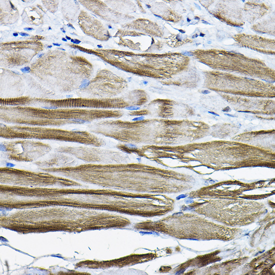
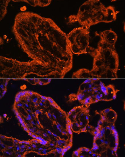
A04633 has pictured LDB3 IHC in mouse stomach paraffin sections and IF in human placenta and rat heart cells (catalog image captions); its listed reactivity includes human, mouse and rat (catalog reactivity).
A04633 renders with mouse stomach paraffin-section IHC at 1:200 after citrate retrieval at pH 6.0 (A04633 IHC image caption). Its IF captions show human placenta cells and rat heart cells at 1:100, with DAPI nuclear staining (A04633 IF image captions).
Which to pick: Choose A04633 for tissue IHC when the pictured mouse stomach paraffin-section result is relevant; its caption reports citrate retrieval but does not report the fixative (A04633 IHC image caption). Choose A04633 for IF/ICC because both applications are listed and IF images are supplied; A04633-2 also lists IF/ICC but has no supplied IF figure (catalog applications and image captions). For work across human, mouse and rat, A04633 lists all three species and is identified as a rabbit polyclonal antibody, although its pictured IHC result is in mouse only (catalog reactivity; A04633 IHC image caption).