LGR5 / Leucine-rich repeat-containing G protein-coupled receptor 5 · IHC design guide

Design Immunohistochemistry for LGR5

Plan LGR5 paraffin IHC around cytoplasmic and membranous staining, with prominent motile cilia in fallopian tube and respiratory epithelia (HPA tissue IHC). Use the catalog antibody’s documented IHC workflow (datasheet A00239-2), and interpret staining cautiously because antibody staining has low RNA agreement and presumed off-target binding (HPA tissue IHC).

Evidence assembled Oct 2026 · For research use; verify linked source records and product datasheet before use
Immunohistochemistry protocol sheet for LGR5 (IHC for LGR5): expected localisation Cytoplasmic and membranous tissue staining (HPA tissue IHC), antibody A00239-2, validated IHC image, and IHC protocol steps
Printable LGR5 IHC protocol sheet — expected localisation Cytoplasmic and membranous tissue staining (HPA tissue IHC), antibody A00239-2, controls and protocol steps. Open the full LGR5 IHC guide →

LGR5 Immunohistochemistry Experimental Design Guide

Expected localisation, validated protocols, controls and antibodies — the at-a-glance facts below, then the full design guide.

Must know before staining
Expected localisation Cytoplasmic and membranous tissue staining (HPA tissue IHC)
Staining pattern Motile cilia in fallopian tube and respiratory epithelia (HPA tissue IHC)
Antigen retrieval EDTA pH 8.0 HIER, heat-mediated (datasheet A00239-2)
Positive control ⓘ Bronchus+4 more · see all
Negative control ⓘ Adipose tissue+4 more · see all
Important caveats
Reasons your staining may differ from the expected pattern.
Fixation Keep paraffin-section fixation consistent (standard IHC practice; not target-specific); Matched tissue-IHC evidence does not establish the fixation claim. Validate the specimen-specific method before use. (selected-SKU IHC image A00239-2)
Caveat Presumed off-target staining and low RNA agreement (HPA tissue IHC)
Regulation Higher in ovary, colon and liver cancers (UniProt)
Isoform / epitope 3 isoforms; map the epitope to extracellular or cytoplasmic regions (UniProt)
Section 1

Recommended LGR5 IHC & IF Protocols

The catalog antibody’s IHC-P protocol is provided separately (datasheet A00239-2). The published protocols below describe LGR5 staining in esophageal, gastric, rectal, and colorectal tissue (PMC3058063; PMC5207613; PMC5727605; PMC10383310).

Recommended immunohistochemistry (IHC-P) protocol parameters
SampleParaffin-embedded mouse small intestine tissue; fixative not specified (datasheet A00239-2)
FixationImage fixative and duration unreported (datasheet A00239-2); verify before use.
Sectioning4–5 µm sections on charged slides (standard)
DeparaffinisationXylene, graded ethanol series to water (standard)
Antigen retrievalHeat retrieval: EDTA pH 8.0 (datasheet A00239-2); 20 min, 95–100 °C (standard)
Peroxidase block3% H2O2, 10 min, room temperature (standard)
Blocking10% goat serum (datasheet A00239-2)
Primary antibodyRabbit anti-LGR5, 2-5 μg/ml (datasheet A00239-2)
Primary incubationOvernight at 4 °C (datasheet A00239-2)
DetectionHRP-conjugated secondary, DAB chromogen (datasheet A00239-2)
CounterstainHematoxylin, blue, dehydrate and mount (standard)
Expected resultLGR5-positive staining in ciliated cells (cilia axoneme) of bronchus (HPA tissue IHC: High). HPA tissue profile: Cytoplasmic and membranous expression in several tissues, mainly in motile cilia in fallopian tube, uterus and respiratory epithelia. No signal in the no-primary control.
💡Decision noteStart with heat-mediated EDTA retrieval at pH 8.0 for the catalog antibody (datasheet A00239-2); use each published retrieval condition with its corresponding published antibody (PMC5727605; PMC10383310).
Section 2

What Is the Expected LGR5 Staining Pattern?

LGR5 is a seven-pass cell-membrane receptor that also localizes to the trans-Golgi network (UniProt O75473 topology and subcellular location). In tissue IHC, HPA reports cytoplasmic and membranous staining, especially in motile cilia of fallopian tube, uterine and respiratory epithelia (HPA tissue IHC). Its tissue assessment is Approved but notes low agreement with RNA data and presumed off-target binding (HPA tissue IHC reliability).

What am I looking at on my slide?
Strong staining along the cilia of bronchial or fallopian-tube ciliated cells (HPA: High in cilia axonemes).This matches the reported tissue pattern; assess the ciliated-cell compartment, not just whether the section contains brown signal (HPA tissue IHC).
Membranous or cytoplasmic staining in duodenal or small-intestinal glandular cells (HPA: Medium).This is compatible with HPA tissue IHC and with membrane and trans-Golgi localization (UniProt O75473). Record the cell type and compartment when scoring.
Nuclear-only staining in a paraffin section, without the expected cell-associated pattern (HPA tissue IHC; UniProt O75473).Treat it as discordant tissue IHC and investigate artefact. HPA separately reports approved nucleoplasmic ICC-IF localization, so nuclear IF alone cannot settle tissue-IHC specificity (HPA subcellular; HPA tissue IHC).
Prominent signal in adipocytes or bone-marrow hematopoietic cells (HPA: Not detected in those cells).Question antibody specificity or endogenous detection activity before assigning LGR5 expression (HPA tissue IHC reliability; standard IHC practice).
No ciliary signal in bronchus or fallopian tube (HPA: High in ciliated cells).Check tissue preservation, retrieval and detection controls using the selected IHC-P workflow (standard IHC practice). A single negative section cannot establish absent LGR5.
💡Expected LGR5 appearanceA convincing chromogenic positive shows strong, cell-restricted ciliary staining in bronchial or fallopian-tube ciliated cells (HPA: High); widespread staining of cell types reported as undetected is a specificity warning (HPA tissue IHC reliability).
How each factor affects the staining
Compartment and epitope locationLGR5 has an extracellular region, seven membrane spans and a cytoplasmic tail (UniProt O75473 topology). Interpret staining against the antibody's documented epitope if available; its epitope is not supplied here.
Intracellular distributionLGR5 is rapidly and constitutively internalized to the trans-Golgi network (UniProt O75473 subcellular location). Cytoplasmic signal need not exclude a membrane receptor, but morphology alone does not prove identity.
Tissue and cell selectionHPA reports High ciliary staining in bronchus and fallopian tube, and Medium glandular staining in duodenum and small intestine (HPA tissue IHC). Compare the same cell types across controls.
Strength of tissue-IHC evidenceThe HPA antibody HPA012530 is IHC Approved, while the tissue summary reports low staining–RNA consistency and presumed off-target binding (HPA antibodies; HPA tissue IHC reliability). Interpret unexpected positives cautiously.
IF/ICC Q&A: should its pattern match paraffin IHC?No direct match is established: HPA reports approved nucleoplasm, vesicles and primary-cilium transition-zone ICC-IF localization, while HPA012530 is ICC Uncertain (HPA subcellular; HPA antibodies).
Why is my staining missing, weak or wrong?
SituationLikely causeNext action
The expected ciliary positive is absent (HPA: High in bronchus and fallopian tube).The section may lack intact ciliated epithelium, or an IHC workflow step may have failed (standard IHC practice).Confirm ciliated cells are present; review the selected IHC-P retrieval and detection steps, then compare a known-positive section processed alongside it (standard IHC practice).
Signal covers most cells or the whole section rather than defined compartments (HPA tissue IHC pattern).Nonspecific primary or detection background is possible (standard IHC practice); HPA also flags presumed off-target binding (HPA tissue IHC reliability).Inspect a no-primary control and review blocking, washing and detection conditions (standard IHC practice); do not score diffuse background as LGR5.
Cells reported as undetected stain strongly (HPA: adipocytes and bone-marrow hematopoietic cells Not detected).Cross-reactivity or endogenous detection activity is possible (HPA tissue IHC reliability; standard IHC practice).Compare cell-resolved staining with a no-primary control and the reported HPA distribution; investigate unexpected signal before calling it positive (standard IHC practice).
Only nuclei stain in tissue IHC (HPA tissue IHC; UniProt O75473).This is discordant with the reported tissue pattern, although nucleoplasm is an approved ICC-IF location (HPA tissue IHC; HPA subcellular).Check control sections and detection background; report the tissue-IHC discrepancy rather than transferring an ICC-IF localization claim to paraffin sections (standard IHC practice; HPA subcellular).
Intestinal crypt-base signal seems broader than expected (UniProt O75473 tissue specificity).UniProt places expression at normal intestinal crypt bases but reports that premalignant cancer expression need not stay there (UniProt O75473 tissue specificity).Record tissue context and cell distribution before judging the pattern; compare with HPA's Medium glandular staining in duodenum and small intestine (HPA tissue IHC).
IHC and IF images appear inconsistent (HPA tissue IHC; HPA subcellular).The assays report different observed patterns, and HPA012530 is IHC Approved but ICC Uncertain (HPA antibodies; HPA subcellular).Interpret each assay against its own controls and HPA record; do not use the ICC-IF image alone to validate a paraffin-IHC result (HPA antibodies; standard IHC practice).

Sample controls for LGR5 IHC & IF

🧪Run bronchus first and assess the cilia axoneme of ciliated cells, where staining is High (HPA: High in bronchus ciliated cells). Use adipose tissue adipocytes as the negative tissue (HPA: Not detected in adipocytes). On the bronchus slide, cells without cilia should lack the specific ciliary staining pattern; evaluate any staining in those cells as background against the negative controls (HPA: cilia axoneme staining in bronchus ciliated cells).
Positive control tissue: Bronchus (Ciliated cells (cilia axoneme), HPA High)
Negative control tissue: Adipose tissue (HPA Not detected)
ICC-IF cell lines (HPA subcellular resource): HPA ICC-IF images show LGR5 in A-431, Hep-G2, U-251MG, ASC52telo, RPTEC/TERT1, hTERT-RPE1 (serum starved), with annotated localisation: Nucleoplasm (approved), Vesicles (approved), Primary cilium transition zone (approved) (HPA subcellular).
Technical controls: Include no-primary (secondary-only) and concentration-matched rabbit IgG isotype controls for the rabbit catalog antibody, plus LGR5-knockout tissue as a biological negative (catalog caption: rabbit anti-LGR5). For chromogenic bronchus sections, block endogenous peroxidase and check for nonspecific signal along the ciliated surface before scoring (catalog caption: peroxidase secondary and DAB; HPA: cilia axoneme staining in bronchus).
⚠️Feasibility: A target-specific fixation window or fixation effect is unreported, and the selected A00239-2 paraffin-section caption does not state the fixative (catalog caption: fixative not stated). The documented IHC procedure uses heat retrieval in EDTA at pH 8.0, followed by 2 μg/ml primary antibody overnight at 4°C; whether other retrieval conditions work or frozen sections or IF are easier is unreported (catalog caption: mouse small intestine paraffin section and retrieval conditions). In bronchus, assess ciliary staining carefully because HPA reports a cilia axoneme signal, while UniProt places LGR5 at the cell and trans-Golgi network membranes; this localization difference warrants confirmation with the biological negative (HPA: bronchus cilia axoneme; UniProt O75473 subcellular location).

HPA tissue IHC evidence for LGR5

Comprehensive Human Protein Atlas IHC scoring per tissue (reliability: Approved — Low consistency between antibody staining and RNA expression data. Presumed off target binding observed and disregarded.). Rows are taken directly from the HPA tissue chart — click any row's HPA link to view the source.

Positive expression · recommended positive controls

TissueCell typeLevelEvidenceSource
Bronchus Ciliated cells (cilia axoneme) High Protein (IHC) HPA →
Fallopian tube Ciliated cells (cilia axoneme) High Protein (IHC) HPA →
Nasopharynx Ciliated cells (cilia axoneme) High Protein (IHC) HPA →
Cerebral cortex Endothelial cells Medium Protein (IHC) HPA →
Cervix Glandular cells Medium Protein (IHC) HPA →

Undetected expression · recommended negative controls

TissueCell typeLevelEvidenceSource
Adipose tissue Adipocytes Not detected Protein (IHC) HPA →
Appendix Glandular cells Not detected Protein (IHC) HPA →
Bone marrow Hematopoietic cells Not detected Protein (IHC) HPA →
Breast Adipocytes Not detected Protein (IHC) HPA →
Caudate Glial cells Not detected Protein (IHC) HPA →
Section 3

Advanced LGR5 IHC Tips

Troubleshoot chromogenic LGR5 IHC in paraffin sections using the catalog antibody’s tissue example, receptor topology, and tissue staining evidence.

Which retrieval conditions should I try first for weak LGR5 staining in paraffin sections?
Start with heat mediated retrieval in EDTA at pH 8.0 (datasheet A00239-2). The catalog antibody’s mouse small intestine example used that condition, followed by 2 μg/ml primary antibody overnight at 4°C (datasheet A00239-2). If staining remains weak, compare retrieval duration on adjacent sections while holding antibody concentration, detection, and development constant (standard IHC practice). Include a section processed without primary antibody to assess retrieval related background, and interpret any new signal against crypt anatomy rather than treating stronger DAB alone as better specificity (UniProt O75473 tissue specificity; standard IHC practice).
Can I attribute weak LGR5 staining to fixation, and how should I troubleshoot it?
Target specific LGR5 fixation sensitivity is unknown: the selected paraffin section caption does not state a fixative (datasheet A00239-2). Record the actual fixative and fixation duration for each specimen, then compare matched sections using the same EDTA pH 8.0 retrieval and detection workflow (datasheet A00239-2; standard IHC practice). If archival specimens differ in handling, evaluate morphology and staining together before attributing a negative result to absent LGR5 (standard IHC practice). Keep an internal positive tissue region or a separately processed positive section in each run, and document any change in background alongside signal (standard IHC practice).
Should LGR5 appear only at the cell surface in chromogenic IHC?
Expect membrane associated staining, but assess intracellular staining because LGR5 also localizes to the trans Golgi network and undergoes constitutive internalization (UniProt O75473 subcellular location). Its 7 transmembrane segments place much of the receptor at membranes, while the extracellular region spans residues 22–561 (UniProt O75473 topology). The HPA tissue survey describes cytoplasmic and membranous staining, although its antibody staining has low consistency with RNA data and presumed off target binding (HPA tissue IHC). Compare the distribution with cell borders, cytoplasmic compartments, and tissue architecture; a compartment assignment based on DAB alone remains provisional (standard IHC practice).
How do unknown epitope and LGR5 isoforms affect my interpretation?
LGR5 has 3 reported isoforms, and the supplied evidence does not identify the catalog antibody’s epitope or establish which isoforms it detects (UniProt O75473 isoforms; datasheet A00239-2). The receptor contains an extracellular region at residues 22–561, several extracellular loops, and a cytoplasmic tail at 824–907 (UniProt O75473 topology). It also has annotated glycosylation sites at 63, 77, 208, 500, and 792, but their effects on this antibody’s staining are untested here (UniProt O75473 glycosylation; datasheet A00239-2). Obtain epitope and isoform coverage information before claiming that staining detects every LGR5 isoform (standard IHC practice).
How should I assess LGR5 by IF alongside this chromogenic IHC workflow?
For multiplex IF, pair LGR5 with a marker identifying the expected cell population, then compare their positions within the same structures (UniProt O75473 tissue specificity; standard IF practice). Choose fluorophores in channels with low measured tissue autofluorescence and include single stain controls for spectral bleed through (standard IF practice). Set permeabilisation according to the antibody’s mapped epitope: an intracellular epitope requires access across the membrane, whereas surface staining of an extracellular epitope can be assessed without permeabilisation (UniProt O75473 topology; standard IF practice). The selected antibody evidence documents paraffin section chromogenic IHC, so establish IF performance independently with appropriate positive and no primary controls (datasheet A00239-2; standard IF practice).
How can I distinguish excess DAB background from plausible LGR5 staining?
First compare a no primary section with the stained section to identify signal from detection reagents or endogenous peroxidase (standard IHC practice). Apply a peroxidase block before HRP detection, and keep DAB development time identical across compared sections (standard IHC practice). The selected example used 10% goat serum blocking, 2 μg/ml primary antibody overnight at 4°C, and an HRP based DAB workflow (datasheet A00239-2). If diffuse staining persists, titrate primary antibody and review washing and section edges; interpret tissue patterns cautiously because presumed off target staining was disregarded in the HPA survey (standard IHC practice; HPA tissue IHC).
What should I measure when comparing LGR5 staining between paraffin sections? ⚠ ANSWER MARKED FOR VERIFICATION
Define the tissue compartment and scoring threshold before examining group assignments, then report the percentage of positive cells and an intensity weighted H-score within that compartment (standard IHC practice). For crypt centered analysis, count positive cells per crypt or per mm² of evaluable epithelium, and state which denominator was used (UniProt O75473 tissue specificity; standard IHC practice). Normalize comparisons to the same cell population or tissue area, section quality, and acquisition and DAB development conditions (standard IHC practice). Record membranous and cytoplasmic patterns separately, because both appear in tissue staining evidence and intensity alone cannot establish receptor location (HPA tissue IHC; standard IHC practice).
Which findings support a true LGR5 positive signal rather than artefact?
A credible result follows tissue architecture and appears in an appropriate cell population, such as cells at the base of intestinal crypts (UniProt O75473 tissue specificity; standard IHC practice). Membranous or plausible intracellular staining can fit receptor localization, including constitutive movement to the trans Golgi network (UniProt O75473 subcellular location). Treat isolated nuclear staining, section edge enhancement, necrotic areas, or staining reproduced in a no primary control as reasons to investigate artefact or endogenous enzyme activity (UniProt O75473 subcellular location; standard IHC practice). Corroborate uncertain patterns with a separate specificity control, particularly because HPA reports low staining to RNA consistency and presumed off target binding (HPA tissue IHC; standard IHC practice).
Boster reagents

Best LGR5 / Leucine-rich repeat-containing G protein-coupled receptor 5 IHC Antibodies

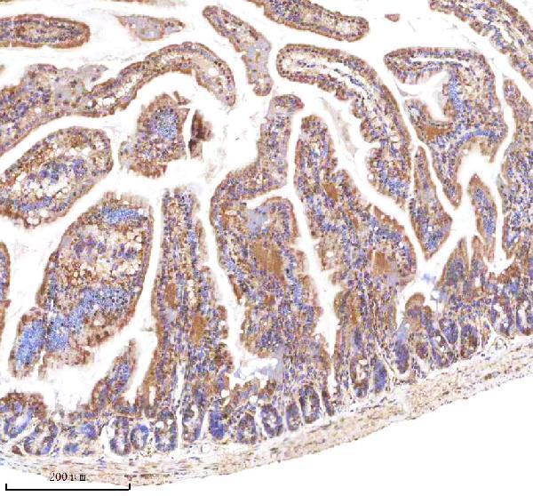
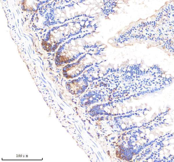
The IHC-validated antibody has paraffin-section images from mouse and rat small intestine and human kidney (catalog image captions); no IF data are supplied (catalog payload).

Real IHC data IHC analysis of LGR5 using anti-LGR5 antibody (A00239-2). LGR5 was detected in a paraffin-embedded section of mouse small intestine tissue. Heat mediated antigen retrieval was performed in EDTA buffer (pH 8.0, epitope retrieval solution). The tissue section was blocked with 10% goat serum. The tissue section was then incubated with 2 μg/ml rabbit anti-LGR5 Antibody (A00239-2) overnight at 4°C. Peroxidase Conjugated Goat Anti-rabbit IgG was used as secondary antibody and incubated for 30 minutes at 37°C. The tissue section was developed using HRP Conjugated Rabbit IgG Super Vision Assay Kit (Catalog # SV0002) with DAB as the chromogen.
Anti-LGR5 Antibody ®
Cat # A00239-2

A00239-2 is listed for IHC and reacts with human, mouse and rat samples (catalog applications and reactivity). Its IHC captions show paraffin sections of mouse and rat small intestine and human kidney (catalog image captions).

Which to pick: Choose A00239-2 for paraffin-section IHC across human, mouse and rat samples (catalog reactivity and IHC image captions). There is no IF/ICC-validated SKU in this payload, so no IF/ICC selection is supported (catalog applications and image captions). The A00239-2 captions specify paraffin sections but do not report the fixative (catalog IHC image captions).

Each figure is that product's own IHC / IF validation image from its datasheet.

References

  1. UniProt Consortium. UniProt entry O75473 (LGR5_HUMAN, Leucine-rich repeat-containing G protein-coupled receptor 5).
  2. Human Protein Atlas. LGR5 tissue IHC expression (reliability: Approved).
  3. Human Protein Atlas. LGR5 subcellular location (ICC-IF): Mainly localized to the nucleoplasm, vesicles and primary cilium transition zone. In addition localized to the centrosome and basal body..
  4. Human Protein Atlas. LGR5 antibody validation summary (1 antibodies).
  5. LgR5 expression and cancer stem cell hypothesis: clue to define the true origin of esophageal adenocarcinomas with and without Barrett's esophagus?. Journal of experimental & clinical cancer research : CR 2011 — PMC3058063.
  6. Expression of Leucine-rich Repeat-containing G-protein Coupled Receptor 5 and CD44: Potential Implications for Gastric Cancer Stem Cell Marker. Journal of cancer prevention 2016 — PMC5207613.
  7. Leucine-rich repeat-containing G protein-coupled receptor 5 and CD133 expression is associated with tumor progression and resistance to preoperative chemoradiotherapy in low rectal cancer. Oncology letters 2017 — PMC5727605.
  8. The Expression of Stem Cell Marker LGR5 and Its Coexpression with Β-Catenin in Sporadic Colorectal Carcinoma and Adenoma: A Comparative Immunohistochemical Study. Medicina (Kaunas, Lithuania) 2023 — PMC10383310.
  9. PubMed PMID:9642114 — UniProt-cited evidence.
  10. PubMed PMID:9849958 — UniProt-cited evidence.
  11. PubMed PMID:16541075 — UniProt-cited evidence.