This website uses cookies to ensure you get the best experience on our website.


- Table of Contents
Plan chromogenic LIAS IHC in paraffin sections using the reported cytoplasmic tissue pattern (HPA tissue IHC). Compare stained cell populations with controls, bearing in mind the low consistency between antibody staining and RNA expression (HPA tissue IHC).

Expected localisation, validated protocols, controls and antibodies — the at-a-glance facts below, then the full design guide.
| Expected localisation | Cytoplasmic tissue staining (HPA tissue IHC); mitochondrial LIAS (UniProt) | |
| Staining pattern | Cytoplasmic signal in glandular, neuronal and tubular cells (HPA tissue IHC) | |
| Antigen retrieval | EDTA pH 8.0 HIER, heat-mediated (datasheet A08974-1) | |
| Positive control | Cerebellum+4 more · see all | |
| Negative control | Adipose tissue+4 more · see all |
| Fixation | Matched tissue-IHC evidence does not establish the fixation claim. Validate the specimen-specific method before use. (selected-SKU IHC image A08974-1) | |
| Caveat | Staining shows low consistency with RNA expression (HPA tissue IHC) | |
| Regulation | No expression regulator annotated (UniProt) | |
| Isoform / epitope | 3 isoforms; mature chain spans residues 28–372 (UniProt) |
The catalog antibody’s IHC-P protocol is paired with one published LIAS IHC method for cartilage and knee-joint sections (PMC12283560).
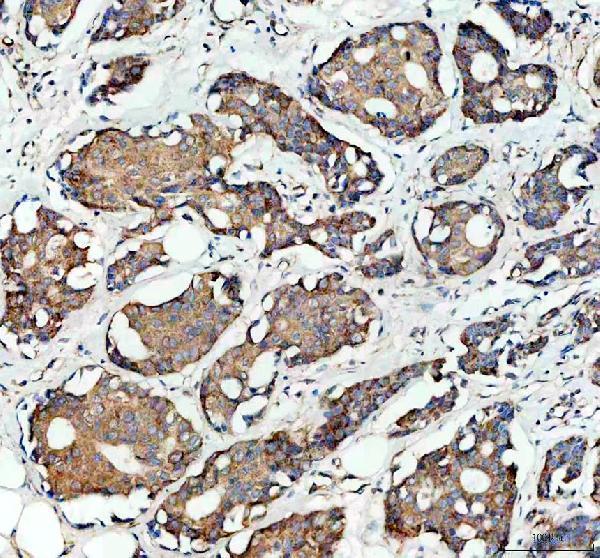
| Sample | Paraffin-embedded human breast cancer tissue; fixative not specified (datasheet A08974-1) |
| Fixation | Image fixative and duration unreported (datasheet A08974-1); verify before use. |
| Sectioning | 4–5 µm sections on charged slides (standard) |
| Deparaffinisation | Xylene, graded ethanol series to water (standard) |
| Antigen retrieval | Heat retrieval: EDTA pH 8.0 (datasheet A08974-1); 20 min, 95–100 °C (standard) |
| Peroxidase block | 3% H2O2, 10 min, room temperature (standard) |
| Blocking | 10% goat serum (datasheet A08974-1) |
| Primary antibody | Rabbit anti-LIAS, 1:50 recommended; image 1:100 (datasheet A08974-1) |
| Primary incubation | Overnight at 4 °C (datasheet A08974-1) |
| Detection | HRP-conjugated secondary, DAB chromogen (datasheet A08974-1) |
| Counterstain | Hematoxylin, blue, dehydrate and mount (standard) |
| Expected result | LIAS-positive staining in purkinje cells of cerebellum (HPA tissue IHC: Medium). HPA tissue profile: Cytoplasmic expression in several tissues. No signal in the no-primary control. |
LIAS is a mitochondrial protein with no transmembrane segment (UniProt O43766 topology). In paraffin IHC, expect cytoplasmic staining in several tissues, including Purkinje cells, neurons, glandular cells, and kidney tubule cells (HPA tissue IHC). HPA rates the tissue staining Approved but reports low consistency with RNA expression and pending external verification (HPA tissue IHC). Interpret the pattern as provisional, especially when comparing tissues.
| Cytoplasmic staining in Purkinje cells, cortical neurons, colon glands, or kidney tubules. | These cells show Medium staining in HPA tissue IHC. Cytoplasmic chromogen agrees with HPA’s tissue profile and LIAS’s mitochondrial location (UniProt O43766 subcellular location). Routine chromogenic IHC cannot resolve individual mitochondria reliably (general IHC practice). |
| Predominantly nuclear staining in a paraffin section, with little cytoplasmic signal. | This differs from the HPA tissue IHC profile and warrants an artefact check (HPA tissue IHC; general IHC practice). Nuclear staining is not categorically impossible: HPA ICC-IF reports approved nucleoplasmic localisation as its main signal, with additional mitochondrial localisation (HPA subcellular ICC-IF). |
| Strong staining in adipocytes or esophageal squamous epithelium, especially across many cells. | HPA lists those cell types as Not detected in tissue IHC (HPA tissue IHC). Check for antibody cross-reactivity or endogenous chromogenic activity before assigning the signal to LIAS (general IHC practice). A negative HPA result is a comparison point, not proof of biological absence. |
| Weak, even colour over cells and tissue spaces, without a clear cellular pattern. | This does not resemble the reported cytoplasmic pattern (HPA tissue IHC). Diffuse colour may reflect nonspecific antibody binding, inadequate blocking, or detection background (general IHC practice); it cannot establish LIAS distribution. |
| No staining in an adequately preserved section containing HPA-positive cells. | Purkinje cells, colon glandular cells, and kidney tubule cells are reported at Medium intensity (HPA tissue IHC). First check the assay’s positive control, antibody conditions, retrieval, and detection steps (general IHC practice). HPA’s reported low RNA–staining consistency limits how strongly one negative section can be interpreted (HPA tissue IHC). |
| Tissue and cell selection | HPA reports Medium staining in selected neurons, glandular cells, and kidney tubule cells, but Not detected staining in specified adipocytes, hematopoietic cells, and squamous cells (HPA tissue IHC). Compare the named cell population within a tissue, rather than treating a whole organ as uniformly positive or negative. |
| Compartment evidence across assays | UniProt places LIAS in mitochondria; HPA tissue IHC describes cytoplasmic expression, while approved HPA ICC-IF shows mainly nucleoplasm and additional mitochondria (UniProt O43766 subcellular location; HPA tissue IHC; HPA subcellular ICC-IF). This difference calls for assay-specific interpretation of a nuclear result. |
| Antibody and tissue evidence | Both listed antibodies, HPA018842 and HPA019076, have Approved IHC status; HPA nevertheless reports low consistency between staining and RNA expression and says external verification is pending (HPA antibodies; HPA tissue IHC). Approved status supports using the images as a guide, not treating every stain as confirmed LIAS. |
| Protein processing and variants | UniProt annotates a mature chain spanning residues 28–372 and 3 isoforms (UniProt O43766 processing; isoforms). No epitope position or isoform coverage is supplied, so those annotations cannot predict which variant the catalog antibody stains or justify a retrieval setting. |
| Retrieval conditions | No LIAS-specific retrieval condition or fixation sensitivity is supplied in the UniProt and HPA records. If signal is weak, review retrieval against the antibody’s actual instructions and an on-slide control (general IHC practice); do not infer a target-specific fixation effect from HPA staining levels. |
| Situation | Likely cause | Next action |
|---|---|---|
| A reported positive cell population has no chromogen. | Possible assay failure or weak detection; HPA’s Medium result is a reference observation, not a guarantee for every specimen (HPA tissue IHC; general IHC practice). | Check an on-slide positive control, antibody preparation, retrieval, and detection sequence; compare the same cell population in the HPA tissue images (general IHC practice; HPA tissue IHC). |
| The slide shows mainly nuclear staining. | The result conflicts with HPA’s cytoplasmic tissue IHC profile, although HPA ICC-IF reports approved nucleoplasmic signal (HPA tissue IHC; HPA subcellular ICC-IF). | Review morphology and controls, then repeat or confirm with an independent antibody before assigning the nuclear IHC signal to LIAS (general IHC practice; HPA antibodies). |
| HPA Not detected cells stain strongly. | Cross-reactivity or endogenous detection activity may explain staining in adipocytes or squamous cells (HPA tissue IHC; general IHC practice). | Inspect no-primary and detection controls, assess endogenous activity, and compare adjacent HPA-positive cell populations processed in the same run (general IHC practice; HPA tissue IHC). |
| Brown colour is widespread and obscures cell boundaries. | Nonspecific binding or detection background can produce a pattern unlike HPA’s cellular cytoplasmic staining (general IHC practice; HPA tissue IHC). | Check blocking, washing, antibody concentration, and no-primary control; score LIAS only after cell-associated signal can be separated from background (general IHC practice). |
| Two sections give different staining intensities. | Cell composition, assay variation, or genuine variation may contribute; HPA also flags low staining–RNA consistency (general IHC practice; HPA tissue IHC). | Compare matched cell types and run controls before interpreting the difference as a LIAS change (general IHC practice). Record the cell population and compartment alongside intensity. |
| Q: Should an IF/ICC image showing nucleoplasmic signal make nuclear IHC staining expected? | A: HPA ICC-IF reports mainly approved nucleoplasmic and additional mitochondrial localisation, whereas its tissue IHC profile is cytoplasmic (HPA subcellular ICC-IF; HPA tissue IHC). | Treat IF/ICC as a separate assay and confirm a nuclear paraffin IHC result independently; the IF/ICC pattern alone does not validate that IHC signal (general IHC practice). |


Comprehensive Human Protein Atlas IHC scoring per tissue (reliability: Approved — Low consistency between antibody staining and RNA expression data. Pending external verification.). Rows are taken directly from the HPA tissue chart — click any row's HPA link to view the source.
| Tissue | Cell type | Level | Evidence | Source |
|---|---|---|---|---|
| Cerebellum | Purkinje cells | Medium | Protein (IHC) | HPA → |
| Cerebral cortex | Neuronal cells | Medium | Protein (IHC) | HPA → |
| Colon | Glandular cells | Medium | Protein (IHC) | HPA → |
| Duodenum | Glandular cells | Medium | Protein (IHC) | HPA → |
| Epididymis | Glandular cells | Medium | Protein (IHC) | HPA → |
| Tissue | Cell type | Level | Evidence | Source |
|---|---|---|---|---|
| Adipose tissue | Adipocytes | Not detected | Protein (IHC) | HPA → |
| Bone marrow | Hematopoietic cells | Not detected | Protein (IHC) | HPA → |
| Breast | Adipocytes | Not detected | Protein (IHC) | HPA → |
| Cervix | Glandular cells | Not detected | Protein (IHC) | HPA → |
| Esophagus | Squamous epithelial cells | Not detected | Protein (IHC) | HPA → |
Use the catalog antibody’s tissue IHC conditions as a starting point, then assess compartment, cell type, and staining controls before scoring LIAS.
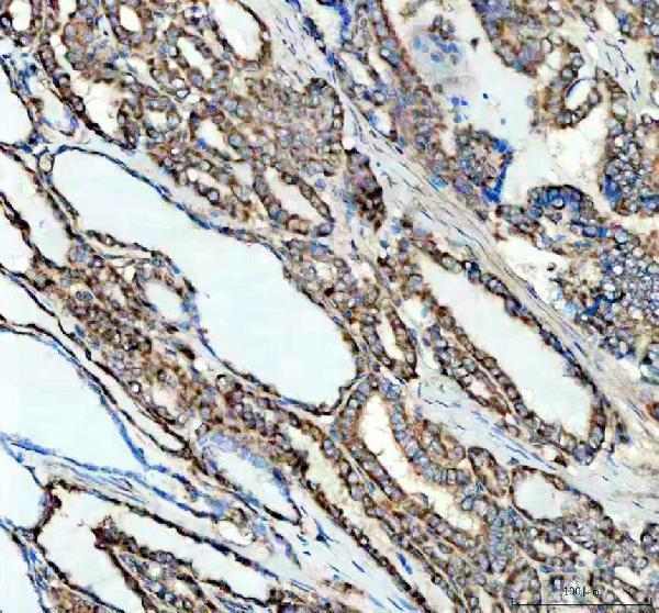
The IHC-validated anti-LIAS antibody has IHC images from paraffin sections of human breast and thyroid cancer and mouse and rat heart (A08974-1 IHC captions). No IF/ICC data are supplied (catalog applications).
A08974-1 will render with an IHC image of a human breast cancer paraffin section (A08974-1 IHC caption). Its additional IHC captions show human thyroid cancer and mouse and rat heart paraffin sections (A08974-1 IHC captions).
Which to pick: Choose A08974-1 for tissue IHC: it is a rabbit polyclonal antibody listed for IHC, with paraffin-section images and human, mouse and rat reactivity (catalog applications, dilution data and reactivity; A08974-1 IHC captions). The catalog lists 1:50 for IHC, while the pictured sections used 1:100 after EDTA retrieval at pH 8.0; their fixative is unreported (catalog IHC dilution; A08974-1 IHC captions). No IF/ICC-validated SKU is provided, so A08974-1 has no supported IF/ICC recommendation here (catalog applications and IF images).