This website uses cookies to ensure you get the best experience on our website.


- Table of Contents
Plan LILRB5 chromogenic IHC in paraffin sections using bone marrow hematopoietic cells and spleen red-pulp cells as medium-staining references (HPA tissue IHC). Start the catalog antibody at 1:100–1:300 (datasheet), and treat membrane staining as a molecular expectation (UniProt).

Expected localisation, validated protocols, controls and antibodies — the at-a-glance facts below, then the full design guide.
| Expected localisation | Membrane expected molecularly; tissue compartment unreported (UniProt; HPA tissue IHC) | |
| Staining pattern | Immune-cell subsets in several tissues; compartment unreported (HPA tissue IHC) | |
| Antigen retrieval | Citrate pH 6.0 HIER, 95–98 °C, 20 min (rule: cytoplasmic / membrane antigen) | |
| Positive control | Bone marrow+1 more · see all | |
| Negative control | Adipose tissue+4 more · see all |
| Fixation | Keep formalin fixation consistent across sections (standard IHC practice; not target-specific) | |
| Caveat | Low antibody–RNA agreement complicates interpretation (HPA tissue IHC) | |
| Regulation | NK-cell expression; induction unreported (UniProt) | |
| Isoform / epitope | 3 isoforms; extracellular versus cytoplasmic epitope needs verification (UniProt) |
The catalog antibody’s IHC-P protocol is accompanied by a published LILRB5 IHC protocol using paraffin-embedded human liver cancer tissue (PMC8943947).
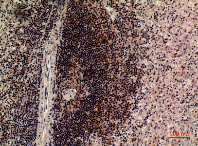
| Sample | Paraffin-embedded human spleen tissue; fixative not specified (datasheet A10961) |
| Fixation | Image fixative and duration unreported (datasheet A10961); verify before use. |
| Sectioning | 4–5 µm sections on charged slides (standard) |
| Deparaffinisation | Xylene, graded ethanol series to water (standard) |
| Antigen retrieval | Heat-induced epitope retrieval in citrate buffer, pH 6.0, 20 min at 95–98 °C (standard rule: cytoplasmic / membrane antigen) |
| Peroxidase block | 3% H2O2, 10 min, room temperature (standard) |
| Blocking | 10% normal serum of the secondary host, 30 min, room temperature (standard) |
| Primary antibody | Rabbit anti-LILRB5, 1:100-1:300 (datasheet A10961) |
| Primary incubation | Overnight at 4 °C (standard) |
| Detection | HRP-polymer secondary, DAB chromogen 5–10 min (standard) |
| Counterstain | Hematoxylin, blue, dehydrate and mount (standard) |
| Expected result | LILRB5-positive staining in hematopoietic cells of bone marrow (HPA tissue IHC: Medium). HPA tissue profile: Distinct expression in subsets of immune cells in several tissues. No signal in the no-primary control. |
LILRB5 is a membrane receptor with an extracellular region at residues 24–458 and a transmembrane segment at 459–479 (UniProt O75023 topology). In paraffin sections, expect staining in subsets of immune cells, especially bone marrow hematopoietic cells and spleen red pulp cells (HPA tissue IHC). HPA rates the tissue staining “Approved” but reports low consistency between antibody staining and RNA expression (HPA tissue IHC reliability).
| Discrete staining in some bone marrow hematopoietic cells or spleen red pulp cells. | This matches the reported medium tissue staining; assess the stained cells against neighboring unstained cells (HPA tissue IHC). Membrane localization is expected from receptor topology, although chromogenic sections may not cleanly resolve the cell edge (UniProt O75023 topology; standard IHC practice). |
| Predominantly nuclear staining, with little staining at the cell boundary. | A nuclear pattern conflicts with the membrane annotation and transmembrane topology (HPA subcellular; UniProt O75023 topology). Treat it as suspect and review morphology and detection controls before assigning it to LILRB5 (standard IHC practice). |
| Strong staining in adipocytes or glandular cells while expected immune cell staining is absent. | HPA reports adipocytes in adipose tissue and glandular cells in adrenal gland and appendix as not detected (HPA tissue IHC). Consider nonspecific antibody binding or endogenous detection activity; the staining alone does not identify which cause applies (standard IHC practice). |
| Diffuse color across cells and extracellular areas, without a distinct stained subset. | That distribution is difficult to reconcile with HPA’s distinct immune cell subset pattern (HPA tissue IHC). Review blocking, antibody concentration, washing, and detection controls as general IHC background checks (standard IHC practice). |
| No discernible staining in bone marrow or spleen. | These are reported medium staining contexts, so an absent result warrants a technical check (HPA tissue IHC). It does not by itself prove that the specimen lacks LILRB5; tissue composition and assay performance need review (standard IHC practice). |
| Cell population and tissue choice | Bone marrow hematopoietic cells and spleen red pulp cells are reported at medium, while lymph node and tonsil non-germinal center cells are low (HPA tissue IHC). Compare like cell populations when judging intensity; a whole-section average can obscure a restricted subset (standard IHC practice). |
| Antibody evidence | The listed HPA antibody HPA012069 is a rabbit polyclonal antibody with IHC status “Approved”; no IHC “Enhanced” status is supplied (HPA antibodies). HPA also flags low consistency with RNA expression, so unexpected staining deserves control-based review (HPA tissue IHC reliability). |
| Sequence features and epitope interpretation | The mature chain begins at residue 24, with extracellular and cytoplasmic regions separated by a transmembrane segment (UniProt O75023 processing and topology). The antibody epitope is not supplied; these features cannot establish antigen retrieval requirements or predict staining after fixation. |
| IF/ICC Q: what localization should be expected? | A: Membrane localization is consistent with UniProt topology and HPA’s subcellular summary (UniProt O75023 topology; HPA subcellular). HPA supplies no ICC-IF image-bearing cell lines or main-location annotation here, so there is no image-based IF pattern to compare (HPA subcellular). |
| Situation | Likely cause | Next action |
|---|---|---|
| Expected immune cell staining is absent. | The chosen field may lack the relevant subset, or the IHC run may have failed (HPA tissue IHC; standard IHC practice). | Inspect cell morphology, then check a bone marrow or spleen section known to contain the reported cells and verify the run controls (HPA tissue IHC; standard IHC practice). |
| Only faint signal appears in lymph node or tonsil. | Non-germinal center cells have a reported low staining level in those tissues (HPA tissue IHC). | Judge the relevant cell population rather than requiring the medium intensity reported in bone marrow and spleen (HPA tissue IHC). |
| Adipocytes stain strongly. | Adipocytes are reported as not detected in adipose tissue; nonspecific staining or detection activity is possible (HPA tissue IHC; standard IHC practice). | Compare with a no-primary control and inspect whether any immune cell subset retains a distinct pattern (standard IHC practice; HPA tissue IHC). |
| Brown signal fills the section or follows tissue edges. | Diffuse signal may reflect background from blocking, washing, or detection rather than the reported subset pattern (standard IHC practice; HPA tissue IHC). | Review the no-primary control and detection steps, then reassess discrete staining in morphologically identified cells (standard IHC practice). |
| A strong nuclear pattern dominates. | This conflicts with the supplied membrane localization and transmembrane topology (HPA subcellular; UniProt O75023 topology). | Check controls and tissue morphology before scoring it as LILRB5; seek independent support for any unexpected compartment assignment (standard IHC practice). |
| Protein staining and tissue RNA expectations disagree. | HPA explicitly reports low consistency between antibody staining and RNA expression (HPA tissue IHC reliability). | Report the observed cell-level staining and antibody status separately from RNA expression; avoid using RNA alone to reclassify an IHC-positive or IHC-negative cell (HPA tissue IHC; standard IHC practice). |


Comprehensive Human Protein Atlas IHC scoring per tissue (reliability: Approved — Low consistency between antibody staining and RNA expression data. Antibody staining in cells/structures not annotated, view images.). Rows are taken directly from the HPA tissue chart — click any row's HPA link to view the source.
| Tissue | Cell type | Level | Evidence | Source |
|---|---|---|---|---|
| Bone marrow | Hematopoietic cells | Medium | Protein (IHC) | HPA → |
| Spleen | Cells in red pulp | Medium | Protein (IHC) | HPA → |
| Tissue | Cell type | Level | Evidence | Source |
|---|---|---|---|---|
| Adipose tissue | Adipocytes | Not detected | Protein (IHC) | HPA → |
| Adrenal gland | Glandular cells | Not detected | Protein (IHC) | HPA → |
| Appendix | Glandular cells | Not detected | Protein (IHC) | HPA → |
| Breast | Adipocytes | Not detected | Protein (IHC) | HPA → |
| Bronchus | Respiratory epithelial cells | Not detected | Protein (IHC) | HPA → |
Troubleshoot LILRB5 staining by checking immune cell context, membrane localisation and controls before interpreting signal intensity (UniProt O75023 topology; HPA tissue IHC).
A10961 is human-reactive and listed for IHC and IF (catalog: reactivity and applications). Its IHC image shows paraffin-embedded human spleen; no IF image is supplied (catalog: image captions).
A10961 is listed for human IHC and IF (catalog: reactivity and applications). Its supplied image shows IHC on paraffin-embedded human spleen at 1:100; no IF image is supplied (catalog: image captions).
Which to pick: For paraffin-section IHC, choose A10961: its own image shows human spleen at 1:100, within its listed 1:100–1:300 IHC range (catalog: IHC image caption and dilution). The fixative is unreported in that caption (catalog: IHC image caption). For IF, A10961 is listed at 1:50, but the payload supplies no IF image or ICC validation; no cross-species option is supported because A10961 lists Human reactivity only (catalog: IF dilution, image captions, applications and reactivity).