This website uses cookies to ensure you get the best experience on our website.


- Table of Contents
Use the catalog antibody at 1:50–1:200 for chromogenic IHC on paraffin sections (datasheet: A01323). Compare high staining in cerebral cortex neurons with undetected staining in skeletal muscle myocytes (HPA tissue IHC), and account for the antibody’s LIMK1/2 recognition (datasheet: A01323).

Expected localisation, validated protocols, controls and antibodies — the at-a-glance facts below, then the full design guide.
| Expected localisation | Cytoplasm and nucleus (HPA tissue IHC) | |
| Staining pattern | Cytoplasmic and nuclear staining in most tissues (HPA tissue IHC) | |
| Antigen retrieval | Tris-EDTA pH 9.0 HIER, 95–98 °C, 20 min (rule: nuclear antigen) | |
| Positive control | Appendix+4 more · see all | |
| Negative control | Skeletal muscle |
| Fixation | Keep formalin fixation consistent across samples. (standard IHC practice; not target-specific) | |
| Caveat | The catalog antibody recognizes LIMK1/2 (datasheet: A01323) | |
| Regulation | Highest expression in nervous system (UniProt) | |
| Isoform / epitope | 4 isoforms; check epitope coverage (UniProt) |
The catalog antibody’s IHC-P protocol (datasheet) appears alongside three published LIMK1 IHC protocols (PMC7176864; PMC5768095; PMC8020211).
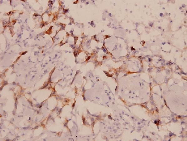
| Sample | Paraffin-embedded human breast carcinoma tissue; fixative not specified (datasheet A01323) |
| Fixation | Image fixative and duration unreported (datasheet A01323); verify before use. |
| Sectioning | 4–5 µm sections on charged slides (standard) |
| Deparaffinisation | Xylene, graded ethanol series to water (standard) |
| Antigen retrieval | Heat-induced epitope retrieval in Tris-EDTA buffer, pH 9.0, 20 min at 95–98 °C (standard rule: nuclear antigen) |
| Peroxidase block | 3% H2O2, 10 min, room temperature (standard) |
| Blocking | 10% normal serum of the secondary host, 30 min, room temperature (standard) |
| Primary antibody | Rabbit anti-LIMK1, 1:50-1:200 (datasheet A01323) |
| Primary incubation | Overnight at 4 °C (standard) |
| Detection | HRP-polymer secondary, DAB chromogen 5–10 min (standard) |
| Counterstain | Hematoxylin, blue, dehydrate and mount (standard) |
| Expected result | LIMK1-positive staining in glandular cells of appendix (HPA tissue IHC: High). HPA tissue profile: Cytoplasmic and nuclear expression in most tissues. No signal in the no-primary control. |
LIMK1 staining should be predominantly cytoplasmic, with nuclear staining possible (UniProt P53667 localisation; HPA: cytoplasmic and nuclear expression in most tissues). Look for strong signal in cerebral cortex neurons, cerebellar molecular-layer cells, and several epithelial or glandular cell populations (HPA: High). LIMK1 has no transmembrane segment (UniProt P53667 topology). HPA rates its tissue IHC profile Approved, with medium consistency between staining and RNA data (HPA: reliability).
| Clear cytoplasmic staining, sometimes accompanied by nuclear staining, in the expected cells. | This fits the reported tissue pattern (HPA: cytoplasmic and nuclear expression in most tissues). Cytoplasm should generally predominate (UniProt P53667 localisation). Assess the cell population as well as the compartment: cerebral cortex neurons and bronchial respiratory epithelial cells are examples with High HPA staining (HPA: tissue IHC). |
| Predominantly crisp cell-surface staining, with little cytoplasmic or nuclear signal. | Treat this as an unexpected compartment pattern and investigate an artefact or off-target signal. LIMK1 has no transmembrane segment (UniProt P53667 topology), while the reported tissue pattern is cytoplasmic and nuclear (HPA: tissue IHC). Nuclear signal alone is not grounds for rejection because both sources permit nuclear localisation (UniProt P53667 localisation; HPA: tissue IHC). |
| Strong staining in skeletal myocytes, especially if the expected positive cells are unstained. | Check for cross-reactivity or endogenous detection activity before assigning the signal to LIMK1 (general IHC practice). HPA reports skeletal myocytes as Not detected by tissue IHC (HPA: skeletal muscle), although UniProt reports lower expression in skeletal muscle than in the nervous system (UniProt P53667 tissue specificity). That distinction makes an isolated strong result worth checking, rather than conclusive evidence of a false positive. |
| Diffuse colour across tissue, empty spaces, or many unrelated cell populations. | Interpret localisation only after background is controlled (general IHC practice). Widespread colour that obscures cell borders and nuclei cannot reliably establish the cytoplasmic and nuclear distribution reported for LIMK1 (HPA: tissue IHC). Review blocking, washes, antibody concentration, and the detection control as general chromogenic IHC checks. |
| No convincing staining in a cerebral cortex section with identifiable neuronal cells. | A negative result in that comparison tissue is unexpected: HPA reports High staining in cerebral cortex neurons, and UniProt reports its highest adult brain expression in cerebral cortex (HPA: cerebral cortex; UniProt P53667 tissue specificity). First check tissue preservation and the general IHC workflow. An absent signal alone does not identify which step failed. |
| Tissue and cell choice | For a positive comparison, use a reported High population, such as cerebral cortex neurons or bronchial respiratory epithelial cells (HPA: tissue IHC). Skeletal myocytes are reported Not detected and offer a contrasting population (HPA: skeletal muscle). These are observed staining levels, not guaranteed outcomes with every section or antibody. |
| Compartment and topology | Expect predominantly cytoplasmic localisation, with nuclear and cytoskeletal localisation also reported (UniProt P53667 localisation). HPA describes cytoplasmic and nuclear tissue expression (HPA: tissue IHC). The absence of a transmembrane segment makes a dominant cell-surface outline difficult to reconcile with the supplied target record (UniProt P53667 topology). |
| Strength of the tissue evidence | HPA labels the tissue IHC profile Approved and describes medium consistency between antibody staining and RNA expression; external characterization supports staining (HPA: reliability). Both listed antibodies are IHC Approved (HPA: HPA028516, HPA073571). Use the reported pattern as a comparison, while checking unexpected staining with controls. |
| Which pattern applies to IF/ICC? | For IF/ICC, HPA reports supported localisation to nuclear speckles and cytosol (HPA: subcellular). HPA lists ICC support for HPA028516; no ICC status is supplied for HPA073571 (HPA: antibody validation). Nuclear speckles are an IF/ICC observation and should not be required as a distinct feature in chromogenic tissue IHC. |
| Epitope and isoform limits | UniProt lists four LIMK1 isoforms and LIM, PDZ, and kinase domains (UniProt P53667 isoforms and domains). The supplied evidence gives no antibody epitope or isoform coverage, so it cannot predict whether a particular isoform will stain. Likewise, it gives no target-specific fixation sensitivity or antigen-retrieval requirement. |
| Situation | Likely cause | Next action |
|---|---|---|
| No signal in a reported High cell population. | The comparison conflicts with reported staining, but does not identify a failed step (HPA: cerebral cortex neurons, High). | Check that the expected cells are present, then review antibody application, retrieval, detection, and counterstaining as general IHC workflow checks. Compare a same-run positive control before interpreting the section as LIMK1-negative. |
| Strong staining appears only in skeletal myocytes. | HPA reports skeletal myocytes as Not detected by tissue IHC (HPA: skeletal muscle); cross-reactivity or endogenous detection activity is possible (general IHC practice). | Compare a reported High population and inspect a detection control without primary antibody. Review the signal's cellular distribution before assigning it to LIMK1. |
| Cell surfaces dominate the staining. | That distribution differs from the reported cytoplasmic and nuclear tissue pattern (HPA: tissue IHC) and the no-transmembrane topology (UniProt P53667 topology). | Recheck localisation against a positive comparison section and a detection control. If cell-surface colour persists without matching intracellular signal, treat target attribution as uncertain. |
| Diffuse chromogen obscures the cells. | Background can prevent a reliable compartment call (general IHC practice). | Review blocking, washes, primary antibody concentration, and detection timing as general IHC checks. Reassess whether cytoplasm and nuclei can be distinguished before scoring the LIMK1 pattern. |
| Nuclear staining is present alongside cytoplasmic staining. | Both compartments are reported in tissue IHC (HPA: tissue IHC), although cytoplasmic localisation predominates in UniProt (UniProt P53667 localisation). | Evaluate whether the signal follows the expected cell population and has manageable background. Do not reject the section solely because nuclei stain; compare compartment balance with the positive tissue. |
| Results vary between antibody preparations or sections. | The supplied record does not establish epitope coverage or target-specific fixation sensitivity; HPA describes only medium staining-to-RNA consistency (HPA: reliability). | Compare matched tissue and cell populations, include same-run controls, and record retrieval and detection conditions as general IHC practice. Avoid attributing the difference to LIMK1 epitope masking without supporting evidence. |


Comprehensive Human Protein Atlas IHC scoring per tissue (reliability: Approved — Medium consistency between antibody staining and RNA expression data. External characterization data supports antibody staining.). Rows are taken directly from the HPA tissue chart — click any row's HPA link to view the source.
| Tissue | Cell type | Level | Evidence | Source |
|---|---|---|---|---|
| Appendix | Glandular cells | High | Protein (IHC) | HPA → |
| Breast | Glandular cells | High | Protein (IHC) | HPA → |
| Bronchus | Respiratory epithelial cells | High | Protein (IHC) | HPA → |
| Cerebellum | Cells in molecular layer | High | Protein (IHC) | HPA → |
| Cerebral cortex | Neuronal cells | High | Protein (IHC) | HPA → |
| Tissue | Cell type | Level | Evidence | Source |
|---|---|---|---|---|
| Skeletal muscle | Myocytes | Not detected | Protein (IHC) | HPA → |
Troubleshoot LIMK1 staining in paraffin sections by checking retrieval, compartment, cell type and antibody specificity before comparing chromogenic signals.
A01323 has real IHC data from paraffin-embedded human breast carcinoma (A01323 IHC image caption). Its listed reactivity covers human, mouse and rat (A01323 catalog reactivity).
A01323 is the sole card and lists IHC among its applications (A01323 catalog applications). Its image shows paraffin-embedded human breast carcinoma stained at 1:100 (A01323 IHC image caption).
Which to pick: Choose A01323 for paraffin-section IHC based on its human breast carcinoma image; the fixative is unreported (A01323 IHC image caption). No listed SKU has an IF/ICC application or IF image, so there is no validated IF/ICC pick here (A01323 catalog applications and image alts). A01323 is the cross-species candidate because it is listed as reactive with human, mouse and rat, though its IHC image documents only human tissue (A01323 catalog reactivity; A01323 IHC image caption).