This website uses cookies to ensure you get the best experience on our website.


- Table of Contents
Plan LIPE chromogenic IHC on paraffin sections using adipocytes as a high-staining reference (HPA tissue IHC). This guide covers fixation, controls and interpretation of staining, with cytosol, caveolae and lipid droplets as molecular localisation expectations (UniProt).

Expected localisation, validated protocols, controls and antibodies — the at-a-glance facts below, then the full design guide.
| Expected localisation | Cytosol, caveolae and lipid droplets (UniProt) | |
| Staining pattern | High staining in adipocytes; compartment unspecified (HPA tissue IHC) | |
| Antigen retrieval | Citrate pH 6.0 HIER, 95–98 °C, 20 min (rule: cytoplasmic / membrane antigen) | |
| Positive control | Adipose tissue+4 more · see all | |
| Negative control | Adrenal gland+4 more · see all |
| Fixation | Keep fixation consistent across sections (standard IHC practice; not target-specific); Matched tissue-IHC evidence does not establish the fixation claim. Validate the specimen-specific method before use. (selected-SKU IHC image A06762) | |
| Caveat | Presumed off-target staining was observed and disregarded (HPA tissue IHC) | |
| Regulation | Insulin shifts LIPE from caveolae to cytoplasm (UniProt) | |
| Isoform / epitope | 2 isoforms; epitope differences are unspecified (UniProt) |
Compare the catalog antibody’s IHC-P protocol with the published LIPE IHC protocol for mouse sciatic nerve (PMC9893521).
| Sample | Tissue sections; selected-image fixative not specified (standard IHC workflow) |
| Fixation | Image fixative and duration unreported (datasheet A06762); verify before use. |
| Sectioning | 4–5 µm sections on charged slides (standard) |
| Deparaffinisation | Xylene, graded ethanol series to water (standard) |
| Antigen retrieval | Heat-induced epitope retrieval in citrate buffer, pH 6.0, 20 min at 95–98 °C (standard rule: cytoplasmic / membrane antigen) |
| Peroxidase block | 3% H2O2, 10 min, room temperature (standard) |
| Blocking | 10% normal serum of the secondary host, 30 min, room temperature (standard) |
| Primary antibody | Rabbit anti-LIPE, 2.5 μg/mL (datasheet A06762) |
| Primary incubation | Overnight at 4 °C (standard) |
| Detection | HRP-polymer secondary, DAB chromogen 5–10 min (standard) |
| Counterstain | Hematoxylin, blue, dehydrate and mount (standard) |
| Expected result | LIPE-positive staining in adipocytes of adipose tissue (HPA tissue IHC: High). HPA tissue profile: High expression in adipocytes. No signal in the no-primary control. |
LIPE is found in the cytosol, at caveolae and on lipid droplets, with no annotated transmembrane segment (UniProt Q05469). In paraffin-section IHC, expect high staining in adipocytes and elongated or late spermatids; intestinal endocrine cells show medium staining (HPA tissue IHC). HPA rates the tissue profile Enhanced, while noting medium consistency with RNA data and presumed off-target binding that was disregarded (HPA tissue IHC).
| Strong staining in adipocytes in adipose tissue or breast, with a predominantly cytoplasmic distribution. | This fits the high adipocyte signal reported in both tissues (HPA tissue IHC). Cytosolic localization is supported by ICC-IF, while UniProt also places LIPE at caveolae and lipid droplets (HPA subcellular; UniProt Q05469). Do not require a sharply defined droplet rim to call a paraffin-section result positive. |
| Strong staining in elongated or late spermatids in testis, or moderate staining in intestinal endocrine cells. | These match distinct reported cell populations: high signal in elongated or late spermatids, and medium signal in endocrine cells of the appendix, colon, duodenum, rectum and small intestine (HPA tissue IHC). Compare the stained cells with the section's morphology before scoring intensity. |
| Predominantly nuclear staining, or a pattern confined to another compartment across the positive cells. | Reassess specificity: supported ICC-IF localization is cytosolic, and UniProt lists cytosol, caveolae and lipid droplets for LIPE (HPA subcellular; UniProt Q05469). An unexpected compartment is a warning sign, though it does not alone identify the artefact or prove every stained cell negative. |
| Strong signal in unrelated cell populations, including cells listed as not detected in HPA tissue IHC. | Consider cross-reactivity or endogenous chromogen-generating activity; use a no-primary control and review cell identity (general IHC practice). HPA reports presumed off-target binding despite its Enhanced tissue reliability rating (HPA tissue IHC). A single unexpected positive cell type needs corroboration before assignment to LIPE. |
| No convincing staining in adipocytes of an otherwise intact adipose tissue section. | This conflicts with HPA's high adipocyte staining and makes a technical failure or an unsuitable antibody application worth checking (HPA tissue IHC; general IHC practice). Verify tissue preservation, retrieval and detection controls before interpreting absence as biological; HPA does not provide LIPE-specific fixation sensitivity. |
| Cell identity and reference tissue | Adipocytes in adipose tissue and breast, and elongated or late spermatids in testis, are high-staining references (HPA tissue IHC). Endocrine cells in the listed intestinal tissues have medium signal (HPA tissue IHC); avoid judging them against an adipocyte-level threshold. |
| Compartment and topology | Cytosol is the supported ICC-IF location; UniProt also lists caveolae and lipid droplets and annotates no transmembrane segment (HPA subcellular; UniProt Q05469). Interpret a cytoplasmic pattern in the appropriate cells without requiring membrane-only staining. |
| Evidence and antibody choice | The tissue profile is Enhanced but has medium agreement with RNA and notes disregarded presumed off-target binding (HPA tissue IHC). CAB017700 has Enhanced IHC validation; HPA006567 lists Supported ICC validation but no IHC status in this payload (HPA antibodies). |
| Isoforms and modified residues | UniProt lists two isoforms and several phosphoserines for LIPE (UniProt Q05469). Neither record identifies the IHC antibody's epitope or shows that these features change paraffin-section staining; do not infer an isoform-specific or phosphorylation-specific pattern. |
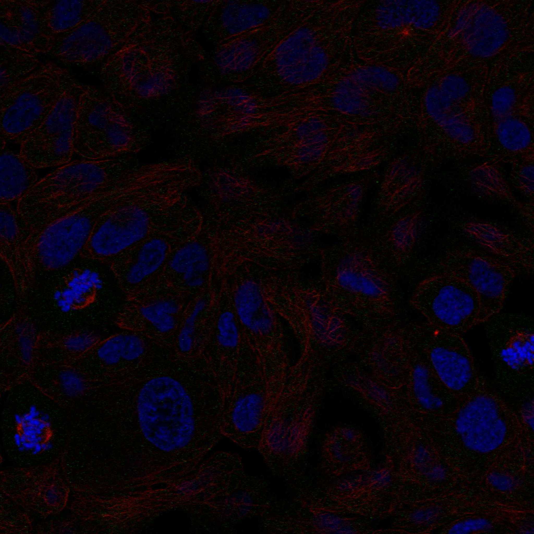
| IF/ICC Q: What localization is expected? | A: Cytosolic staining is supported by HPA ICC-IF, with images listed for RT-4, SH-SY5Y and U2OS (HPA subcellular). UniProt also lists caveolae and lipid droplets (UniProt Q05469). This localization answer does not establish an IF/ICC protocol for the IHC application. |
| Situation | Likely cause | Next action |
|---|---|---|
| Positive adipose tissue has no adipocyte signal. | The result disagrees with HPA's high adipocyte staining; a failed retrieval or detection step is possible (HPA tissue IHC; general IHC practice). | Check a known-positive section and run-level detection controls, then review retrieval against the catalog antibody's IHC instructions (general IHC practice). Do not assume a LIPE-specific fixation effect. |
| Staining is weak in the expected adipocytes. | Weak signal can reflect a general IHC sensitivity issue; HPA reports high adipocyte signal but supplies no LIPE-specific retrieval or fixation response (HPA tissue IHC; general IHC practice). | Confirm adipocyte morphology, antibody application, retrieval and detection performance with controls before changing the interpretation threshold (general IHC practice). |
| Broad brown haze obscures cell boundaries. | Nonspecific binding, incomplete blocking or detection background can obscure a cellular pattern (general IHC practice). | Compare a no-primary control; review blocking, washes and chromogen development, then score only interpretable cells (general IHC practice). |
| Unexpected nuclear staining dominates. | This differs from supported cytosolic localization and may indicate nonspecific staining or misread morphology (HPA subcellular; general IHC practice). | Check the counterstain and a no-primary control, then compare with cytoplasmic staining in a known-positive tissue (general IHC practice; HPA tissue IHC). |
| Cells outside the expected populations stain strongly. | Cross-reactivity or endogenous detection activity is possible; HPA notes presumed off-target binding in its tissue assessment (HPA tissue IHC; general IHC practice). | Identify the cells morphologically, inspect a no-primary control and corroborate an unexpected population independently before reporting LIPE positivity (general IHC practice). |
| A reported HPA-negative cell population stains. | HPA lists several cell populations as not detected, including adrenal glandular cells and bone-marrow hematopoietic cells (HPA tissue IHC). Unexpected staining alone does not establish its source. | Check the exact cell type and controls; treat a discordant result as unresolved until independently corroborated (HPA tissue IHC; general IHC practice). |


Comprehensive Human Protein Atlas IHC scoring per tissue (reliability: Enhanced — Medium consistency between antibody staining and RNA expression data. Presumed off target binding observed and disregarded.). Rows are taken directly from the HPA tissue chart — click any row's HPA link to view the source.
| Tissue | Cell type | Level | Evidence | Source |
|---|---|---|---|---|
| Adipose tissue | Adipocytes | High | Protein (IHC) | HPA → |
| Breast | Adipocytes | High | Protein (IHC) | HPA → |
| Testis | Elongated or late spermatids | High | Protein (IHC) | HPA → |
| Appendix | Endocrine cells | Medium | Protein (IHC) | HPA → |
| Colon | Endocrine cells | Medium | Protein (IHC) | HPA → |
| Tissue | Cell type | Level | Evidence | Source |
|---|---|---|---|---|
| Adrenal gland | Glandular cells | Not detected | Protein (IHC) | HPA → |
| Bone marrow | Hematopoietic cells | Not detected | Protein (IHC) | HPA → |
| Bronchus | Respiratory epithelial cells | Not detected | Protein (IHC) | HPA → |
| Caudate | Glial cells | Not detected | Protein (IHC) | HPA → |
| Cerebellum | Cells in granular layer | Not detected | Protein (IHC) | HPA → |
Troubleshoot LIPE staining in paraffin sections by checking retrieval, cell type, cytoplasmic localisation and controls before comparing signal intensity.
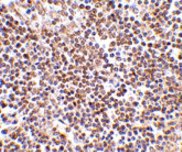
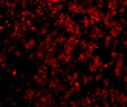
A06762 has IHC data from human lymph node tissue and IF data from human lymph node cells; Human, Mouse and Rat are listed as reactive species (A06762 image captions; catalog reactivity).
A06762 is the only SKU with a rendered card; its IHC image shows human lymph node tissue at 2.5 μg/mL (A06762 IHC image caption). Its IF image shows human lymph node cells at 20 μg/mL (A06762 IF image caption).
Which to pick: Choose A06762 for paraffin-section IHC: IHC-P is listed, and its own IHC image shows human lymph node tissue; the fixative is unreported (A06762 applications; A06762 IHC image caption). Choose A06762 for IF/ICC evaluation because IF is listed and its own IF image shows human lymph node cells (A06762 applications; A06762 IF image caption). For Mouse or Rat samples, both A06762 and the monoclonal P06762-1 list reactivity, but the supplied images demonstrate only human samples, and P06762-1 has no supplied IHC image (catalog reactivity; image captions; P06762-1 clone and image fields).