This website uses cookies to ensure you get the best experience on our website.


- Table of Contents
Plan chromogenic IHC for LITAF in paraffin sections using its cytoplasmic tissue pattern and strong lymphoid staining (HPA tissue IHC). This guide covers positive controls, antibody dilution, and interpretation of macrophage-rich regions (HPA tissue IHC; datasheet).

Expected localisation, validated protocols, controls and antibodies — the at-a-glance facts below, then the full design guide.
| Expected localisation | Cytoplasmic tissue staining; endosomal membranes are expected (HPA tissue IHC; UniProt) | |
| Staining pattern | Cytoplasmic staining, most abundant in lymphoid tissues (HPA tissue IHC) | |
| Antigen retrieval | Tris-EDTA pH 9.0 HIER, 95–98 °C, 20 min (rule: nuclear antigen) | |
| Positive control | Bone marrow+4 more · see all | |
| Negative control | Esophagus+3 more · see all |
| Fixation | Keep fixation consistent across paraffin sections. (standard IHC practice; not target-specific) | |
| Caveat | Macrophage-rich regions may dominate the signal (HPA tissue IHC) | |
| Regulation | Expression regulation not specified (UniProt) | |
| Isoform / epitope | 3 isoforms; extracellular versus cytoplasmic epitope topology is unassigned (UniProt) |
The catalog antibody IHC-P protocol is accompanied by four published LITAF IHC procedures covering pancreatic specimens, tonsil follicles, and mouse brain (PMC5790452; PMC4111142; PMC10447466; PMC11705406).
| Sample | Paraffin-embedded human prostate tissue; fixative not specified (datasheet A03043) |
| Fixation | Image fixative and duration unreported (datasheet A03043); verify before use. |
| Sectioning | 4–5 µm sections on charged slides (standard) |
| Deparaffinisation | Xylene, graded ethanol series to water (standard) |
| Antigen retrieval | Heat-induced epitope retrieval in Tris-EDTA buffer, pH 9.0, 20 min at 95–98 °C (standard rule: nuclear antigen) |
| Peroxidase block | 3% H2O2, 10 min, room temperature (standard) |
| Blocking | 10% normal serum of the secondary host, 30 min, room temperature (standard) |
| Primary antibody | Rabbit anti-LITAF, 1:50-1:200 (datasheet A03043) |
| Primary incubation | Overnight at 4 °C (standard) |
| Detection | HRP-polymer secondary, DAB chromogen 5–10 min (standard) |
| Counterstain | Hematoxylin, blue, dehydrate and mount (standard) |
| Expected result | LITAF-positive staining in hematopoietic cells of bone marrow (HPA tissue IHC: High). HPA tissue profile: General cytoplasmic expression, most abundant in lymphoid tissues. No signal in the no-primary control. |
LITAF is membrane-associated in endosomal and lysosomal compartments and can enter the nucleus despite having no transmembrane segment (UniProt Q99732 localization and topology). In tissue IHC, expect mainly cytoplasmic staining, especially in lymphoid tissue, lung macrophages, and bone marrow hematopoietic cells (HPA tissue IHC). HPA rates its tissue IHC evidence Approved, with medium consistency between staining and RNA expression (HPA tissue IHC).
| Cytoplasmic staining is strong in lung macrophages, bone marrow hematopoietic cells, or lymph node non-germinal center cells (HPA tissue IHC: High). | This fits the reported tissue pattern (HPA tissue IHC). Compare cell populations within the section: the expected result is cellular staining with recognizable boundaries, while reported intensity varies by tissue and cell type (HPA tissue IHC; general IHC practice). |
| Punctate or perinuclear cytoplasmic staining accompanies a broader cytoplasmic signal (UniProt Q99732 localization; HPA tissue IHC). | An endosomal or lysosomal distribution is plausible because LITAF associates with those membranes (UniProt Q99732 localization). Tissue IHC alone cannot identify the stained organelle; reserve that assignment for localization evidence beyond chromogenic morphology (general IHC practice). |
| Strong staining appears only in an unexpected compartment, such as extracellular material (UniProt Q99732 localization). | Review it as possible nonspecific signal because extracellular localization is absent from the supplied record (UniProt Q99732 localization). Nuclear staining alone is inconclusive: UniProt reports nuclear translocation, and HPA ICC-IF supports nucleoplasmic localization (UniProt Q99732; HPA subcellular ICC-IF). |
| Squamous epithelium or placental trophoblasts stain strongly while expected positive cells remain faint (HPA tissue IHC: Not detected in those cell types). | The distribution conflicts with HPA tissue IHC and warrants checks for antibody cross-reactivity or endogenous detection activity (HPA tissue IHC; general IHC practice). Interpret placenta by cell type: UniProt reports placental expression, whereas HPA reports trophoblasts as undetected (UniProt Q99732 tissue specificity; HPA tissue IHC). |
| Color spreads across many cell types or obscures tissue architecture, including areas expected to be weak (HPA tissue IHC: variable cell-type levels). | Diffuse background limits compartment and cell-type scoring (general IHC practice). Compare the test section with an appropriate detection control before attributing widespread color to LITAF; low or absent HPA staining is a useful comparison, not proof of a universally negative tissue (HPA tissue IHC; general IHC practice). |
| Compartment and topology (UniProt Q99732; HPA subcellular ICC-IF) | LITAF associates with endosome and lysosome membranes but has no transmembrane segment (UniProt Q99732). HPA tissue IHC describes general cytoplasmic expression; HPA ICC-IF additionally supports nucleoplasm, nuclear speckles, Golgi apparatus, and plasma membrane localization (HPA tissue IHC; HPA subcellular ICC-IF). |
| Cell-type variation (HPA tissue IHC) | HPA reports High staining in lung macrophages, spleen red-pulp cells, and tonsil non-germinal center cells, but Medium staining in adipocytes and Low staining in several neuronal or glandular populations (HPA tissue IHC). Score the relevant cells rather than averaging an entire mixed tissue (general IHC practice). |
| Placental evidence (UniProt Q99732; HPA tissue IHC) | UniProt describes placenta among predominant expression sites, while HPA reports trophoblastic cells as Not detected by tissue IHC (UniProt Q99732 tissue specificity; HPA tissue IHC). Neither statement identifies every placental cell population; avoid treating trophoblasts as an assured positive control (HPA tissue IHC). |
| Isoforms and processing (UniProt Q99732) | UniProt lists 3 isoforms and a chain spanning residues 1–161, with no signal peptide or propeptide (UniProt Q99732). The supplied evidence gives no antibody epitope or isoform coverage, so a staining difference cannot be assigned to a particular isoform (UniProt Q99732; supplied antibody record). |
| Antibody validation (HPA antibodies; HPA tissue IHC) | Target-specific fixation effects are not established by the supplied assay evidence. Verify with a matched IHC source before attributing a result to fixation. |
| IF/ICC Q: Should the same compartment rule apply? (HPA subcellular ICC-IF) | A: Evaluate IF/ICC against its own localization evidence: HPA supports nucleoplasm and also reports nuclear speckles, Golgi apparatus, vesicles, and plasma membrane (HPA subcellular ICC-IF). This tissue IHC pattern is a separate readout (HPA tissue IHC). |
| Situation | Likely cause | Next action |
|---|---|---|
| No staining in lung macrophages or bone marrow hematopoietic cells (HPA tissue IHC: High). | The result conflicts with reported high staining; the cause is undetermined from the tissue profile (HPA tissue IHC). | Check section integrity, antibody application, retrieval, and detection controls using the catalog antibody’s IHC-P instructions where available (general IHC practice); do not infer LITAF-specific retrieval sensitivity (HPA tissue IHC). |
| Strong color occurs throughout a section, including HPA-undetected squamous epithelium (HPA tissue IHC). | Nonspecific binding or endogenous detection activity could contribute (general IHC practice). | Review a no-primary control and the detection system’s appropriate blocking step; compare staining by cell type with the HPA profile (general IHC practice; HPA tissue IHC). |
| Nuclear staining accompanies cytoplasmic staining (UniProt Q99732; HPA tissue IHC). | Nuclear localization is possible, although tissue IHC chiefly reports cytoplasmic expression (UniProt Q99732; HPA tissue IHC). | Record nuclear and cytoplasmic scores separately and inspect morphology and controls before interpreting the nuclear component (general IHC practice); HPA ICC-IF supports nucleoplasmic localization (HPA subcellular ICC-IF). |
| Trophoblasts are negative in an otherwise stained placental section (HPA tissue IHC: Not detected). | That cell-type result matches HPA, even though UniProt lists placenta among predominant expression sites (HPA tissue IHC; UniProt Q99732 tissue specificity). | Identify the cells being scored and choose a reported high-staining population for a positive comparison (HPA tissue IHC; general IHC practice). |
| Signal is patchy across lymphoid or mixed-cell tissue (HPA tissue IHC: cell-type variation). | Different populations can have different reported levels; HPA highlights non-germinal center cells in lymph node and tonsil (HPA tissue IHC). | Score named cell populations separately and compare like regions across sections (HPA tissue IHC; general IHC practice). |
| Fine vesicle-like staining cannot be assigned confidently on chromogenic IHC (UniProt Q99732 localization). | LITAF has several reported membrane-associated locations, and chromogenic morphology alone may not resolve them (UniProt Q99732; general IHC practice). | Report the visible cytoplasmic pattern without naming an organelle; use the separate IF/ICC guide when subcellular localization is the question (HPA subcellular ICC-IF; general IHC practice). |
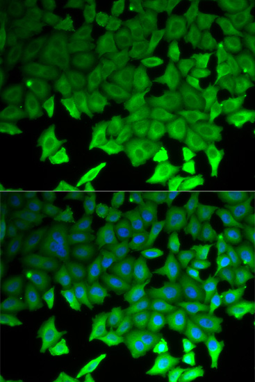


Comprehensive Human Protein Atlas IHC scoring per tissue (reliability: Approved — Medium consistency between antibody staining and RNA expression data.). Rows are taken directly from the HPA tissue chart — click any row's HPA link to view the source.
| Tissue | Cell type | Level | Evidence | Source |
|---|---|---|---|---|
| Bone marrow | Hematopoietic cells | High | Protein (IHC) | HPA → |
| Lung | Macrophages | High | Protein (IHC) | HPA → |
| Lymph node | Non-germinal center cells | High | Protein (IHC) | HPA → |
| Prostate | Glandular cells | High | Protein (IHC) | HPA → |
| Smooth muscle | Smooth muscle cells | High | Protein (IHC) | HPA → |
| Tissue | Cell type | Level | Evidence | Source |
|---|---|---|---|---|
| Esophagus | Squamous epithelial cells | Not detected | Protein (IHC) | HPA → |
| Oral mucosa | Squamous epithelial cells | Not detected | Protein (IHC) | HPA → |
| Placenta | Trophoblastic cells | Not detected | Protein (IHC) | HPA → |
| Vagina | Squamous epithelial cells | Not detected | Protein (IHC) | HPA → |
Troubleshoot LITAF staining by comparing compartment, cell type, and control sections with the documented paraffin IHC image and tissue expression data.
The catalog lists human- and mouse-reactive LITAF antibodies for IHC (catalog: A03043, A03043-2), with a paraffin-section image for A03043 (A03043 image caption). A03043-2 also lists rat reactivity and IF/ICC (catalog: A03043-2).
A03043 lists human and mouse IHC reactivity and has a human prostate paraffin-section IHC image at 1:100 (catalog: A03043; A03043 image caption). A03043-2 lists human, mouse and rat IHC reactivity plus human IF/ICC, but has no IHC or IF image in the supplied catalog (catalog: A03043-2).
Which to pick: For tissue IHC, choose A03043 when a matching paraffin-section example matters: its human prostate caption specifies microwave retrieval in 10 mM PBS at pH 7.2, but does not report the fixative (A03043 image caption). For IF/ICC, choose A03043-2, which lists human IF/ICC at 5 μg/ml without an IF image (catalog: A03043-2). For rat tissue IHC, A03043-2 lists rat reactivity and IHC at 2–5 μg/ml; both catalog antibodies are rabbit polyclonals (catalog: A03043, A03043-2).