This website uses cookies to ensure you get the best experience on our website.


- Table of Contents
Plan chromogenic IHC for LMNB2 around the nuclear membranous tissue pattern, with strong staining reported in CNS cells (HPA tissue IHC). Use the catalog antibody’s paraffin-section protocol and compare nuclear rim staining across consistently fixed samples (datasheet M05348; standard IHC practice).
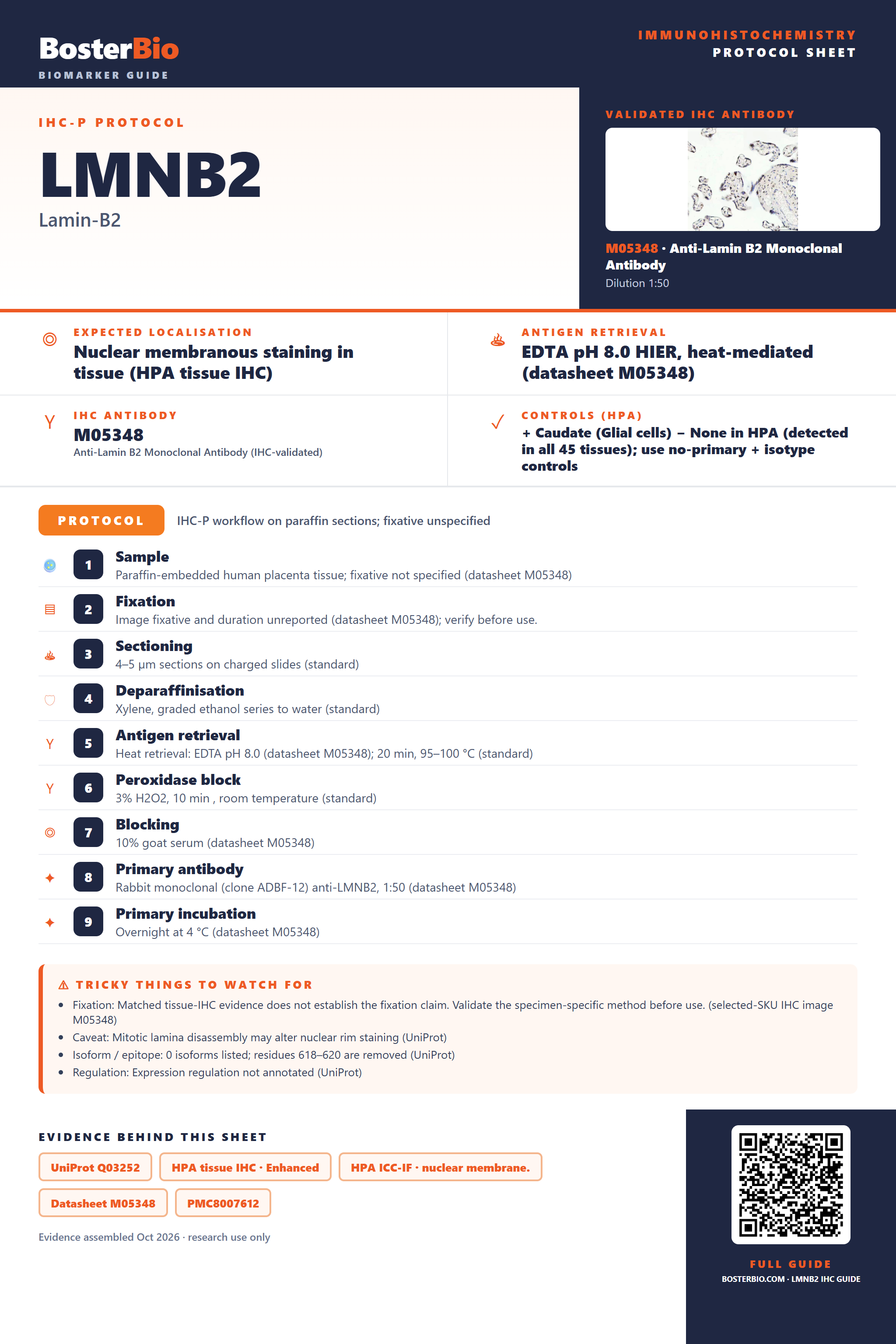
Expected localisation, validated protocols, controls and antibodies — the at-a-glance facts below, then the full design guide.
| Expected localisation | Nuclear membranous staining in tissue (HPA tissue IHC) | |
| Staining pattern | Nuclear rims in glia and cerebellar granular-layer cells (HPA tissue IHC) | |
| Antigen retrieval | EDTA pH 8.0 HIER, heat-mediated (datasheet M05348) | |
| Positive control | Caudate+4 more · see all | |
| Negative control | None in HPA (detected in all 45 tissues); use no-primary + isotype controls |
| Fixation | Matched tissue-IHC evidence does not establish the fixation claim. Validate the specimen-specific method before use. (selected-SKU IHC image M05348) | |
| Caveat | Mitotic lamina disassembly may alter nuclear rim staining (UniProt) | |
| Regulation | Expression regulation not annotated (UniProt) | |
| Isoform / epitope | 0 isoforms listed; residues 618–620 are removed (UniProt) |
The catalog antibody protocol uses paraffin sections; the published IHC protocols below report LMNB2 staining in colorectal, breast, and bladder cancer tissues (PMC8007612; PMC7846963; PMC9282643).
| Sample | Paraffin-embedded human placenta tissue; fixative not specified (datasheet M05348) |
| Fixation | Image fixative and duration unreported (datasheet M05348); verify before use. |
| Sectioning | 4–5 µm sections on charged slides (standard) |
| Deparaffinisation | Xylene, graded ethanol series to water (standard) |
| Antigen retrieval | Heat retrieval: EDTA pH 8.0 (datasheet M05348); 20 min, 95–100 °C (standard) |
| Peroxidase block | 3% H2O2, 10 min, room temperature (standard) |
| Blocking | 10% goat serum (datasheet M05348) |
| Primary antibody | Rabbit monoclonal (clone ADBF-12) anti-LMNB2, 1:50 (datasheet M05348) |
| Primary incubation | Overnight at 4 °C (datasheet M05348) |
| Detection | HRP-conjugated secondary, DAB chromogen (datasheet M05348) |
| Counterstain | Hematoxylin, blue, dehydrate and mount (standard) |
| Expected result | LMNB2-positive staining in glial cells of caudate (HPA tissue IHC: High). HPA tissue profile: Nuclear membranous expression, most abundant in CNS. No signal in the no-primary control. |
LMNB2 is a nuclear lamina protein, so a convincing IHC signal should outline nuclei at their perimeter (UniProt Q03252; HPA: nuclear membranous expression). HPA reports high staining in several CNS cell populations, as well as selected glandular, collecting duct, and endothelial cells (HPA: tissue IHC, Enhanced reliability). LMNB2 has no transmembrane segment; its rim pattern reflects its place in the lamina (UniProt Q03252 topology and function).
| A crisp chromogenic rim surrounds nuclei in glial cells of caudate, cerebral cortex, or hippocampus. | This matches the expected compartment and documented high staining in those cells (UniProt Q03252: nuclear lamina; HPA: High in glial cells at these sites). Score the nuclear rim separately from any diffuse staining. |
| The strongest deposit is cytoplasmic, extracellular, or spread uniformly through nuclei without a discernible rim. | That distribution does not match the reported nuclear membranous pattern (HPA: tissue IHC; UniProt Q03252: nuclear lamina). Check section morphology and detection controls before interpreting it as LMNB2. |
| Strong staining appears mainly in a cell population reported as low, while the expected nuclear rim is absent. | HPA reports low staining in, for example, duodenal glandular cells and colonic endothelial cells (HPA: tissue IHC). Review cell identification and controls; cross-reactivity or endogenous detection activity is possible, but HPA does not establish the cause. |
| Color forms a diffuse haze across tissue, including spaces outside cells. | A tissue-wide haze obscures the nuclear rim used to recognize LMNB2 (HPA: nuclear membranous expression). Assess reagent background and washing with routine IHC controls; haze alone cannot establish target expression. |
| No nuclear rim is visible in a known high-staining compartment, such as kidney collecting ducts. | Collecting ducts are reported as High by HPA (HPA: kidney collecting ducts). Check whether nuclei are preserved and whether a validated positive control stained; one blank section cannot distinguish assay failure from a sample-specific result. |
| Cell and tissue context | HPA records High staining in CNS glial cells, cerebellar granular-layer cells, kidney collecting ducts, lung endothelial cells, and epididymal or fallopian-tube glandular cells (HPA: tissue IHC). Its Low entries provide comparison sites, not proven negative controls; HPA lists no negative tissue here. |
| Antibody evidence | The tissue profile has Enhanced reliability, meaning high consistency between staining and RNA data (HPA: tissue IHC). HPA047863 and HPA062477 have Enhanced IHC validation; CAB022593 has Supported IHC validation (HPA: antibody records). These labels do not validate every staining event in a new specimen. |
| Epitope and processing | UniProt lists a lamin-B2 chain spanning residues 1–617 and a propeptide at 618–620, plus modified residues (UniProt Q03252). No antibody epitope is supplied, so these annotations cannot predict which reagent will stain a given section or explain signal loss. |
| Retrieval and detection | No LMNB2-specific antigen-retrieval condition or fixation-sensitivity result is supplied (UniProt Q03252; HPA: supplied records). Treat retrieval and chromogenic detection settings as assay variables to optimize with controls under general IHC practice; do not infer a target-specific fixation effect from HPA staining levels. |
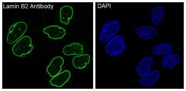
| IF/ICC Q&A: where should signal appear? | A nuclear-membrane rim is the supported IF/ICC location (HPA: subcellular, nuclear membrane supported). HPA lists ICC-IF images in A-431, U-251MG, and U2OS, and Supported ICC validation for HPA062477 (HPA: subcellular and antibody records). These observations do not specify an IF protocol for this IHC guide. |
| Situation | Likely cause | Next action |
|---|---|---|
| No staining in the test section or an HPA High positive-control tissue. | The result is indeterminate: the expected population may be missed, nuclear preservation may be poor, or the staining run may have failed (HPA: High tissue populations). | Confirm the relevant cells on the counterstain, then review the positive control and run records. Recheck routine IHC retrieval, reagent, and detection steps without assuming an LMNB2-specific fixation effect. |
| A faint rim appears in a tissue listed as Low. | Low is a reported staining level, not an assertion that LMNB2 is absent (HPA: tissue IHC Low entries). | Compare the same run with an HPA High population and assess whether the faint signal remains confined to nuclear perimeters. Record intensity and cell type rather than calling every faint rim nonspecific. |
| Brown deposit fills cytoplasm or lies outside nuclei. | This conflicts with the reported nuclear membranous location (HPA: tissue IHC; UniProt Q03252: nuclear lamina); the source of the deposit is unresolved. | Inspect morphology and the no-primary control, then review blocking, washing, and chromogen development using routine IHC practice. Require a discernible nuclear rim before assigning LMNB2 positivity. |
| Unexpected cells stain strongly while the anticipated high-staining cells do not. | Cell identification, cross-reactivity, or endogenous detection activity may account for the mismatch; HPA documents specific High and Low populations but cannot diagnose this section (HPA: tissue IHC). | Reidentify the cells on the counterstain and compare a known HPA High tissue on the same run. Use detection controls to investigate background before attributing the unexpected pattern to LMNB2. |
| Diffuse background makes nuclear boundaries hard to score. | Background may arise from routine chromogenic detection or incomplete washing; the haze does not reproduce the reported nuclear rim (HPA: nuclear membranous expression). | Review the no-primary control, blocking and wash steps, and development time. Score only cells whose nuclear edge can be distinguished from surrounding color; document any areas that remain uninterpretable. |
| An IF/ICC image shows a rim but the IHC section lacks one. | HPA supports a nuclear-membrane IF/ICC location, while its tissue IHC profile separately reports nuclear membranous staining (HPA: subcellular and tissue IHC). Agreement in compartment does not establish that both assays will perform alike. | Interpret each preparation against its own controls. For the IHC section, verify the relevant HPA High cells and chromogenic run controls; use the IF/ICC observation only as a location check. |


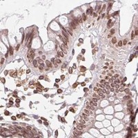


Comprehensive Human Protein Atlas IHC scoring per tissue (reliability: Enhanced — High consistency between antibody staining and RNA expression data.). Rows are taken directly from the HPA tissue chart — click any row's HPA link to view the source.
| Tissue | Cell type | Level | Evidence | Source |
|---|---|---|---|---|
| Caudate | Glial cells | High | Protein (IHC) | HPA → |
| Cerebellum | Cells in granular layer | High | Protein (IHC) | HPA → |
| Cerebral cortex | Glial cells | High | Protein (IHC) | HPA → |
| Epididymis | Glandular cells | High | Protein (IHC) | HPA → |
| Fallopian tube | Glandular cells | High | Protein (IHC) | HPA → |
| Tissue | Cell type | Level | Evidence | Source |
|---|---|---|---|---|
| None in HPA: LMNB2 is detected in all 45 scored tissues. Use a no-primary (secondary-only) and an isotype control instead. | ||||
Troubleshoot LMNB2 staining by checking nuclear rim localisation, tissue context and the documented conditions for the IHC-validated antibody.
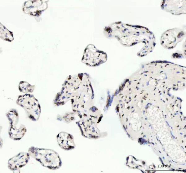
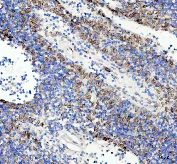
LMNB2 antibodies have paraffin-section IHC images from human placenta and rat intestine, and an IF image from HeLa cells (M05348 and PA1636 IHC captions; M05348 IF caption).
M05348 has IHC images from human placenta, spleen, breast cancer and lung cancer paraffin sections, plus an IF image from HeLa cells (M05348 image captions). PA1636 has IHC images from rat intestine paraffin and frozen sections and a human brain paraffin section (PA1636 image captions).
Which to pick: For tissue IHC across human and rat samples, choose PA1636: its images cover human brain and rat intestine paraffin sections, and it lists Human, Mouse and Rat reactivity (PA1636 IHC captions; PA1636 catalog reactivity). For IF/ICC, choose the rabbit monoclonal M05348: IF and ICC are listed applications, and its IF caption shows HeLa cells (M05348 catalog applications and clone; M05348 IF caption). M05348 also has human paraffin-section IHC images and lists Human, Mouse and Rat reactivity; neither SKU’s paraffin-section caption reports a fixative (M05348 catalog reactivity and IHC captions; PA1636 IHC captions).