This website uses cookies to ensure you get the best experience on our website.


- Table of Contents
Plan LMO2 paraffin-section IHC around nuclear staining in bone marrow hematopoietic cells and cytoplasmic staining in several tissues (HPA tissue IHC). Use the tissue pattern and the reported staining uncertainty when selecting controls and scoring results (HPA tissue IHC).

Expected localisation, validated protocols, controls and antibodies — the at-a-glance facts below, then the full design guide.
| Expected localisation | Nuclear in marrow; cytoplasmic in several tissues (HPA tissue IHC) | |
| Staining pattern | Marrow hematopoietic nuclei; cytoplasm in several tissues (HPA tissue IHC) | |
| Antigen retrieval | EDTA pH 8.0 HIER, heat-mediated (datasheet M03502) | |
| Positive control | Adrenal gland+4 more · see all | |
| Negative control | Adipose tissue+4 more · see all |
| Fixation | Keep fixation consistent across sections. (standard IHC practice; not target-specific) | |
| Caveat | Presumed off-target staining; verification pending (HPA tissue IHC) | |
| Regulation | Expression regulators are not specified (UniProt) | |
| Isoform / epitope | 3 isoforms; epitope differences unreported (UniProt) |
The catalog antibody has an IHC-P protocol (datasheet: M03502). The published protocols below cover lymph node, bone marrow, and whole tissue or microarray sections (PMC8234657; PMC9427670; PMC4305438).
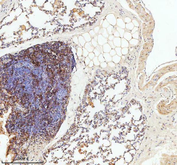
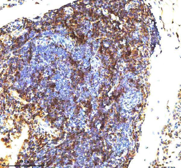
| Sample | Paraffin-embedded lymph node of rat lung tissue; fixative not specified (datasheet M03502) |
| Fixation | Image fixative and duration unreported (datasheet M03502); verify before use. |
| Sectioning | 4–5 µm sections on charged slides (standard) |
| Deparaffinisation | Xylene, graded ethanol series to water (standard) |
| Antigen retrieval | Heat retrieval: EDTA pH 8.0 (datasheet M03502); 20 min, 95–100 °C (standard) |
| Peroxidase block | 3% H2O2, 10 min, room temperature (standard) |
| Blocking | 10% goat serum (datasheet M03502) |
| Primary antibody | Rabbit monoclonal (clone ACOA-12) anti-LMO2, 1:50 (datasheet M03502) |
| Primary incubation | Overnight at 4 °C (datasheet M03502) |
| Detection | HRP-conjugated secondary, DAB chromogen (datasheet M03502) |
| Counterstain | Hematoxylin, blue, dehydrate and mount (standard) |
| Expected result | LMO2-positive staining in glandular cells of adrenal gland (HPA tissue IHC: High). HPA tissue profile: Nuclear expression in bone marrow and cytoplasmic expression in several tissues. No signal in the no-primary control. |
LMO2 is a nuclear protein with no transmembrane segment or signal peptide (UniProt P25791). In paraffin-section IHC, expect strong nuclear staining in bone marrow hematopoietic cells (HPA tissue IHC: High; nuclear expression). HPA also reports cytoplasmic expression in several tissues; its tissue IHC reliability is Approved, with presumed off-target binding disregarded and external verification pending (HPA tissue IHC). Interpret staining by cell type and compartment together.
| Distinct, strong staining of hematopoietic cell nuclei in bone marrow, with nuclei identifiable by counterstain. | This is the clearest tissue-level positive pattern in the supplied record: hematopoietic cells are High, and the bone marrow pattern is nuclear (HPA tissue IHC). Nuclear localisation agrees with UniProt P25791. Record the fraction of stained cells as well as intensity; intensity alone can obscure whether the expected cells carry the signal (general IHC practice). |
| Predominantly cytoplasmic staining, with little or no convincing nuclear signal. | Treat this as uncertain LMO2 localisation, especially in bone marrow, where HPA describes nuclear expression (HPA tissue IHC; UniProt P25791). HPA also describes cytoplasmic expression in several tissues and notes presumed off-target binding, so cytoplasmic colour alone cannot establish which signal represents LMO2 (HPA tissue IHC). Compare compartments within the same cell type and seek independent validation before scoring it as specific. |
| Strong staining in adipocytes, smooth muscle cells or oral mucosal squamous epithelial cells. | These cell types are recorded as Not detected, making them useful comparisons for unexpected staining, not absolute biological negatives (HPA tissue IHC). Check whether colour persists without primary antibody and whether it tracks tissue structures; cross-reactivity or detection activity becomes more plausible if the expected positive-cell pattern is absent (general IHC practice). |
| Diffuse colour across cells and extracellular space, without crisp cellular boundaries. | This pattern cannot support a nuclear LMO2 call: UniProt places LMO2 in the nucleus, and HPA describes nuclear bone marrow staining (UniProt P25791; HPA tissue IHC). Uneven reagent distribution, excessive detection signal or inadequate washing can create background in chromogenic IHC (general IHC practice). Judge specificity only after the diffuse component is reduced. |
| No visible staining in bone marrow hematopoietic cells. | This conflicts with the High, nuclear bone marrow pattern reported by HPA, but a blank slide does not establish true absence of LMO2 (HPA tissue IHC). First confirm that hematopoietic cells are present and assess whether the staining run worked on its controls (general IHC practice). HPA's Approved designation remains pending external verification (HPA tissue IHC). |
| Compartment and tissue context | UniProt assigns LMO2 to the nucleus, while HPA reports nuclear bone marrow staining and cytoplasmic expression in several tissues (UniProt P25791; HPA tissue IHC). Use the marrow nuclear pattern as the clearest reference; do not assume every HPA High tissue shares it. |
| Strength of tissue evidence | The tissue IHC profile is Approved, but HPA says presumed off-target binding was disregarded and external verification is pending (HPA tissue IHC). The IHC antibody CAB016258 is Approved; the supplied antibody list gives no Enhanced IHC status (HPA antibodies). Treat unexpected compartments as provisional. |
| Protein architecture and variants | LMO2 has two LIM zinc-binding domains and three listed isoforms; the supplied record does not locate this assay's epitope within them (UniProt P25791). Isoform-specific staining differences therefore cannot be predicted from these data. |
| Processing and topology | UniProt lists no signal peptide, propeptide, transmembrane segment or glycosylation sites, and a single chain spanning residues 1–158 (UniProt P25791). These annotations support interpretation as a non-membrane protein; they do not establish antigen-retrieval needs or fixation sensitivity. |
| Situation | Likely cause | Next action |
|---|---|---|
| Bone marrow nuclei are blank or much weaker than expected. | The result conflicts with HPA's High nuclear staining in hematopoietic cells (HPA tissue IHC). A failed staining run, unsuitable tissue section or missed target-cell population are practical possibilities (general IHC practice). | Verify hematopoietic cells on the counterstained section, inspect run controls, and check that retrieval, antibody use and detection followed the chosen IHC-P assay instructions (general IHC practice). Do not assign an LMO2-specific retrieval or fixation effect from this record. |
| Colour appears only in cytoplasm. | HPA reports cytoplasmic expression in several tissues but nuclear expression in bone marrow, and notes presumed off-target binding (HPA tissue IHC). Cytoplasmic-only colour therefore has uncertain specificity. | Record tissue and cell type before scoring. Compare bone marrow nuclear staining and a no-primary control, then seek independent confirmation for a cytoplasmic LMO2 claim (HPA tissue IHC; general IHC practice). |
| Cells expected to be unstained show strong colour. | HPA lists adipocytes, smooth muscle cells and oral mucosal squamous epithelial cells as Not detected (HPA tissue IHC). Cross-reactivity or background is possible, though Not detected does not guarantee universal absence. | Compare the suspect cells with nearby expected-positive cells and a no-primary control; lower nonspecific staining through standard blocking and washing checks before interpreting the result (general IHC practice). |
| Diffuse colour obscures nuclei and cell boundaries. | The slide cannot resolve the reported nuclear bone marrow pattern (HPA tissue IHC). Background from the detection workflow is a general IHC possibility (general IHC practice). | Review reagent coverage, blocking, washes and detection development with run controls; reassess only when nuclei and cell boundaries can be distinguished (general IHC practice). |
| Colour persists in a no-primary-antibody control. | Primary-antibody-independent colour can arise from endogenous detection activity or other background in chromogenic IHC (general IHC practice). That control cannot identify LMO2. | Address endogenous activity with a detection-system-appropriate block, check secondary and chromogen controls, and repeat the comparison with primary antibody (general IHC practice). |
| Q: What should an IF/ICC image show? | HPA's ICC-IF summary places LMO2 mainly in the nucleoplasm, with an Approved main location; three listed antibodies have Approved ICC status (HPA subcellular; HPA antibodies). | A: Look for nucleoplasmic signal and compare it with nuclear counterstain (HPA subcellular; general IF practice). Use the separate IF/ICC guide for its workflow; the IHC tissue pattern alone does not validate an IF/ICC staining run. |
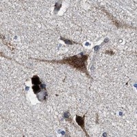


Comprehensive Human Protein Atlas IHC scoring per tissue (reliability: Approved — Presumed off target binding observed and disregarded. Pending external verification.). Rows are taken directly from the HPA tissue chart — click any row's HPA link to view the source.
| Tissue | Cell type | Level | Evidence | Source |
|---|---|---|---|---|
| Adrenal gland | Glandular cells | High | Protein (IHC) | HPA → |
| Bone marrow | Hematopoietic cells | High | Protein (IHC) | HPA → |
| Bronchus | Respiratory epithelial cells | High | Protein (IHC) | HPA → |
| Caudate | Neuronal cells | High | Protein (IHC) | HPA → |
| Cerebellum | Cells in molecular layer | High | Protein (IHC) | HPA → |
| Tissue | Cell type | Level | Evidence | Source |
|---|---|---|---|---|
| Adipose tissue | Adipocytes | Not detected | Protein (IHC) | HPA → |
| Fallopian tube | Glandular cells | Not detected | Protein (IHC) | HPA → |
| Liver | Cholangiocytes | Not detected | Protein (IHC) | HPA → |
| Oral mucosa | Squamous epithelial cells | Not detected | Protein (IHC) | HPA → |
| Ovary | Ovarian stroma cells | Not detected | Protein (IHC) | HPA → |
Troubleshoot LMO2 staining in paraffin sections by checking retrieval, compartment, cell identity, controls and scoring before interpreting a chromogenic signal.
M03502 has real IHC images from paraffin sections of mouse lung and a rat specimen described as a lymph node from lung tissue; no IF image is supplied (M03502 image captions; catalog IF images).
M03502 is listed for IHC, with paraffin-section images from mouse lung and a rat specimen described as a lymph node from lung tissue (catalog applications; M03502 image captions). Human, mouse and rat reactivity is listed, while the supplied IHC images show mouse and rat specimens (catalog reactivity; M03502 image captions).
Which to pick: For tissue IHC, choose M03502, a rabbit monoclonal shown at 1:50 on paraffin sections; the fixative is unreported (catalog host and clone; M03502 image captions). No IF/ICC pick is supported because M03502 has no listed IF/ICC application or IF image (catalog applications; catalog IF images). For cross-species work, M03502 lists human, mouse and rat reactivity, with IHC images for mouse and rat specimens (catalog reactivity; M03502 image captions).