This website uses cookies to ensure you get the best experience on our website.


- Table of Contents
Plan LMOD1 paraffin-section IHC around cytoplasmic staining in smooth muscle and myoepithelial cells (HPA tissue IHC). Start with the catalog antibody at 1:50–1:200 and score staining by cell type (datasheet: IHC dilution; HPA tissue IHC).

Expected localisation, validated protocols, controls and antibodies — the at-a-glance facts below, then the full design guide.
| Expected localisation | Cytoplasmic tissue staining (HPA tissue IHC) | |
| Staining pattern | Selective cytoplasmic staining in smooth muscle and myoepithelial cells (HPA tissue IHC) | |
| Antigen retrieval | Citrate pH 6.0 HIER, 95–98 °C, 20 min (rule: cytoplasmic / membrane antigen) | |
| Positive control | Adrenal gland+3 more · see all | |
| Negative control | Appendix+4 more · see all |
| Fixation | Keep paraffin-section fixation consistent across samples. (standard IHC practice; not target-specific) | |
| Caveat | Adrenal glandular and placental endothelial cells also stain (HPA tissue IHC) | |
| Regulation | Staining-linked regulation is not established (UniProt) | |
| Isoform / epitope | 2 isoforms; isoform-specific epitope coverage is unreported (UniProt; datasheet) |
The catalog antibody’s IHC-P protocol is accompanied by published LMOD1 IHC methods for brain and visceral tissues, C. elegans, and leiomyosarcoma (PMC10439352; PMC5594508; PMC4526352).
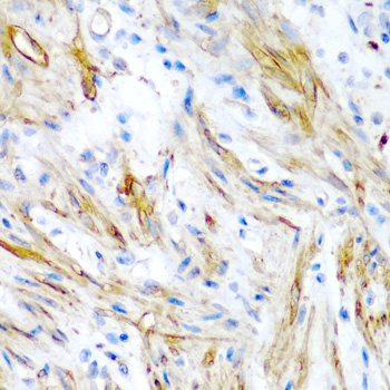
| Sample | Paraffin-embedded human colon tissue; fixative not specified (datasheet A08666) |
| Fixation | Image fixative and duration unreported (datasheet A08666); verify before use. |
| Sectioning | 4–5 µm sections on charged slides (standard) |
| Deparaffinisation | Xylene, graded ethanol series to water (standard) |
| Antigen retrieval | Heat-induced epitope retrieval in citrate buffer, pH 6.0, 20 min at 95–98 °C (standard rule: cytoplasmic / membrane antigen) |
| Peroxidase block | 3% H2O2, 10 min, room temperature (standard) |
| Blocking | 10% normal serum of the secondary host, 30 min, room temperature (standard) |
| Primary antibody | Rabbit anti-LMOD1, 1:50-1:200 (datasheet A08666) |
| Primary incubation | Overnight at 4 °C (standard) |
| Detection | HRP-polymer secondary, DAB chromogen 5–10 min (standard) |
| Counterstain | Hematoxylin, blue, dehydrate and mount (standard) |
| Expected result | LMOD1-positive staining in glandular cells of adrenal gland (HPA tissue IHC: High). HPA tissue profile: Highly selective cytoplasmic expression in smooth muscle and myoepithelial cells. No signal in the no-primary control. |
LMOD1 should stain chiefly in the cytoplasm of smooth muscle and myoepithelial cells (HPA tissue IHC: highly selective cytoplasmic profile). It is a cytoplasmic, actin-associated protein with no transmembrane segment (UniProt P29536: subcellular location and topology). HPA rates the tissue IHC pattern Enhanced, citing high consistency between antibody staining and RNA expression (HPA tissue IHC: reliability).
| Cytoplasmic staining in smooth muscle cells, with little staining in adjacent unrelated cells (HPA tissue IHC: smooth muscle, Medium; selective profile). | This fits the expected pattern. Breast myoepithelial cells are another supported positive population (HPA tissue IHC: breast myoepithelial cells, Medium). Judge the relevant cell population, not the whole section, as positive. |
| Predominantly nuclear or cell-surface staining, without a convincing cytoplasmic component. | Treat this as a localisation mismatch: UniProt places LMOD1 in the cytoplasm and cytoskeleton, and HPA describes cytoplasmic tissue staining (UniProt P29536: subcellular location; HPA tissue IHC: profile). Review the staining pattern before scoring it as LMOD1. |
| Strong staining in cells listed as undetected, such as bone marrow hematopoietic cells or bronchial respiratory epithelial cells (HPA tissue IHC: Not detected). | Check for cross-reactivity or endogenous detection activity (standard IHC practice). Cell identity matters: HPA also reports High staining in placental endothelial and adrenal glandular cells, so those populations are not automatically unexpected (HPA tissue IHC: positive cells). |
| Diffuse chromogen across tissue and background, with no clear cytoplasmic cell pattern. | A diffuse deposit does not establish LMOD1 expression. Assess blocking, detection background and chromogen development with appropriate controls (standard chromogenic IHC practice); compare against HPA’s selective cytoplasmic profile (HPA tissue IHC: profile). |
| No signal in a section containing identifiable smooth muscle cells. | A negative result in a supported positive cell population needs a control check (HPA tissue IHC: smooth muscle cells, Medium). Confirm that the cells are present and that the detection run worked before interpreting the absence as biological (standard IHC practice). |
| Cell population and intensity | HPA reports Medium staining in smooth muscle and breast myoepithelial cells, but High staining in placental endothelial and adrenal glandular cells (HPA tissue IHC: positive cells). Interpret intensity with the identified cell type; a single intensity threshold cannot describe every listed positive population. |
| Subcellular position | UniProt places LMOD1 in the cytoplasm, myofibril and cytoskeleton and notes colocalisation with actin filaments in sarcomeres (UniProt P29536: subcellular location). HPA’s tissue summary is cytoplasmic (HPA tissue IHC: profile). Neither source supports a nuclear or membrane-only IHC call. |
| Antibody validation | Three listed rabbit polyclonal antibodies have Enhanced IHC status: HPA028325, HPA028435 and HPA030097 (HPA antibodies: IHC Enhanced). This supports the reported tissue pattern; it does not establish that every reagent or staining run will reproduce it (HPA: Enhanced reliability; standard IHC practice). |
| Isoforms and modifications | UniProt lists 2 isoforms and four example phosphoserine sites (UniProt P29536: isoforms and modified residues). The supplied sources do not map an IHC antibody epitope or show an isoform- or phosphorylation-dependent staining difference, so do not assign one. |
| Situation | Likely cause | Next action |
|---|---|---|
| Smooth muscle cells are unstained in an otherwise interpretable section (HPA tissue IHC: smooth muscle, Medium). | The target population may be absent from the examined field, or the IHC run may have failed (standard IHC practice); the supplied sources do not identify an LMOD1-specific fixation sensitivity. | Locate smooth muscle on the section and inspect a suitable positive control and detection controls before scoring the sample as negative (standard IHC practice). |
| The entire section has diffuse brown staining instead of selective cytoplasmic signal (HPA tissue IHC: profile). | Nonspecific background or endogenous detection activity can obscure a cell-specific pattern (standard chromogenic IHC practice). HPA’s tissue profile does not identify which workflow step caused it. | Compare with omission and detection controls; review blocking, washes and chromogen development under the laboratory’s IHC workflow (standard IHC practice). |
| Only nuclei or cell borders stain strongly. | That distribution conflicts with the documented cytoplasmic location (UniProt P29536: subcellular location; HPA tissue IHC: profile). It may reflect nonspecific staining (standard IHC interpretation). | Check morphology and controls, then repeat interpretation using cytoplasmic staining in a supported positive cell population as the reference (HPA tissue IHC: smooth muscle or myoepithelial cells). |
| Bronchial respiratory epithelium or bone marrow hematopoietic cells stain strongly (HPA tissue IHC: Not detected). | Cross-reactivity or endogenous detection activity is plausible (standard IHC practice); HPA reports these specific cell populations as undetected (HPA tissue IHC: negative cells). | Confirm the stained cell identity and compare with detection controls and a supported positive population before attributing that signal to LMOD1 (standard IHC practice; HPA tissue IHC: positive cells). |
| The slide contains strong placental endothelial or adrenal glandular staining. | These are reported High positive populations, despite HPA’s overall selective smooth muscle and myoepithelial summary (HPA tissue IHC: positive cells and profile). | Score the identified endothelial or glandular cells against the tissue-specific HPA observations; do not label them cross-reactive solely because they are outside the summary profile (HPA tissue IHC: positive cells). |
| IF/ICC Q: What intracellular pattern should be expected? | HPA approves cytosol as the main ICC-IF location; HPA028435 has Supported ICC status (HPA subcellular: Cytosol approved; HPA antibodies: ICC Supported). | A: Look for cytosolic localisation when interpreting IF/ICC images (HPA subcellular: Cytosol approved). Use the separate IF/ICC guide for its workflow. |


![HPA ICC-IF BJ [Human fibroblast] — LMOD1 (HPA028435)](https://images.proteinatlas.org/28435/1446_H1_1_blue_red_green.jpg)
![HPA ICC-IF BJ [Human fibroblast] — LMOD1 (HPA028435)](https://images.proteinatlas.org/28435/1446_H1_6_blue_red_green.jpg)
Comprehensive Human Protein Atlas IHC scoring per tissue (reliability: Enhanced — High consistency between antibody staining and RNA expression data.). Rows are taken directly from the HPA tissue chart — click any row's HPA link to view the source.
| Tissue | Cell type | Level | Evidence | Source |
|---|---|---|---|---|
| Adrenal gland | Glandular cells | High | Protein (IHC) | HPA → |
| Placenta | Endothelial cells | High | Protein (IHC) | HPA → |
| Breast | Myoepithelial cells | Medium | Protein (IHC) | HPA → |
| Smooth muscle | Smooth muscle cells | Medium | Protein (IHC) | HPA → |
| Tissue | Cell type | Level | Evidence | Source |
|---|---|---|---|---|
| Appendix | Glandular cells | Not detected | Protein (IHC) | HPA → |
| Bone marrow | Hematopoietic cells | Not detected | Protein (IHC) | HPA → |
| Bronchus | Respiratory epithelial cells | Not detected | Protein (IHC) | HPA → |
| Caudate | Glial cells | Not detected | Protein (IHC) | HPA → |
| Cerebellum | Cells in granular layer | Not detected | Protein (IHC) | HPA → |
Use cell type and cytoplasmic localisation to assess LMOD1 staining, with the paraffin colon image as the catalog antibody’s direct tissue example.
The catalog includes an anti-LMOD1 antibody with real IHC data from paraffin-embedded human colon (A08666 IHC image caption). No IF image is supplied (A08666 IF image list).
A08666 is listed for human IHC (catalog applications/reactivity). Its IHC image shows paraffin-embedded human colon at 1:200 (A08666 IHC image caption).
Which to pick: Choose A08666 for human paraffin-section IHC: its own image documents that preparation, while the fixative is unreported (A08666 IHC image caption). No listed SKU has IF/ICC validation or an IF image, so there is no supported IF/ICC pick (catalog applications/IF image list). A08666 is rabbit-hosted with clonality unreported and human-only listed reactivity; the payload supports no cross-species pick (catalog host/clone/reactivity fields).