This website uses cookies to ensure you get the best experience on our website.


- Table of Contents
Plan LOXL1 paraffin IHC around matrix and plasma staining, with additional cytoplasmic signal (HPA tissue IHC). Use the catalog antibody at 0.5–1 μg/mL with chromogenic detection (datasheet PB9719; standard IHC practice).

Expected localisation, validated protocols, controls and antibodies — the at-a-glance facts below, then the full design guide.
| Expected localisation | Extracellular matrix; additional cytoplasmic staining (HPA tissue IHC) | |
| Staining pattern | Matrix and plasma staining; some cytoplasm; glandular cells high (HPA tissue IHC) | |
| Antigen retrieval | EDTA pH 8.0 HIER, heat-mediated (datasheet PB9719) | |
| Positive control | Appendix+4 more · see all | |
| Negative control | Adipose tissue+4 more · see all |
| Fixation | Keep formalin fixation consistent across sections (standard IHC practice; not target-specific) | |
| Caveat | As a secreted protein, staining may differ from tissue RNA (HPA tissue IHC) | |
| Regulation | Regulation not specified (UniProt) | |
| Isoform / epitope | No isoforms annotated; map epitopes across the 95/96 cleavage (UniProt) |
The catalog antibody’s IHC-P protocol is followed by two published LOXL1 IHC protocols with reported retrieval and detection details (PMC9087977; PMC9455728: Methods).
| Sample | Paraffin-embedded human prostatic cancer tissue; fixative not specified (datasheet PB9719) |
| Fixation | Image fixative and duration unreported (datasheet PB9719); verify before use. |
| Sectioning | 4–5 µm sections on charged slides (standard) |
| Deparaffinisation | Xylene, graded ethanol series to water (standard) |
| Antigen retrieval | Heat retrieval: EDTA pH 8.0 (datasheet PB9719); 20 min, 95–100 °C (standard) |
| Peroxidase block | 3% H2O2, 10 min, room temperature (standard) |
| Blocking | 10% goat serum (datasheet PB9719) |
| Primary antibody | Rabbit anti-LOXL1, 0.5-1μg/ml (datasheet PB9719) |
| Primary incubation | Overnight at 4 °C (datasheet PB9719) |
| Detection | Streptavidin-biotin complex (SABC), DAB chromogen (datasheet PB9719) |
| Counterstain | Hematoxylin, blue, dehydrate and mount (standard) |
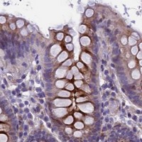
| Expected result | LOXL1-positive staining in glandular cells of appendix (HPA tissue IHC: High). HPA tissue profile: Extracellular matrix and plasma positivity in several tissues with additional cytoplasmic expression. No signal in the no-primary control. |
LOXL1 is a secreted protein found in extracellular space and matrix, with no transmembrane segment (UniProt Q08397 localisation and topology). In paraffin IHC, expect extracellular matrix and plasma positivity in several tissues, sometimes with cytoplasmic staining (HPA: tissue IHC, Approved). Reported positive cells include glandular cells in the appendix and colon and squamous epithelial cells in the esophagus (HPA: High in each).
| Extracellular staining accompanies glandular staining in appendix or colon. | This fits the reported matrix and plasma pattern and High glandular staining in those tissues (HPA: tissue IHC). Score extracellular and cellular signal separately so their distribution remains clear (standard IHC practice). |
| Strong staining appears confined to nuclei, without the expected extracellular or glandular pattern. | A nucleus-only pattern does not fit LOXL1's reported extracellular location or the HPA tissue profile (UniProt Q08397 localisation; HPA: tissue IHC). Treat it as suspect and compare with controls before assigning specificity (standard IHC practice). |
| A cell population reported as negative stains strongly, such as respiratory epithelial cells in bronchus. | HPA reports LOXL1 as Not detected in those cells (HPA: bronchus). Consider cross-reactivity or endogenous detection activity; inspect the no-primary control and the expected positive tissue before interpreting the signal (standard IHC practice). |
| Diffuse chromogen covers cells and empty areas with little anatomical structure. | This is less convincing than HPA's cell-specific and extracellular pattern (HPA: tissue IHC). Check the no-primary control for detection background and review blocking, washes and development time (standard IHC practice). |
| No signal appears in appendix glandular cells despite intact tissue morphology. | Appendix glandular cells are reported High (HPA: appendix). A negative run there calls for checking the IHC-validated antibody, detection reagents and run controls before concluding that another specimen lacks LOXL1 (standard IHC practice). |
| Secreted protein and compartment | LOXL1 has a signal peptide and is annotated in extracellular space and matrix (UniProt Q08397 topology and localisation). HPA also reports cytoplasmic expression, so extracellular and cellular staining can coexist (HPA: tissue IHC). |
| Processing | UniProt annotates signal peptide residues 1–25, propeptide residues 26–95 and a chain beginning at residue 96 (UniProt Q08397 processing). The supplied record gives no antibody epitope, so it cannot predict which processed forms the stain detects. |
| Tissue and cell selection | Appendix and colon glandular cells and esophageal squamous cells are High; stomach glandular cells are Medium (HPA: tissue IHC). Bronchial respiratory epithelium is Not detected (HPA: bronchus). Use the specified cells when comparing sections. |
| Evidence and antibody scope | HPA rates the tissue profile Approved and notes that RNA and protein locations can differ for a secreted protein (HPA: tissue IHC). HPA042111 is IHC Approved; that status alone does not establish the performance of another antibody (HPA: antibody validation). |
| IF/ICC Q&A: Is endoplasmic reticulum staining expected? | Yes, HPA reports approved endoplasmic reticulum localisation in ICC-IF (HPA: subcellular). The listed ICC-IF antibody is HPA063583, while HPA042111 is IHC Approved (HPA: antibody validation). Interpret each modality using its own evidence. |
| Situation | Likely cause | Next action |
|---|---|---|
| Appendix or colon glandular cells are negative. | These are High reference cells (HPA: appendix and colon); a failed run or weak detection is possible (standard IHC practice). | Review run controls, chromogen development and the IHC-validated antibody's stated IHC-P conditions before calling the specimen negative (standard IHC practice). |
| Bronchial respiratory epithelial cells stain strongly. | Those cells are Not detected in the HPA profile (HPA: bronchus); cross-reactivity or endogenous detection activity is possible (standard IHC practice). | Compare a no-primary control and a known-positive section, then judge whether the signal follows the reported cell pattern (HPA: tissue IHC; standard IHC practice). |
| The entire section has diffuse brown background. | Broad background obscures the anatomical distribution reported by HPA (HPA: tissue IHC). Detection background or overdevelopment may contribute (standard IHC practice). | Check the no-primary control; review blocking, washing and chromogen development using the same run conditions (standard IHC practice). |
| Staining is predominantly nuclear. | Nuclear-only staining conflicts with extracellular LOXL1 localisation and the reported tissue profile (UniProt Q08397 localisation; HPA: tissue IHC). | Compare compartment and cell type against a positive control and inspect no-primary staining before accepting specificity (standard IHC practice). |
| Extracellular signal and cell-associated signal receive one score. | HPA reports extracellular matrix and plasma positivity with additional cytoplasmic expression (HPA: tissue IHC). A single score can hide which component changed. | Record compartment, stained cell type and intensity separately in each section (standard IHC practice). |
| An ICC-IF endoplasmic reticulum image seems inconsistent with extracellular IHC. | HPA reports endoplasmic reticulum in ICC-IF and extracellular matrix or plasma staining in tissue IHC (HPA: subcellular; HPA: tissue IHC). | Compare each result with its modality-specific HPA pattern and antibody validation, without treating an ICC-IF image as an IHC control (HPA: antibody validation; standard IHC practice). |



![HPA ICC-IF BJ [Human fibroblast] — LOXL1 (HPA063583)](https://images.proteinatlas.org/63583/1866_E10_10_cr5b69379151755_blue_red_green.jpg)
![HPA ICC-IF BJ [Human fibroblast] — LOXL1 (HPA063583)](https://images.proteinatlas.org/63583/1866_E10_20_cr5b69379151cce_blue_red_green.jpg)
Comprehensive Human Protein Atlas IHC scoring per tissue (reliability: Approved — Secreted protein, tissue location of RNA and protein is expected to differ.). Rows are taken directly from the HPA tissue chart — click any row's HPA link to view the source.
| Tissue | Cell type | Level | Evidence | Source |
|---|---|---|---|---|
| Appendix | Glandular cells | High | Protein (IHC) | HPA → |
| Colon | Glandular cells | High | Protein (IHC) | HPA → |
| Esophagus | Squamous epithelial cells | High | Protein (IHC) | HPA → |
| Rectum | Glandular cells | High | Protein (IHC) | HPA → |
| Small intestine | Glandular cells | High | Protein (IHC) | HPA → |
| Tissue | Cell type | Level | Evidence | Source |
|---|---|---|---|---|
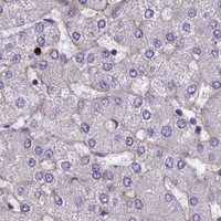
| Adipose tissue | Adipocytes | Not detected | Protein (IHC) | HPA → |
| Breast | Adipocytes | Not detected | Protein (IHC) | HPA → |
| Bronchus | Respiratory epithelial cells | Not detected | Protein (IHC) | HPA → |
| Caudate | Glial cells | Not detected | Protein (IHC) | HPA → |
| Endometrium | Cells in endometrial stroma | Not detected | Protein (IHC) | HPA → |
Troubleshoot LOXL1 chromogenic IHC by checking retrieval, compartment-specific staining and extracellular matrix signal before comparing sections.
PB9719 has real IHC data from a human paraffin-embedded prostatic cancer section (PB9719 image caption); no IF image is provided (catalog image list).
PB9719 is listed for human IHC and WB (catalog applications and reactivity). Its IHC image shows staining in a human paraffin-embedded prostatic cancer section; the fixative is unreported (PB9719 image caption).
Which to pick: Choose PB9719 for human paraffin-section IHC, supported by its own IHC image caption and application listing (PB9719 image caption; catalog applications). No listed SKU is validated for IF/ICC or other species, and PB9719’s clonality is unreported (catalog applications, reactivity and clone field).