This website uses cookies to ensure you get the best experience on our website.


- Table of Contents
Plan LPP staining in paraffin sections using the IHC-validated antibody at 0.5–1 µg/mL (datasheet A01240-2). Assess cytoplasmic staining across cell types, with smooth muscle cells as a strong reference population (HPA tissue IHC).

Expected localisation, validated protocols, controls and antibodies — the at-a-glance facts below, then the full design guide.
| Expected localisation | Predominantly cytoplasmic in tissue (HPA tissue IHC) | |
| Staining pattern | Cytoplasmic staining, strongest in smooth muscle cells (HPA tissue IHC) | |
| Antigen retrieval | EDTA pH 8.0 HIER, heat-mediated (datasheet A01240-2) | |
| Positive control | Kidney+4 more · see all | |
| Negative control | Adipose tissue+4 more · see all |
| Fixation | Matched tissue-IHC evidence does not establish the fixation claim. Validate the specimen-specific method before use. (selected-SKU IHC image A01240-2) | |
| Caveat | Adipocytes show no detected staining despite the LPP name (HPA tissue IHC) | |
| Regulation | High in smooth muscle cells (HPA tissue IHC) | |
| Isoform / epitope | No annotated isoforms; one chain with no transmembrane region (UniProt) |
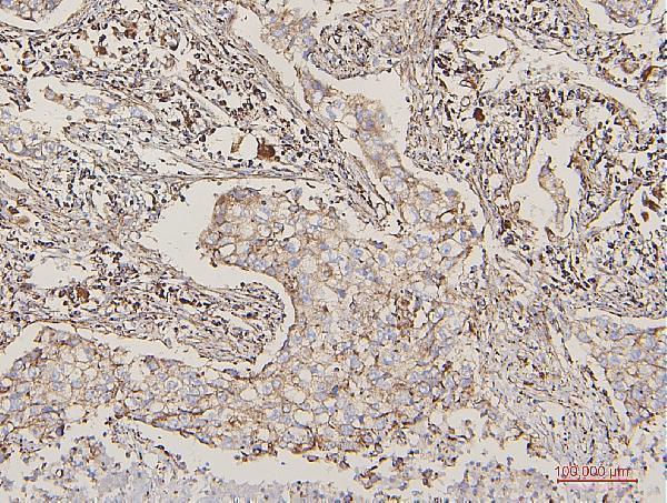
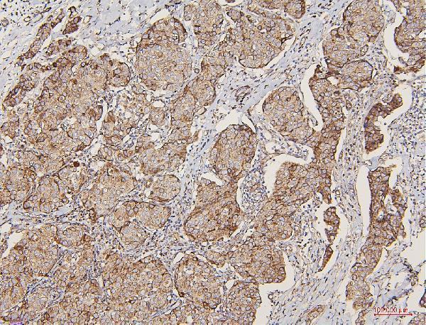
The catalog antibody has an IHC-P protocol (datasheet: A01240-2); a published LPP protocol adds a tissue microarray workflow for Paget disease (PMC7748372).
| Sample | Paraffin-embedded human lung cancer tissue; fixative not specified (datasheet A01240-2) |
| Fixation | Image fixative and duration unreported (datasheet A01240-2); verify before use. |
| Sectioning | 4–5 µm sections on charged slides (standard) |
| Deparaffinisation | Xylene, graded ethanol series to water (standard) |
| Antigen retrieval | Heat retrieval: EDTA pH 8.0 (datasheet A01240-2); 20 min, 95–100 °C (standard) |
| Peroxidase block | 3% H2O2, 10 min, room temperature (standard) |
| Blocking | 10% goat serum (datasheet A01240-2) |
| Primary antibody | Rabbit anti-LPP, 0.5-1μg/ml (datasheet A01240-2) |
| Primary incubation | Overnight at 4 °C (datasheet A01240-2) |
| Detection | Streptavidin-biotin complex (SABC), DAB chromogen (datasheet A01240-2) |
| Counterstain | Hematoxylin, blue, dehydrate and mount (standard) |
| Expected result | LPP-positive staining in cells in glomeruli of kidney (HPA tissue IHC: High). HPA tissue profile: Cytoplasmic expression in several cell types, most abundant in smooth muscle cells. No signal in the no-primary control. |
LPP is found in the cytoplasm, nucleus and cell adhesion sites; it has no transmembrane segment (UniProt Q93052). In tissue IHC, expect predominantly cytoplasmic staining, most abundant in smooth muscle cells, with high staining also reported in kidney glomerular cells and the skin granular layer (HPA tissue IHC). HPA rates the tissue profile Enhanced, while reporting medium consistency between antibody staining and RNA expression (HPA tissue IHC).
| Smooth muscle cells show strong cytoplasmic staining; cells in kidney glomeruli or the skin granular layer also stain strongly (HPA tissue IHC). | This matches the reported tissue distribution (HPA tissue IHC). LPP can occupy cell adhesion sites, but fine focal adhesion staining may be difficult to resolve in chromogenic paraffin sections (UniProt Q93052; general IHC practice). |
| A normal tissue section shows predominantly nuclear staining with little cytoplasmic signal. | Review the pattern before calling it LPP: HPA describes tissue staining as cytoplasmic (HPA tissue IHC). UniProt also places LPP in nuclei and notes that certain fusion proteins are mainly nuclear, so nuclear signal alone is not proof of an artefact (UniProt Q93052). |
| Adipocytes or bone marrow hematopoietic cells stain strongly, while expected positive cells do not. | HPA reports LPP as undetected in those cell populations (HPA tissue IHC). Consider antibody cross-reactivity or endogenous detection activity; compare an appropriate positive section and detection controls before assigning the signal to LPP (general IHC practice). |
| Color is diffuse across tissue and lacks a recognizable cell-specific pattern. | That appearance does not match the reported cytoplasmic, cell-dependent profile (HPA tissue IHC). Check background with a primary-antibody omission control, then review blocking, antibody concentration and washing (general IHC practice). |
| Smooth muscle cells and other expected positive cells show no visible signal. | This conflicts with the high smooth muscle staining reported by HPA (HPA tissue IHC). First establish that the section and detection run worked; then assess retrieval and antibody concentration as general IHC variables, without assuming a known LPP-specific fixation effect (general IHC practice). |
| Tissue and cell selection (HPA tissue IHC) | Smooth muscle cells are the clearest reported high-staining population; glomerular cells and the skin granular layer are additional high-staining examples, while adipocytes and bone marrow hematopoietic cells are reported as undetected (HPA tissue IHC). |
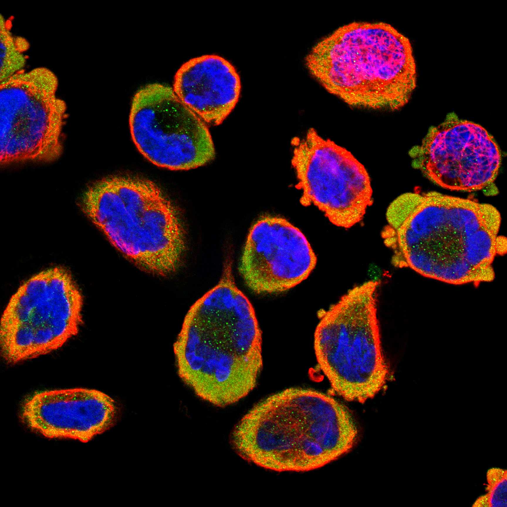
| Compartment and resolution (UniProt Q93052; HPA subcellular ICC-IF) | LPP occurs at adhesion sites and in the cytoplasm, with nuclear localization also documented (UniProt Q93052). HPA ICC-IF supports cytosol and focal adhesion sites as main locations; those fine structures may be less distinct by chromogenic IHC (HPA subcellular ICC-IF; general IHC practice). |
| Evidence strength (HPA tissue IHC; HPA antibodies) | HPA calls the tissue profile Enhanced but describes medium consistency with RNA expression; HPA011133 and HPA017342 each have Enhanced IHC validation (HPA tissue IHC; HPA antibodies). Interpret unexpected staining with controls rather than treating validation as proof for every cell type (general IHC practice). |
| Topology and processing (UniProt Q93052) | LPP has no transmembrane segment or signal peptide, and UniProt lists a single chain spanning residues 1–612 (UniProt Q93052). Membrane-adjacent staining can reflect adhesion-site localization; these features provide no basis to predict extracellular staining or a fixation-specific effect (UniProt Q93052). |
| Antigen retrieval and epitope (general IHC practice) | Target-specific fixation effects are not established by the supplied assay evidence. Verify with a matched IHC source before attributing a result to fixation. |
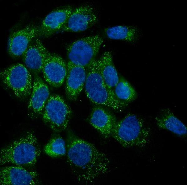
| IF/ICC Q&A (HPA subcellular ICC-IF) | What pattern should IF/ICC show? Mainly cytosol and focal adhesion sites, with additional plasma membrane and cell junction staining (HPA subcellular ICC-IF). These ICC-IF observations help interpret localization; they do not supply an IHC-P protocol (HPA subcellular ICC-IF). |
| Situation | Likely cause | Next action |
|---|---|---|
| No staining in smooth muscle cells (HPA tissue IHC) | The expected high-staining population is negative; the cause cannot be assigned from the image alone (HPA tissue IHC; general IHC practice). | Check a concurrently stained positive section and detection controls, then review the validated IHC conditions and assess retrieval or antibody concentration if needed (general IHC practice). |
| Signal appears only in nuclei of otherwise normal tissue. | HPA describes the tissue profile as cytoplasmic, although UniProt also records nuclear LPP and mainly nuclear fusion forms (HPA tissue IHC; UniProt Q93052). | Compare cytoplasmic staining in known positive cells and inspect primary-antibody omission controls before interpreting the nuclear signal (HPA tissue IHC; general IHC practice). |
| Adipocytes or bone marrow hematopoietic cells are strongly colored (HPA tissue IHC). | HPA reports these cell populations as undetected; cross-reactivity or endogenous detection activity is possible (HPA tissue IHC; general IHC practice). | Run primary-antibody omission and appropriate detection controls, then compare the suspect cells with smooth muscle cells in the same run (HPA tissue IHC; general IHC practice). |
| Diffuse color obscures cell boundaries. | Broad background can arise from nonspecific antibody binding, detection activity or inadequate washing (general IHC practice). | Check an omission control, review blocking and washes, and adjust antibody concentration according to the validated IHC workflow (general IHC practice). |
| Membrane-associated color appears without clear focal adhesion puncta. | LPP can occupy adhesion sites and cell junctions, while chromogenic section imaging may not resolve fine focal adhesions (UniProt Q93052; general IHC practice). | Judge the result against the expected cell types and cytoplasmic staining; use the separate IF/ICC localization evidence if fine spatial detail is needed (HPA tissue IHC; HPA subcellular ICC-IF). |
| Different tissue regions give conflicting positive and negative calls. | LPP staining varies by cell population, and HPA reports only medium consistency between antibody staining and RNA expression (HPA tissue IHC). | Score named cell populations separately and compare each with HPA's reported level; repeat ambiguous calls with suitable positive and detection controls (HPA tissue IHC; general IHC practice). |



Comprehensive Human Protein Atlas IHC scoring per tissue (reliability: Enhanced — Medium consistency between antibody staining and RNA expression data.). Rows are taken directly from the HPA tissue chart — click any row's HPA link to view the source.
| Tissue | Cell type | Level | Evidence | Source |
|---|---|---|---|---|
| Kidney | Cells in glomeruli | High | Protein (IHC) | HPA → |
| Skin | Cells in granular layer | High | Protein (IHC) | HPA → |
| Smooth muscle | Smooth muscle cells | High | Protein (IHC) | HPA → |
| Appendix | Glandular cells | Medium | Protein (IHC) | HPA → |
| Breast | Glandular cells | Medium | Protein (IHC) | HPA → |
| Tissue | Cell type | Level | Evidence | Source |
|---|---|---|---|---|
| Adipose tissue | Adipocytes | Not detected | Protein (IHC) | HPA → |
| Adrenal gland | Glandular cells | Not detected | Protein (IHC) | HPA → |
| Bone marrow | Hematopoietic cells | Not detected | Protein (IHC) | HPA → |
| Endometrium | Cells in endometrial stroma | Not detected | Protein (IHC) | HPA → |
| Epididymis | Glandular cells | Not detected | Protein (IHC) | HPA → |
Troubleshoot LPP staining in paraffin sections by checking retrieval, compartment, tissue context, and controls before changing how signal is scored.
Anti-LPP antibodies have IHC images from human cancer, mouse cardiac muscle, and rat intestine paraffin sections, plus IF images from A431 cells and human intestinal cancer sections (catalog IHC/IF captions).
A01240-2 shows IHC in human lung, mammary, and rectal cancers and mouse cardiac muscle, plus IF in A431 cells (A01240-2 IHC/IF captions). A01240-3 shows IHC in human mammary and rectal cancers and rat intestine, plus IF in human intestinal cancer sections (A01240-3 IHC/IF captions).
Which to pick: For tissue IHC, choose A01240-2 for its human and mouse paraffin-section examples at 0.5–1 μg/ml; A01240-3 provides human and rat paraffin-section examples at 2 μg/ml in its images (catalog dilutions and IHC captions). For IF/ICC, A01240-2 lists both applications and has an A431-cell IF image, while A01240-3 lists IF and has an image from human intestinal cancer sections (catalog applications and IF captions). For broader listed species coverage, A01240-3 includes monkey reactivity, though its IHC images show human and rat samples; neither entry reports a clone designation, and the paraffin-section captions do not report the fixative (catalog reactivity, clone fields, and IHC captions).