This website uses cookies to ensure you get the best experience on our website.


- Table of Contents
Plan LRBA staining in paraffin sections using the IHC-validated antibody A04947 (datasheet A04947). Interpret cytoplasmic staining against the variable tissue profile reported by HPA (HPA tissue IHC).

Expected localisation, validated protocols, controls and antibodies — the at-a-glance facts below, then the full design guide.
| Expected localisation | Cytoplasmic staining across tissues (HPA tissue IHC) | |
| Staining pattern | Variable cytoplasmic staining across tissue cell types (HPA tissue IHC) | |
| Antigen retrieval | Tris-EDTA pH 9.0 HIER, heat-mediated (datasheet A04947) | |
| Positive control | Appendix+4 more · see all | |
| Negative control | None in HPA (detected in all 45 tissues); use no-primary + isotype controls |
| Fixation | Keep fixation conditions consistent across sections (standard IHC practice; not target-specific) | |
| Caveat | Staining and RNA show medium consistency (HPA tissue IHC) | |
| Regulation | Expression varies across tissues (HPA tissue IHC) | |
| Isoform / epitope | Two isoforms; check epitope coverage when comparing staining (UniProt) |
The catalog antibody protocol uses Tris-EDTA pH 9.0 retrieval (datasheet A04947). Two published LRBA IHC protocols provide additional tissue-specific examples (PMC12815959; PMC12266018).
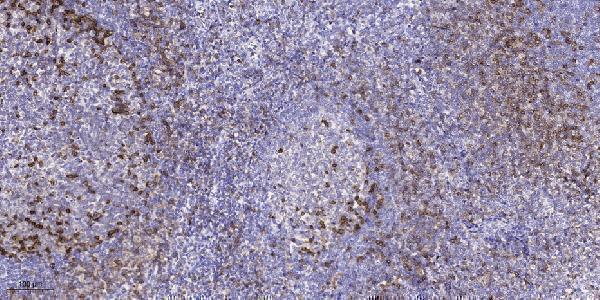
| Sample | Paraffin-embedded human tonsil tissue; fixative not specified (datasheet A04947) |
| Fixation | Image fixative and duration unreported (datasheet A04947); verify before use. |
| Sectioning | 4–5 µm sections on charged slides (standard) |
| Deparaffinisation | Xylene, graded ethanol series to water (standard) |
| Antigen retrieval | Heat retrieval: Tris-EDTA pH 9.0 (datasheet A04947); 20 min, 95–100 °C (standard) |
| Peroxidase block | 3% H2O2, 10 min, room temperature (standard) |
| Blocking | 10% normal serum of the secondary host, 30 min, room temperature (standard) |
| Primary antibody | Rabbit anti-LRBA, 1:50 recommended; image 1:200 (datasheet A04947) |
| Primary incubation | Overnight at 4 °C (standard) |
| Detection | HRP-polymer secondary, DAB chromogen 5–10 min (standard) |
| Counterstain | Hematoxylin, blue, dehydrate and mount (standard) |
| Expected result | LRBA-positive staining in glandular cells of appendix (HPA tissue IHC: High). HPA tissue profile: Cytoplasmic expression in all tissues at variable levels. No signal in the no-primary control. |
LRBA should appear as variable cytoplasmic staining across tissues, with strong staining reported in several epithelial cell populations (HPA tissue IHC). Its membrane associations include the endoplasmic reticulum, trans-Golgi network, lysosome and cell membrane; UniProt annotates one transmembrane segment (UniProt P50851 topology). HPA rates the tissue IHC profile Approved, with medium agreement between staining and RNA data and external verification pending (HPA tissue IHC).
| Strong cytoplasmic staining in bronchial respiratory epithelium or colon glandular cells (HPA tissue IHC). | This matches two reported High cell populations (HPA tissue IHC). Judge the stained cell population and cytoplasmic compartment together; HPA reports variable expression across tissues (HPA tissue IHC). A positive field alone does not establish antibody specificity (standard IHC practice). |
| Predominantly nuclear staining, with little cytoplasmic signal, in a candidate positive section. | A nuclear-dominant pattern conflicts with HPA's cytoplasmic tissue profile and UniProt's membrane locations (HPA tissue IHC; UniProt P50851). Treat it as suspect; review controls and staining conditions before scoring the nuclei as LRBA positive (standard IHC practice). |
| The strongest signal is in cells outside the expected stained population for the selected tissue. | For example, HPA reports High staining in colon glandular cells (HPA tissue IHC). Unexpected staining elsewhere may reflect cross-reactivity or endogenous detection activity (standard IHC practice). Compare cell identity, compartment and negative controls before assigning it to LRBA. |
| A broad brown haze covers tissue and background, obscuring cell boundaries. | That distribution cannot be read as the variable cellular cytoplasmic pattern reported for LRBA (HPA tissue IHC). Background can arise from nonspecific binding or chromogenic detection activity (standard IHC practice); assess the matched negative control before changing the interpretation. |
| No cytoplasmic staining appears in bronchial respiratory epithelial cells or colon glandular cells. | Both are reported High by HPA, so an absent signal warrants a technical check (HPA tissue IHC). HPA's Approved rating has medium staining–RNA consistency and pending external verification; one negative section does not by itself establish that LRBA is absent (HPA tissue IHC). |
| Tissue and cell population | HPA reports High staining in appendix, colon and duodenum glandular cells and bronchial respiratory epithelium, but Low staining in glia and fibroblasts (HPA tissue IHC). Select and interpret controls by cell population, since whole-section intensity can obscure that distinction (standard IHC practice). |
| Compartment and image resolution | HPA tissue IHC describes cytoplasmic staining; HPA ICC-IF supports a mainly Golgi location, with additional cytosol localization marked uncertain (HPA tissue IHC; HPA subcellular). A chromogenic section need not resolve individual organelle membranes to fit that profile (standard IHC practice). |
| Topology and epitope location | UniProt annotates one transmembrane segment at residues 1531–1548 and membrane locations including the trans-Golgi network and lysosome (UniProt P50851 topology). The supplied record gives no antibody epitope, so topology cannot predict whether a particular retrieval condition exposes it. |
| Isoforms and processing | UniProt lists 2 isoforms and a chain spanning residues 2–2863, with no signal peptide or propeptide annotation (UniProt P50851). Without an antibody epitope map, these annotations cannot establish which isoforms the IHC-validated antibody detects. |
| Antibody validation scope | HPA lists HPA019366 and HPA023597 as IHC Approved; HPA019366 is also ICC Supported (HPA antibodies). The tissue profile has medium staining–RNA consistency and awaits external verification (HPA tissue IHC). These ratings support a working expectation, not an absolute pass rule for every specimen. |
| Situation | Likely cause | Next action |
|---|---|---|
| A reported High cell population has no IHC signal (HPA tissue IHC). | The section, detection run or antibody conditions may have failed (standard IHC practice); HPA's High designation does not guarantee every specimen will stain (HPA tissue IHC). | Run a known-positive section in the same batch; confirm the antibody's IHC-P instructions, retrieval and detection steps, then inspect the intended cells before calling the sample negative (standard IHC practice). |
| Signal is nuclear-dominant despite an otherwise clean section. | Nuclear dominance does not match the reported cytoplasmic tissue pattern or annotated membrane locations (HPA tissue IHC; UniProt P50851). | Check the negative control, antibody concentration and staining distribution; require convincing cytoplasmic signal in a reported positive cell population before interpreting the nuclear signal as LRBA (standard IHC practice; HPA tissue IHC). |
| Brown staining appears in unexpected cells or persists in a negative control. | Cross-reactivity, endogenous enzyme activity or nonspecific detection may contribute (standard IHC practice); the location conflicts with the selected tissue's reported cell pattern (HPA tissue IHC). | Compare a matched no-primary control and the known-positive cell population. Check the detection system's endogenous-activity blocking and background controls before scoring unexpected cells (standard IHC practice). |
| Diffuse background makes cytoplasmic boundaries hard to judge. | Nonspecific binding or excessive detection signal can obscure cellular staining (standard IHC practice). The resulting haze cannot establish the HPA cytoplasmic pattern (HPA tissue IHC). | Review the negative control and the catalog antibody's IHC-P working conditions; adjust blocking, antibody dilution or detection time as appropriate to the validated workflow, then reassess cell-specific signal (standard IHC practice). |
| A Low-reference cell population stains faintly, or a High-reference population varies between sections. | HPA reports variable cytoplasmic levels across tissues, including Low staining in glia and fibroblasts and High staining in several epithelia (HPA tissue IHC). Its tissue profile has medium staining–RNA consistency (HPA tissue IHC). | Score the named cell population and compartment rather than whole-section color; compare sections processed together and include a reported High population as a reference (standard IHC practice; HPA tissue IHC). |
| IF/ICC: what localization should an independently designed experiment seek? | HPA supports a mainly Golgi location and marks additional cytosol localization uncertain; HPA019366 has ICC Supported status (HPA subcellular; HPA antibodies). | Use Golgi-enriched signal as the principal localization expectation, with the cytosolic component interpreted cautiously (HPA subcellular). Consult the separate IF/ICC guide for experiment design. |


Comprehensive Human Protein Atlas IHC scoring per tissue (reliability: Approved — Medium consistency between antibody staining and RNA expression data. Pending external verification.). Rows are taken directly from the HPA tissue chart — click any row's HPA link to view the source.
| Tissue | Cell type | Level | Evidence | Source |
|---|---|---|---|---|
| Appendix | Glandular cells | High | Protein (IHC) | HPA → |
| Bronchus | Respiratory epithelial cells | High | Protein (IHC) | HPA → |
| Colon | Glandular cells | High | Protein (IHC) | HPA → |
| Duodenum | Glandular cells | High | Protein (IHC) | HPA → |
| Endometrium | Glandular cells | High | Protein (IHC) | HPA → |
| Tissue | Cell type | Level | Evidence | Source |
|---|---|---|---|---|
| None in HPA: LRBA is detected in all 45 scored tissues. Use a no-primary (secondary-only) and an isotype control instead. | ||||
Troubleshoot LRBA chromogenic IHC in paraffin sections using the A04947 tonsil caption, the tissue staining profile, and known protein localisation.
A04947 has a real IHC image from paraffin-embedded human tonsil (A04947 image caption). IF is listed for human and mouse, but no IF image is supplied (A04947 catalog: applications, reactivity, IF image alts).
A04947 will render with its paraffin-embedded human tonsil IHC image (A04947 image caption). The catalog lists IHC and IF applications and human and mouse reactivity; it supplies no IF image (A04947 catalog: applications, reactivity, IF image alts).
Which to pick: For tissue IHC, choose A04947: its own image caption documents staining in paraffin-embedded human tonsil, using 1:200 primary antibody overnight at 4°C and Tris-EDTA retrieval at pH 9.0 (A04947 image caption). For IF, A04947 is the listed option; ICC is not listed, and no IF image is supplied (A04947 catalog: applications, IF image alts). A04947 lists human and mouse reactivity, but the pictured tissue is human; its host is rabbit, clonality is unreported, and the tissue fixative is unreported (A04947 catalog: host, clone, reactivity; A04947 image caption).