LRFN2 / Leucine-rich repeat and fibronectin type-III domain-containing protein 2 · IHC design guide

Design Immunohistochemistry for LRFN2

Plan LRFN2 IHC-P staining with cerebellar Purkinje cells and cerebral cortical neurons as reference positives (HPA tissue IHC). Assess the observed cytoplasmic signal in a subset of neurons alongside the expected postsynaptic membrane location (HPA tissue IHC; UniProt).

Evidence assembled Oct 2026 · For research use; verify linked source records and product datasheet before use
Immunohistochemistry protocol sheet for LRFN2 (IHC for LRFN2): expected localisation Observed neuronal cytoplasm (HPA tissue IHC); expected postsynaptic membrane (UniProt), antibody A11623, validated IHC image, and IHC protocol steps
Printable LRFN2 IHC protocol sheet — expected localisation Observed neuronal cytoplasm (HPA tissue IHC); expected postsynaptic membrane (UniProt), antibody A11623, controls and protocol steps. Open the full LRFN2 IHC guide →

LRFN2 Immunohistochemistry Experimental Design Guide

Expected localisation, validated protocols, controls and antibodies — the at-a-glance facts below, then the full design guide.

Must know before staining
Expected localisation Observed neuronal cytoplasm (HPA tissue IHC); expected postsynaptic membrane (UniProt)
Staining pattern A subset of neurons shows cytoplasmic staining (HPA tissue IHC)
Antigen retrieval Citrate pH 6.0 HIER, 95–98 °C, 20 min (rule: cytoplasmic / membrane antigen)
Positive control ⓘ Cerebellum+1 more · see all
Negative control ⓘ Adipose tissue+4 more · see all
Important caveats
Reasons your staining may differ from the expected pattern.
Fixation Keep fixation conditions consistent across sections. (standard IHC practice; not target-specific)
Caveat Only some neurons stain; hippocampal signal is low (HPA tissue IHC)
Regulation Expression regulation is not annotated (UniProt)
Isoform / epitope No isoforms annotated; epitope side of the membrane matters (UniProt)
Section 1

Recommended LRFN2 IHC & IF Protocols

Compare the catalog antibody’s IHC-P protocol with the published patient and mouse tumor staining workflow (PMC9530863).

Recommended immunohistochemistry (IHC-P) protocol parameters
SampleTissue sections; selected-image fixative not specified (standard IHC workflow)
FixationImage fixative and duration unreported (datasheet A11623); verify before use.
Sectioning4–5 µm sections on charged slides (standard)
DeparaffinisationXylene, graded ethanol series to water (standard)
Antigen retrievalHeat-induced epitope retrieval in citrate buffer, pH 6.0, 20 min at 95–98 °C (standard rule: cytoplasmic / membrane antigen)
Peroxidase block3% H2O2, 10 min, room temperature (standard)
Blocking10% normal serum of the secondary host, 30 min, room temperature (standard)
Primary antibodyRabbit anti-LRFN2, 2.5 μg/mL (datasheet A11623)
Primary incubationOvernight at 4 °C (standard)
DetectionHRP-polymer secondary, DAB chromogen 5–10 min (standard)
CounterstainHematoxylin, blue, dehydrate and mount (standard)
Expected resultLRFN2-positive staining in purkinje cells - cytoplasm/membrane of cerebellum (HPA tissue IHC: Medium). HPA tissue profile: Cytoplasmic expression in a subset of neuronal cells. No signal in the no-primary control.
💡Decision noteStart with citrate pH 6.0 retrieval at 95–98 °C for 20 min (page retrieval rule; UniProt Q9ULH4: membrane localisation).
Section 2

What Is the Expected LRFN2 Staining Pattern?

LRFN2 is a synaptic, postsynaptic membrane protein with one transmembrane segment and an extracellular region (UniProt Q9ULH4 topology). In tissue IHC, expect staining in a subset of neuronal cells, with cytoplasmic signal and some membrane staining in Purkinje cells (HPA tissue IHC). HPA rates its tissue pattern Enhanced because antibody staining is highly consistent with RNA expression data (HPA tissue IHC).

What am I looking at on my slide?
Medium staining in Purkinje cell cytoplasm and membranes, with neuronal staining in cerebral cortex (HPA tissue IHC).This fits the reported tissue pattern. HPA records medium staining for both sites and describes cytoplasmic expression in a subset of neurons (HPA tissue IHC). Membrane staining is also consistent with LRFN2 topology and postsynaptic localization (UniProt Q9ULH4 topology). Score the stained cells and compartments separately; the expected result does not require every neuron to stain (HPA tissue IHC).
Strong nuclear staining dominates, while the expected neuronal cytoplasmic or membrane pattern is absent.A nuclear dominant result conflicts with the reported tissue IHC pattern and membrane localization (HPA tissue IHC; UniProt Q9ULH4 topology). Treat it as a possible staining artifact until controls and tissue morphology have been reviewed. A visible chromogenic deposit alone does not establish that the antibody has detected LRFN2 (general IHC practice).
Staining concentrates in cells outside the expected neuronal population, including cells reported as not detected by HPA (HPA tissue IHC).Consider cross reactivity or endogenous detection activity, particularly if the signal persists in a no primary control (general IHC practice). HPA reports no detection in adipocytes of adipose tissue and glandular cells of adrenal gland or appendix (HPA tissue IHC). Those observations provide comparisons, not a guarantee that every cell in those tissues is negative.
Weak, diffuse color covers cells and surrounding tissue without a discernible neuronal pattern.This is difficult to interpret as LRFN2 because HPA describes staining in a subset of neuronal cells (HPA tissue IHC). Compare background with a no primary control and inspect whether color follows tissue edges, debris or damaged areas (general IHC practice). Reassess blocking, washing and detection conditions before assigning a positive score (general IHC practice).
No signal appears in Purkinje cells or cortical neurons in an otherwise readable section.This conflicts with the medium staining reported for those cells (HPA tissue IHC). Check that the relevant cells are present, then review primary antibody application, retrieval, detection and counterstain balance (general IHC practice). HPA reports a tissue pattern, not a guarantee that every section or processing run will produce detectable signal (HPA tissue IHC; general IHC practice).
💡Expected LRFN2 appearanceCall positive when a subset of neurons shows cytoplasmic staining, including medium Purkinje cell cytoplasm or membrane staining and medium cortical neuronal staining; diffuse tissue wide or nuclear dominant color is suspect (HPA tissue IHC; UniProt Q9ULH4 topology).
How each factor affects the staining
Compartment and topologyLRFN2 has an extracellular region at residues 21–534, one transmembrane segment at 535–555, and a cytoplasmic region at 556–789 (UniProt Q9ULH4 topology). These features support membrane localization, but they do not identify the catalog antibody's epitope or predict how its staining will change with retrieval (UniProt Q9ULH4 topology).
Tissue and cell selectionHPA reports medium staining in cerebellar Purkinje cells and cerebral cortical neurons; hippocampal and caudate neurons are listed as low (HPA tissue IHC). Choose a section containing a reported medium staining population when judging whether an IHC run worked. A low staining population is less useful for diagnosing an absent signal (HPA tissue IHC; general IHC practice).
Antibody evidenceThe HPA tissue pattern has Enhanced reliability based on consistency between staining and RNA expression; antibody HPA076660 is also rated Enhanced for IHC (HPA tissue IHC; HPA antibodies). This supports the reported tissue pattern, while interpretation still depends on the cells and compartments visible in the actual section (HPA tissue IHC; general IHC practice).
IF/ICC Q: Is the same antibody validated for IF/ICC?A: HPA reports supported plasma membrane localization by ICC-IF, but rates HPA076660 Uncertain for ICC (HPA subcellular; HPA antibodies). The supported localization informs what to inspect in IF images; it does not establish reliable ICC performance for that antibody (HPA subcellular; HPA antibodies).
Why is my staining missing, weak or wrong?
SituationLikely causeNext action
Purkinje cells are unstained.The expected population may be missing from the section, or an IHC step may have failed (HPA tissue IHC; general IHC practice).Confirm Purkinje cells by morphology, then review primary antibody application, retrieval and chromogenic detection records; compare a reported positive tissue processed in the same run (HPA tissue IHC; general IHC practice).
Only faint staining appears in hippocampal or caudate neurons.HPA lists these neuronal populations as low staining (HPA tissue IHC).Use reported medium Purkinje or cortical neuronal staining to judge the run before interpreting a low signal as technical failure (HPA tissue IHC; general IHC practice).
Nuclei are the clearest positive structures.Nuclear dominant staining disagrees with HPA's neuronal cytoplasmic pattern and UniProt's membrane localization (HPA tissue IHC; UniProt Q9ULH4 topology).Review morphology and no primary control staining; withhold an LRFN2 positive score unless the expected neuronal compartment pattern is also credible (general IHC practice; HPA tissue IHC).
Color appears in many unrelated cell types.Cross reactivity or endogenous detection activity may contribute, especially where HPA reports no detection (HPA tissue IHC; general IHC practice).Compare a no primary control and an HPA reported negative cell population in the same run; investigate persistent background before assigning cell specific positivity (HPA tissue IHC; general IHC practice).
Diffuse background obscures neuronal detail.Nonspecific binding, insufficient washing or detection background can reduce contrast (general IHC practice).Check the no primary control, blocking and wash steps, and detection exposure; score LRFN2 only where a distinct neuronal pattern remains visible (general IHC practice; HPA tissue IHC).
Membrane staining and cytoplasmic staining disagree between images.HPA tissue IHC describes neuronal cytoplasmic staining and Purkinje cytoplasm or membrane staining; ICC-IF supports plasma membrane localization (HPA tissue IHC; HPA subcellular).Record method, cell type and compartment for each image. Interpret paraffin section IHC against its reported tissue pattern; treat ICC observations separately because HPA076660 has Uncertain ICC validation (HPA tissue IHC; HPA antibodies).

Sample controls for LRFN2 IHC & IF

🧪Run cerebellum first and score Purkinje cells for cytoplasmic or membrane staining (HPA: Medium in cerebellar Purkinje cells). Run adipose tissue as the negative tissue and assess its adipocytes for background staining (HPA: Not detected in adipocytes); on the cerebellar slide, neighboring unstained cells provide an internal background comparison, though HPA does not establish them as LRFN2-negative.
Positive control tissue: Cerebellum (Purkinje cells - cytoplasm/membrane, HPA Medium)
Negative control tissue: Adipose tissue (HPA Not detected)
ICC-IF cell lines (HPA subcellular resource): HPA ICC-IF images show LRFN2 in REH, SH-SY5Y, with annotated localisation: Plasma membrane (supported) (HPA subcellular).
Technical controls: Include a no-primary (secondary-only) slide, an isotype control matched to the primary antibody’s host species, isotype and concentration, and a biological negative such as knockout tissue or peptide competition when the immunizing peptide is available. For brain sections, quench endogenous peroxidase for chromogenic IHC, account for endogenous biotin if using avidin-based detection, and assess autofluorescence for IF (standard IHC/IF practice).
⚠️Feasibility: A target-specific fixation window or fixation effect is unreported in the supplied evidence; the selected mouse-brain IHC caption also leaves the fixative unreported (A11623 caption). Antigen-retrieval dependence is unreported, so retrieval requires optimization for paraffin IHC; the supplied evidence does not establish frozen sections or IF as easier for LRFN2. Brain autofluorescence can complicate IF interpretation (standard IF practice).

HPA tissue IHC evidence for LRFN2

Comprehensive Human Protein Atlas IHC scoring per tissue (reliability: Enhanced — High consistency between antibody staining and RNA expression data.). Rows are taken directly from the HPA tissue chart — click any row's HPA link to view the source.

Positive expression · recommended positive controls

TissueCell typeLevelEvidenceSource
Cerebellum Purkinje cells - cytoplasm/membrane Medium Protein (IHC) HPA →
Cerebral cortex Neuronal cells Medium Protein (IHC) HPA →

Undetected expression · recommended negative controls

TissueCell typeLevelEvidenceSource
Adipose tissue Adipocytes Not detected Protein (IHC) HPA →
Adrenal gland Glandular cells Not detected Protein (IHC) HPA →
Appendix Glandular cells Not detected Protein (IHC) HPA →
Breast Adipocytes Not detected Protein (IHC) HPA →
Bronchus Respiratory epithelial cells Not detected Protein (IHC) HPA →
Section 3

Advanced LRFN2 IHC Tips

Troubleshoot LRFN2 staining in paraffin sections by checking retrieval, cellular localisation, controls, and scoring before interpreting signal strength.

How should I retrieve LRFN2 in paraffin sections when neuronal staining is weak?
Start with citrate buffer at pH 6.0 for heat induced epitope retrieval at 95–98 °C for 20 min (page retrieval rule for LRFN2). Keep section thickness, cooling time, and antibody incubation consistent while comparing retrieval conditions (standard IHC practice). The extracellular portion spans residues 21–534, while the cytoplasmic portion spans 556–789; retrieval may expose these regions differently, but the antibody’s epitope is not specified (UniProt Q9ULH4 topology; supplied antibody evidence). Compare neuronal staining with a no primary antibody control, because stronger diffuse signal alone does not establish improved specificity (standard IHC practice). Record whether any improvement preserves the expected neuronal and membrane associated pattern (HPA tissue IHC; UniProt Q9ULH4 subcellular annotation).
Could fixation explain inconsistent LRFN2 staining between paraffin sections?
The supplied LRFN2 tissue IHC caption does not state a fixative, so target specific fixation sensitivity is unknown for the catalog antibody (A11623 tissue IHC caption). Record the fixative, fixation duration, tissue thickness, and time before processing for each specimen; compare sections prepared under matched conditions (standard IHC practice). If signal varies, change one preparation variable at a time while keeping retrieval at citrate pH 6.0, 95–98 °C for 20 min (page retrieval rule for LRFN2; standard IHC practice). Include a consistently processed neuronal control section in each run to distinguish batch variation from specimen variation (HPA tissue IHC; standard IHC practice). Do not infer fixation tolerance from LRFN2 topology or observed tissue expression (UniProt Q9ULH4 topology; HPA tissue IHC).
Where should convincing LRFN2 chromogenic staining appear?
Assess staining in neuronal cells, with attention to membrane associated signal and the cytoplasmic staining documented in a neuronal subset (UniProt Q9ULH4 subcellular annotation; HPA tissue IHC). LRFN2 has one transmembrane segment at residues 535–555 and is annotated at the postsynaptic cell membrane (UniProt Q9ULH4 topology and subcellular annotation). HPA reports medium staining in cerebellar Purkinje cells at the cytoplasm or membrane and in cerebral cortical neuronal cells (HPA tissue IHC). Examine cellular outlines and neuronal processes at higher magnification before calling diffuse chromogen deposition a membrane pattern (standard IHC practice). Score cytoplasmic and membrane associated staining separately, since the tissue evidence includes both appearances (HPA tissue IHC; standard IHC practice).
How does epitope position affect interpretation of LRFN2 staining?
Confirm the antibody’s immunogen or mapped epitope before assigning staining to an extracellular or cytoplasmic LRFN2 region; neither position is supplied here (supplied antibody evidence; UniProt Q9ULH4 topology). The extracellular region spans residues 21–534, and the cytoplasmic region spans 556–789 (UniProt Q9ULH4 topology). UniProt lists glycosylation sites at residues 29, 332, 341, and 384, all within the annotated extracellular region (UniProt Q9ULH4 glycosylation and topology). Its supplied record lists 0 isoforms, so the evidence does not support an isoform specific staining explanation (UniProt Q9ULH4 isoforms). If staining changes after retrieval, investigate epitope accessibility experimentally rather than attributing the change to a particular glycosylation site (standard IHC practice).
How can I check LRFN2 by IF alongside chromogenic IHC?
Use IF as a separate assay and first establish a specific LRFN2 signal with a no primary antibody control (standard IF practice). Multiplex with a neuronal marker to check whether LRFN2 signal belongs to the expected cell population, while retaining separate single stain controls (HPA tissue IHC; standard IF practice). In brain sections, inspect autofluorescence in an unstained control and consider a far red fluorophore when shorter wavelengths give excessive background (A11623 tissue IHC caption; standard IF practice). Choose permeabilisation after confirming the epitope: access to the cytoplasmic residues 556–789 generally requires it, whereas an accessible extracellular epitope may be examined without it (UniProt Q9ULH4 topology; standard IF practice). The supplied mouse brain caption documents IHC at 2.5 µg/mL, not an IF dilution or fixation condition (A11623 tissue IHC caption).
What should I check when LRFN2 DAB staining looks widespread?
Run a no primary antibody section to assess detection reagent background, and inspect a negative tissue or cell population processed in the same run (standard IHC practice). Block endogenous peroxidase before chromogenic detection and check whether residual signal persists without primary antibody (standard IHC practice). HPA reports LRFN2 in a subset of neuronal cells and lists adipose tissue adipocytes as not detected, providing expression context rather than proof that every negative section must be blank (HPA tissue IHC). Review section edges, folds, and areas of tissue damage separately because local reagent pooling or damaged tissue can mimic strong staining (standard IHC practice). Titrate the catalog antibody around the documented mouse brain IHC concentration of 2.5 µg/mL only with matched controls; the caption supplies no other working concentration (A11623 tissue IHC caption; standard IHC practice).
How should I quantify heterogeneous LRFN2 staining in neuronal regions? ⚠ ANSWER MARKED FOR VERIFICATION
Define the neuronal region and count eligible cells before viewing treatment groups, then apply the same inclusion rules across sections (HPA tissue IHC; standard IHC practice). Report the percentage of positive neuronal cells and, if intensity is reproducibly graded, an H-score using 0–3 intensity categories (standard IHC practice). Normalise positive counts to the total number of eligible neuronal cells, or report positive cell density per mm² of analysed tissue when cell counts are impractical (standard IHC practice). Keep cytoplasmic and membrane associated scores separate because both appearances occur in the supplied tissue profile (HPA tissue IHC). Use matched staining batches, exposure settings for image capture, and a prespecified positivity threshold so technical variation does not become a biological difference (standard IHC practice).
When is an LRFN2 positive IHC result convincing?
A convincing result places staining in neuronal cells, with a pattern compatible with the membrane association and neuronal cytoplasmic signal described for LRFN2 (UniProt Q9ULH4 subcellular annotation; HPA tissue IHC). Compare cerebellar Purkinje cells or cerebral cortical neuronal cells with the same run’s controls; HPA reports medium staining in those populations (HPA tissue IHC; standard IHC practice). Treat isolated staining in an unexpected cell population or a predominantly unrelated compartment as a finding requiring independent validation, not automatic target identification (HPA tissue IHC; standard IHC practice). Check whether apparent positives cluster at cut edges, folds, or damaged areas, and whether they remain in the no primary antibody control (standard IHC practice). Residual endogenous peroxidase can produce chromogen signal, so interpret DAB deposits only after evaluating the peroxidase block and control section (standard IHC practice).
Boster reagents

Best LRFN2 / Leucine-rich repeat and fibronectin type-III domain-containing protein 2 IHC Antibodies

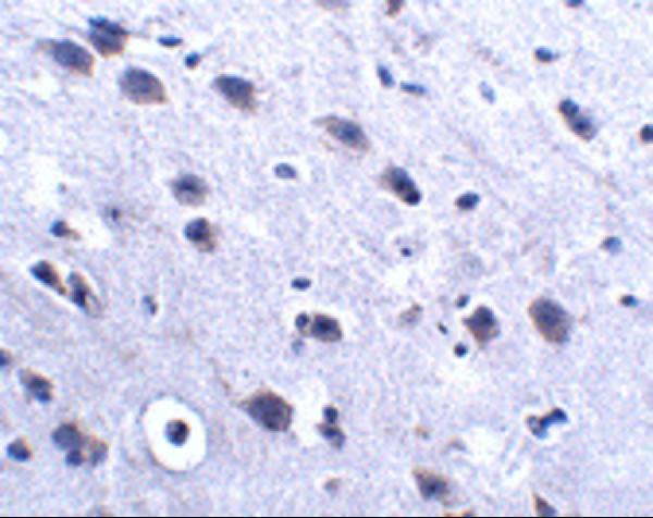
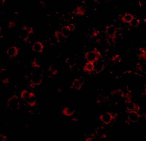
A11623 has IHC and IF images from mouse brain tissue (catalog: image captions) and listed reactivity with human, mouse and rat (catalog: reactivity).

Real IHC data Immunohistochemistry of LRFN2 in mouse brain tissue with LRFN2 antibody at 2.5 μg/mL.
Anti-LRFN2 Antibody
Cat # A11623

A11623 is listed for IHC-P, with an IHC image from mouse brain tissue at 2.5 μg/mL (catalog: applications; IHC image caption). Its IF image shows mouse brain tissue at 20 μg/mL, and its listed reactivity includes human, mouse and rat (catalog: IF image caption; reactivity).

Which to pick: For paraffin-section IHC, choose A11623 based on its IHC-P listing and mouse brain IHC image at 2.5 μg/mL; the fixative is unreported (catalog: applications; IHC image caption). For IF, A11623 has a mouse brain image at 20 μg/mL; separate ICC validation is unreported (catalog: IF image caption; applications). For cross-species planning, A11623 lists human, mouse and rat reactivity, although the supplied IHC and IF images show mouse brain only; clonality is unreported.

Each figure is that product's own IHC / IF validation image from its datasheet.

References

  1. UniProt Consortium. UniProt entry Q9ULH4 (LRFN2_HUMAN, Leucine-rich repeat and fibronectin type-III domain-containing protein 2).
  2. Human Protein Atlas. LRFN2 tissue IHC expression (reliability: Enhanced).
  3. Human Protein Atlas. LRFN2 subcellular location (ICC-IF): Localized to the plasma membrane..
  4. Human Protein Atlas. LRFN2 antibody validation summary (1 antibodies).
  5. Bladder cancer intrinsic LRFN2 drives anticancer immunotherapy resistance by attenuating CD8(+) T cell infiltration and functional transition. Journal for immunotherapy of cancer 2023 — PMC10565151.
  6. LRFN2 binding to NMDAR inhibits the progress of ESCC via regulating the Wnt/β-Catenin and NF-κB signaling pathway. Cancer science 2022 — PMC9530863.
  7. PubMed PMID:10574462 — UniProt-cited evidence.
  8. PubMed PMID:14702039 — UniProt-cited evidence.
  9. PubMed PMID:14574404 — UniProt-cited evidence.