This website uses cookies to ensure you get the best experience on our website.


- Table of Contents
Plan paraffin-section LRG1 IHC around distinct plasma positivity (HPA tissue IHC) and its secreted location (UniProt). Use the high Paneth-cell staining in duodenum as a reference (HPA tissue IHC) alongside the catalog antibody’s documented chromogenic workflow (datasheet A04365-3).

Expected localisation, validated protocols, controls and antibodies — the at-a-glance facts below, then the full design guide.
| Expected localisation | Secreted (UniProt); plasma positivity (HPA tissue IHC) | |
| Staining pattern | Distinct plasma positivity; Paneth cells high (HPA tissue IHC) | |
| Antigen retrieval | EDTA pH 8.0 HIER, heat-mediated (datasheet A04365-3) | |
| Positive control | Duodenum+3 more · see all | |
| Negative control | Adipose tissue+4 more · see all |
| Fixation | Keep fixation consistent across sections (standard IHC practice; not target-specific) | |
| Caveat | Secretion can separate tissue staining from local RNA expression (HPA tissue IHC) | |
| Regulation | Liver-enriched RNA (HPA tissue RNA) | |
| Isoform / epitope | No isoforms annotated; mature chain starts at residue 36 (UniProt) |
The catalog antibody protocol is paired with 4 published LRG1 IHC protocols from hepatobiliary, eye, mouse knee joint, and colon specimens (PMC3241550; PMC8396268; PMC5386532; PMC11436608).
| Sample | Paraffin-embedded human pancreas cancer tissue; fixative not specified (datasheet A04365-3) |
| Fixation | Image fixative and duration unreported (datasheet A04365-3); verify before use. |
| Sectioning | 4–5 µm sections on charged slides (standard) |
| Deparaffinisation | Xylene, graded ethanol series to water (standard) |
| Antigen retrieval | Heat retrieval: EDTA pH 8.0 (datasheet A04365-3); 20 min, 95–100 °C (standard) |
| Peroxidase block | 3% H2O2, 10 min, room temperature (standard) |
| Blocking | 10% goat serum (datasheet A04365-3) |
| Primary antibody | Rabbit anti-LRG1, 2-5 μg/ml recommended; image 1:100 (datasheet A04365-3) |
| Primary incubation | Overnight at 4 °C (datasheet A04365-3) |
| Detection | HRP-conjugated secondary, DAB chromogen (datasheet A04365-3) |
| Counterstain | Hematoxylin, blue, dehydrate and mount (standard) |
| Expected result | LRG1-positive staining in paneth cells of duodenum (HPA tissue IHC: High). HPA tissue profile: Distinct positivity in plasma. No signal in the no-primary control. |
LRG1 is secreted and has no transmembrane segment (UniProt P02750 topology). In paraffin-section IHC, expect distinct plasma positivity, high staining in duodenal Paneth cells, and medium staining in appendix enterocytes, colon goblet cells, and rectal endothelial cells (HPA tissue IHC: Enhanced). Because LRG1 is secreted, tissue protein location can differ from its site of RNA expression (HPA tissue IHC: reliability description).
| Distinct plasma staining, with high staining in duodenal Paneth cells (HPA tissue IHC). | This matches the strongest supplied IHC observations (HPA tissue IHC: Enhanced). Assess plasma and cell staining separately: secretion can make protein location differ from RNA location (HPA tissue IHC: reliability description; UniProt P02750: Secreted). |
| Medium staining in appendix enterocytes, colon goblet cells, or rectal endothelial cells (HPA tissue IHC). | These are reported positive cell populations (HPA tissue IHC: Medium). Compare the named cells within each tissue; the HPA levels describe observed staining and do not establish a universal intensity cutoff (HPA tissue IHC: tissue profile). |
| Strong nuclear staining dominates a section, without the reported plasma or cell pattern (HPA tissue IHC). | Treat this as a possible artefact requiring investigation: LRG1 is annotated as secreted with no transmembrane segment, while the supplied IHC profile emphasizes plasma positivity (UniProt P02750 topology; HPA tissue IHC). |
| Signal appears mainly in an unreported cell population, or in adipocytes listed as not detected (HPA tissue IHC). | Check cell identification, cross-reactivity, and endogenous detection activity (general IHC practice). An unreported cell population alone is not proof of a false positive; the HPA observations are limited to the sampled tissues and cells (HPA tissue IHC). |
| Uniform haze covers cells and empty areas, or duodenal Paneth cells show no signal (HPA tissue IHC: Paneth cells High). | Haze obscures cell-level interpretation and calls for background controls (general IHC practice). Absent signal in the reported high-staining cells calls for a run-control and detection check before interpreting other samples as negative (HPA tissue IHC; general IHC practice). |
| Secretion and tissue context (UniProt P02750; HPA tissue IHC). | LRG1 is secreted, and HPA reports distinct plasma positivity; interpret plasma signal alongside the named positive cells (UniProt P02750: Secreted; HPA tissue IHC). Liver-enriched RNA does not, by itself, predict where protein will stain (HPA tissue IHC: RNA specificity and reliability description). |
| Mature chain and glycosylation (UniProt P02750). | The annotated signal peptide spans residues 1–35; the mature chain spans 36–347 and has five annotated glycosylation sites (UniProt P02750). These annotations do not identify the catalog antibody's epitope or establish an antigen-retrieval condition (UniProt P02750 record scope). |
| IHC evidence and antibody choice (HPA antibodies; HPA tissue IHC). | HPA reports Enhanced IHC status for HPA001888 and HPA001889, and Enhanced reliability for the tissue profile (HPA antibodies; HPA tissue IHC). Use that as support for the reported pattern, while checking the IHC-validated antibody's own controls in the current run (general IHC practice). |
| IF/ICC Q: Where is LRG1 localized? (HPA subcellular ICC-IF). | A: HPA approves vesicular localization and lists Hep-G2 among cell lines with ICC-IF images (HPA subcellular ICC-IF). This is an IF/ICC observation; interpret paraffin-section chromogenic IHC using the tissue profile above (HPA tissue IHC; HPA subcellular ICC-IF). |
| Situation | Likely cause | Next action |
|---|---|---|
| No staining in duodenal Paneth cells (HPA tissue IHC: High). | The positive-control run may have failed, or the detection conditions may be unsuitable (general IHC practice). | Check the positive-control section, antibody dilution, retrieval, detection reagents, and counterstain in the IHC workflow (general IHC practice). No LRG1-specific retrieval condition is established by the supplied sources (HPA tissue IHC; UniProt P02750). |
| Plasma stains but the expected cells are hard to judge (HPA tissue IHC). | Prominent plasma signal can complicate cell-level scoring for a secreted protein (HPA tissue IHC: distinct plasma positivity; UniProt P02750: Secreted). | Score plasma and named cell populations separately, using tissue architecture and matched controls to locate cells (HPA tissue IHC; general IHC practice). |
| Diffuse brown haze obscures tissue boundaries (general IHC practice). | Background can arise from insufficient blocking or washing, or from excess antibody or detection reagent (general IHC practice). | Review blocking, washes, dilution, and a control omitting primary antibody; retain conditions that preserve the expected cell pattern (general IHC practice; HPA tissue IHC). |
| Staining appears in a listed negative cell population (HPA tissue IHC). | Cross-reactivity or endogenous detection activity is possible; a single discrepant section cannot establish either cause (general IHC practice). | Check a control omitting primary antibody and review antibody specificity; compare with the exact HPA cell designation, such as adipocytes in adipose tissue (general IHC practice; HPA tissue IHC: Not detected). |
| Nuclear color dominates the slide (general IHC practice). | The dominant pattern conflicts with secreted LRG1 and the reported plasma-centered IHC profile (UniProt P02750 topology; HPA tissue IHC). | Review morphology, counterstain, detection controls, and antibody specificity before scoring the nuclear color as LRG1 (general IHC practice). |
| IF/ICC vesicles seem inconsistent with IHC plasma staining (HPA subcellular ICC-IF; HPA tissue IHC). | The observations come from different preparations: HPA reports vesicles by ICC-IF and distinct plasma positivity by tissue IHC (HPA subcellular ICC-IF; HPA tissue IHC). | Use the tissue IHC observations to judge paraffin sections; consult the separate IF/ICC guide for fluorescence interpretation (HPA tissue IHC; HPA subcellular ICC-IF). |
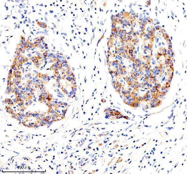
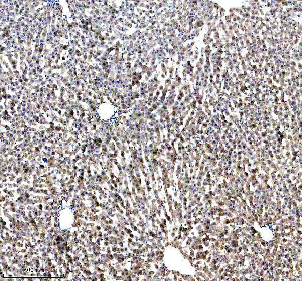
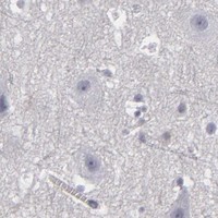


Comprehensive Human Protein Atlas IHC scoring per tissue (reliability: Enhanced — Secreted protein, tissue location of RNA and protein is expected to differ.). Rows are taken directly from the HPA tissue chart — click any row's HPA link to view the source.
| Tissue | Cell type | Level | Evidence | Source |
|---|---|---|---|---|
| Duodenum | Paneth cells | High | Protein (IHC) | HPA → |
| Appendix | Enterocytes | Medium | Protein (IHC) | HPA → |
| Colon | Goblet cells | Medium | Protein (IHC) | HPA → |
| Rectum | Endothelial cells | Medium | Protein (IHC) | HPA → |
| Tissue | Cell type | Level | Evidence | Source |
|---|---|---|---|---|
| Adipose tissue | Adipocytes | Not detected | Protein (IHC) | HPA → |
| Adrenal gland | Glandular cells | Not detected | Protein (IHC) | HPA → |
| Bone marrow | Hematopoietic cells | Not detected | Protein (IHC) | HPA → |
| Breast | Adipocytes | Not detected | Protein (IHC) | HPA → |
| Bronchus | Respiratory epithelial cells | Not detected | Protein (IHC) | HPA → |
Troubleshoot LRG1 staining by checking retrieval and controls, then score tissue compartments separately because LRG1 is secreted (UniProt P02750).
The catalog antibody A04365-3 has IHC images from human pancreas cancer, human liver cancer, human liver, and rat liver paraffin sections (catalog IHC captions); no IF image is supplied (catalog IF images).
A04365-3 has IHC images from human pancreas cancer, human liver cancer, human liver, and rat liver paraffin sections (catalog IHC captions). M04365-1 lists human IHC reactivity but has no IHC image in the payload (catalog applications, reactivity, and IHC images).
Which to pick: Choose A04365-3 for paraffin-section tissue IHC because its own captions document those human and rat sections; the fixative is unreported (A04365-3 IHC captions). For IF/ICC, neither SKU has a listed IF application or IF image, so neither has documented IF/ICC validation here (catalog applications and IF images). For cross-species work, A04365-3 is the rabbit polyclonal option with listed human, mouse, and rat reactivity, although its IHC images document human and rat tissue only; M04365-1 is a rabbit monoclonal listed for human reactivity (catalog dilution_raw, reactivity, and IHC captions; M04365-1 catalog host and clone).